. 2016 Oct 22;13(1):179.
doi: 10.1186/s12985-016-0634-z.

Human cytomegalovirus reactivation from latency: validation of a "switch" model in vitro

Affiliations

Human cytomegalovirus reactivation from latency: validation of a "switch" model in vitro

Maria-Cristina Arcangeletti et al. Virol J. .

Abstract

Background: Human cytomegalovirus (HCMV) is an opportunistic pathogen leading to severe and even fatal diseases in 'at-risk' categories of individuals upon primary infection or the symptomatic reactivation of the endogenous virus. The mechanisms which make the virus able to reactivate from latency are still matter of intense study. However, the very low number of peripheral blood monocytes (an important latent virus reservoir) harbouring HCMV DNA makes it very difficult to obtain adequate viral quantities to use in such studies. Thus, the aim of the present study was to demonstrate the usefulness of human THP-1 monocytes, mostly employed as HCMV latent or lytic infection system, as a reactivation model.

Methods: THP-1 monocytes were infected with HCMV TB40E strain (latency model) at multiplicities of infection (MOI) of 0.5, 0.25 or 0.125. After infection, THP-1 aliquots were differentiated into macrophages (reactivation model). Infections were carried out for 30 h, 4, 6 and 7 days. Viral DNA evaluation was performed with viable and UV-inactivated virus by q-Real-Time PCR. RNA extracted from latency and reactivation models at 7 days post-infection (p.i.) was subjected to RT-PCR to analyse viral latency and lytic transcripts. To perform viral progeny analysis and titration, the culture medium from infected THP-1 latency and reactivation models (7 days p.i.) was used to infect human fibroblasts; it was also checked for the presence of exosomes. For viral progeny analysis experiments, the Towne strain was also used.

Results: Our results showed that, while comparable TB40E DNA amounts were present in both latent and reactivation models at 30 h p.i., gradually increased quantities of viral DNA were only evident in the latter model at 4, 6, 7 days p.i.. The completion of the lytic cycle upon reactivation was also proved by the presence of HCMV lytic transcripts and an infectious viral yield at 7 days p.i.

Conclusions: Our data demonstrate the effectiveness of THP-1 cells as a "switch" model for studying the mechanisms that regulate HCMV reactivation from latency. This system is able to provide adequate quantities of cells harbouring latent/reactivated virus, thereby overcoming the intrinsic difficulties connected to the ex vivo system.

Keywords: HCMV; Latency; Reactivation; THP-1 differentiation; THP-1 monocytes.


Figures
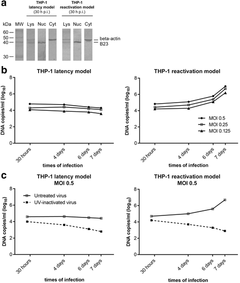
Fig. 1
Fig. 1
Time-course changes in HCMV DNA nuclear amounts in THP-1 latency and reactivation models. a At the indicated time points p.i., undifferentiated (panel “THP-1 latency model”) and differentiated THP-1 cells (panel “THP-1 reactivation model”) were subjected to cellular fractionation in order to obtain purified nuclei (Nuc) and cytoplasmic (Cyt) fractions derived from total cell lysates (Lys). After SDS-PAGE, WB analysis was done using a rabbit polyclonal antibody directed to B23 (40 kDa) and a mouse monoclonal anti-beta-actin (44 kDa) antibody as nuclear and cytoplasmic markers, respectively. The immunoreactions were revealed by AP-conjugated anti-rabbit and anti-mouse antibodies. Molecular weight markers are indicated on the left-hand figure. b Total DNA was extracted from nuclei of undifferentiated THP-1 monocytes (panel “THP-1 latency model”) and THP-1 monocytes differentiated after infection (panel “THP-1 reactivation model”). HCMV DNA amounts were measured by q-Real-time PCR at 30 h, 4, 6 and 7 days p.i. at MOI 0.5, 0.25 and 0.125 in both experimental models. Comparable results were obtained from two independent experiments. c Total DNA was extracted from the nuclei of “viable” HCMV-infected THP-1 monocytes (“untreated virus”) and UV-inactivated HCMV-infected THP-1 monocytes (panel “THP-1 latency model, MOI 0.5”) and THP-1 monocytes differentiated after infection (panel “THP-1 reactivation model, MOI 0.5”). HCMV DNA amounts were measured by q-Real-time PCR at 30 h, 4, 6 and 7 days p.i. at MOI 0.25 in both experimental models
Fig. 2
Fig. 2
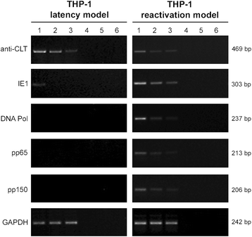
Analysis of HCMV transcripts from THP-1 latency and reactivation models at 7 days p.i.. The expression patterns of the anti-sense CLT (cytomegalovirus latency associated transcript), IE1 (immediate-early), DNA pol (early), pp65 (early-late) and pp150 (late) viral transcripts were analysed in both models (panels “THP-1 latency model” and “THP-1 reactivation model”) after infection at three different MOI. The cellular GAPDH housekeeping gene amplification product was used as a reaction control. Lanes 1, 2 and 3: MOI 0.5, 0.25, 0.125, respectively; lanes 4, 5 and 6: DNA contamination controls (PCR performed for MOI 0.5, 0.25, 0.125, respectively, in the absence of reverse transcription). Molecular weights of the amplified transcript fragments are shown on the right side. The results are representative of two independent experiments
Fig. 3
Fig. 3
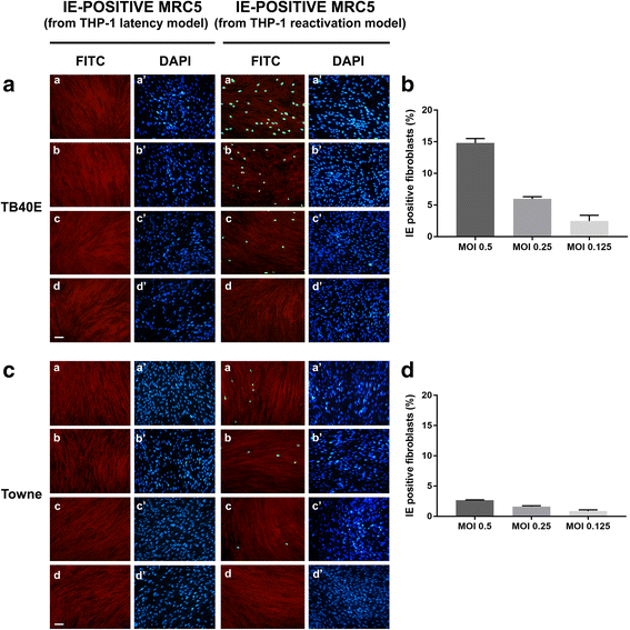
Analysis of HCMV progeny from THP-1 latency and reactivation models: IE antigen immunofluorescence pattern. a and c MRC5 fibroblasts were infected with the cell culture medium derived from THP-1 monocytes (“IE-positive MRC5 from THP-1 latency model”) and THP-1 macrophages (“IE-positive MRC5 from THP-1 reactivation model”) which had been infected with TB40E (a) or Towne (c) HCMV strains at MOI of 0.5 (panels a, a’), 0.25 (panels b, b’) or 0.125 (panels c, c’) for 7 days. At 24 h p.i., MRC5 cells were fixed and labelled with a monoclonal antibody specific for the common epitope encoded by exon 2 of HCMV IE1 and IE2 genes. The immunoreaction was revealed by Alexa-Fluor FITC-conjugated goat anti-mouse IgG (panels a, b, c, d: FITC, green nuclei). Cells were counterstained with Evans blue (panels a, b, c, d: red cells) and DAPI (panels a’, b’, c’, d’: blue nuclei). Panels d, d’: uninfected cells. Images were collected using a conventional fluorescence microscopy. Bar: 25 μm. b and d The quantitative evaluation of IE-positive MRC5 fibroblasts infected with the cell culture medium derived from TB40E (b) or Towne (d) reactivation models. Values were expressed as mean percentages of IE-positive cells per field (ten randomly selected fields per slide were counted) from two independent experiments; error bars indicate standard deviations. Values were processed using GraphPad Prism 7 software
Fig. 4
Fig. 4
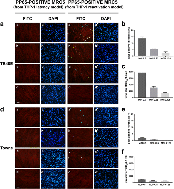
Analysis of HCMV progeny from THP-1 latency and reactivation models: pp65 antigen immunofluorescence pattern and viral yield titration. a and d MRC5 fibroblasts were infected with cell culture medium derived from THP-1 monocytes (“pp65-positive MRC5 from THP-1 latency model”) and THP-1 macrophages (“pp65-positive MRC5 from THP-1 reactivation model”) which had been infected with TB40E (a) or Towne (d) HCMV strains at MOI of 0.5 (panels a, a’), 0.25 (panels b, b’) and 0.125 (panels c, c’) for 7 days. At 48 h p.i., MRC5 cells were fixed and labelled with a monoclonal antibody reacting with the viral matrix phosphoprotein pp65. The immunoreaction was revealed by Alexa-Fluor FITC-conjugated goat anti-mouse IgG (panels a, b, c, d: FITC, green nuclei). Cells were counterstained with Evans blue (panels a, b, c, d: red cells) and DAPI (panels a’, b’, c’, d’: blue nuclei). Panels d, d’: uninfected cells. Images were collected using a conventional fluorescence microscopy. Bar: 25 μm. b and e The quantitative evaluation of pp65-positive MRC5 fibroblasts infected with the cell culture medium derived from TB40E (b) or Towne (e) reactivation models. Values were expressed as mean percentages of pp65-positive cells per field (ten randomly selected fields per slide were counted) from two independent experiments; error bars indicate standard deviations. Values were processed using GraphPad Prism 7 software. c and f Virus yields were evaluated by the TCID50 assay from cell culture medium derived from TB40E (c) or Towne (f) reactivation models, as detailed in the “Methods” section. Two independent experiments were performed; error bars in graphs represent standard deviations. Values were processed by the GraphPad Prism 7 software
Fig. 5
Fig. 5
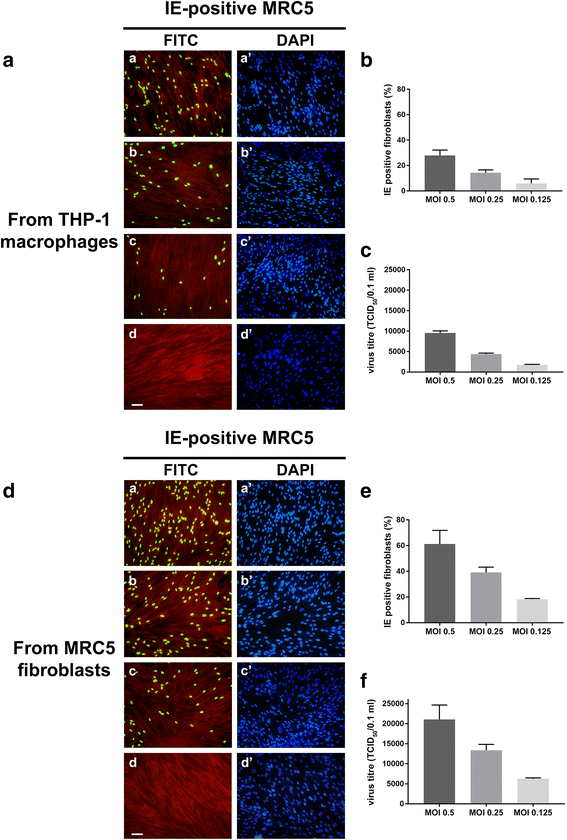
Analysis of TB40E progeny from THP-1 macrophages (lytic model), and MRC5 fibroblasts. a and d MRC5 fibroblasts were infected with cell culture medium derived from THP-1 lytic model (a) or from MRC5 fibroblasts (d) which had been infected with HCMV TB40E at MOI of 0.5 (panels a, a’), 0.25 (panels b, b’) and 0.125 (panels c, c’) for 7 days. At 24 h p.i., MRC5 cells were fixed and labelled with a monoclonal antibody specific for the common epitope encoded by exon 2 of HCMV IE1 and IE2 genes. The immunoreaction was revealed by Alexa-Fluor FITC-conjugated goat anti-mouse IgG (panels a, b, c, d: FITC, green nuclei). Cells were counterstained with Evans blue (panels a, b, c, d: red cells) and DAPI (panels a’, b’, c’, d’: blue nuclei). Panels d, d’: uninfected cells. Images were collected using a conventional fluorescence microscopy. Bar: 25 μm. b and e The quantitative evaluation of IE-positive MRC5 fibroblasts infected with the cell culture medium derived from THP-1 lytic model (b) or MRC5 fibroblasts as already described (see Fig. 3 legend) (e). c and f Virus yields were evaluated by the TCID50 assay from cell culture medium derived from THP-1 lytic model (c) or MRC5 fibroblasts (f), as detailed in the “Methods” section. Two independent experiments were performed; error bars in graphs represent standard deviations. Values were processed by the GraphPad Prism 7 software
Fig. 6
Fig. 6
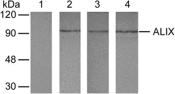
Biochemical analysis of Alix exosomal marker. After a 7 day-infection at an MOI 0.5, exosomes were purified from cell culture supernatants of infected THP-1 derived macrophages (reactivation model) and uninfected THP-1 derived-macrophages. Equal amounts (20 μg) of each sample, as well as the viral inoculum derived from the TB40E reactivation model (MOI 0.5) and a positive control (exosomes purified from COLO-1 cells), were run by SDS-PAGE and transferred to a nitrocellulose membrane, WB analysis was performed using a mouse monoclonal antibody raised against the ALIX (95 kDa) exosomal marker. The immunoreactions were revealed by AP-conjugated anti-mouse antibody. Lane 1: protein fraction obtained from the supernatant of TB40E-infected THP-1 derived-macrophages (reactivation model), as detailed in the Methods section; lane 2: protein fraction obtained from the supernatant of uninfected THP-1 derived-macrophages processed for exosome extraction, as detailed in the Methods section; lane 3: protein fraction obtained from the supernatant of infected THP-1 derived-macrophages (reactivation model) processed for exosome extraction, as detailed in the Methods section; lane 4: purified exosomes derived from the cell culture supernatant of COLO1 cells (positive control). Molecular weight markers are indicated to the left of the figure

Similar articles

Cited by

References

    1. Mocarski ES, Tan Courcelle C. Cytomegaloviruses and their replication. In: Howley DMKPM, editor. Fields Virology. 4. Philadelphia: Lippincott Williams and Wilkins; 2001. pp. 2629–2673.
    1. Kumar A, Herbein G. Epigenetic regulation of human cytomegalovirus latency: an update. Epigenomics. 2014;6:533–546. doi: 10.2217/epi.14.41. - DOI - PubMed
    1. Sissons JG, Carmichael AJ, McKinney N, Sinclair JH, Wills MR. Human cytomegalovirus and immunopathology. Springer Semin Immunopathol. 2002;24:169–185. doi: 10.1007/s00281-002-0104-0. - DOI - PubMed
    1. Malm G, Engman ML. Congenital cytomegalovirus infections. Semin Fetal Neonatal Med. 2007;12:154–159. doi: 10.1016/j.siny.2007.01.012. - DOI - PubMed
    1. Huang MM, Kew VG, Jestice K, Wills MR, Reeves MB. Efficient human cytomegalovirus reactivation is maturation dependent in the Langerhans dendritic cell lineage and can be studied using a CD14+ experimental latency model. J Virol. 2012;86:8507–8515. doi: 10.1128/JVI.00598-12. - DOI - PMC - PubMed

LinkOut - more resources