. 2016 Dec;17(12):1381-1387.
doi: 10.1038/ni.3582. Epub 2016 Oct 17.

A tissue checkpoint regulates type 2 immunity

Affiliations

A tissue checkpoint regulates type 2 immunity

Steven J Van Dyken et al. Nat Immunol. 2016 Dec.

Abstract

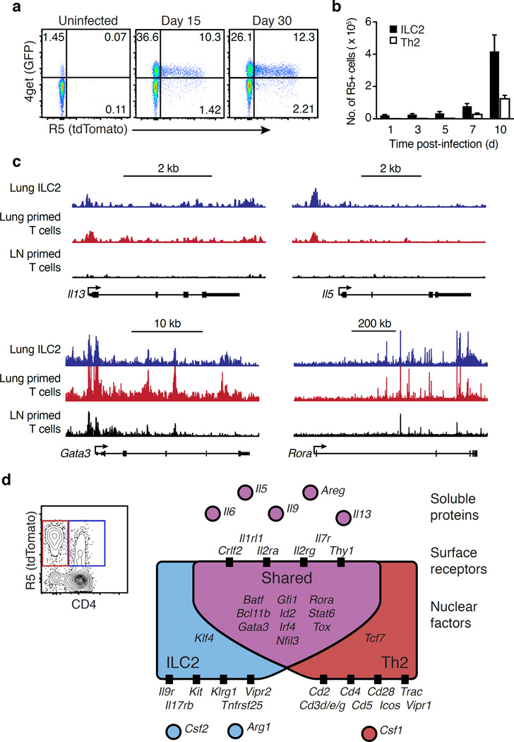
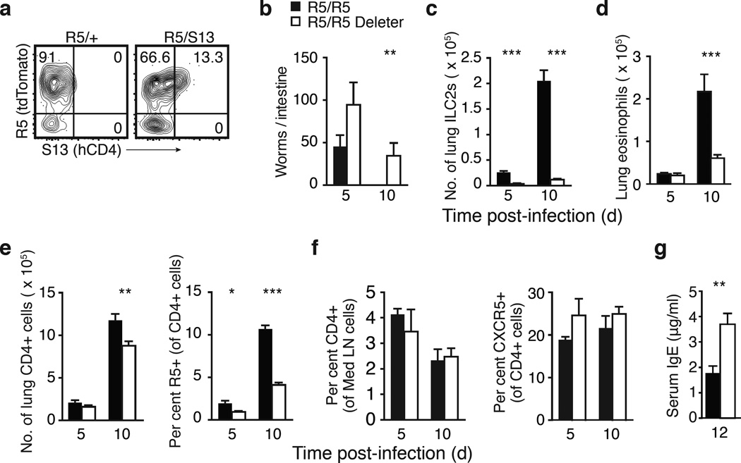
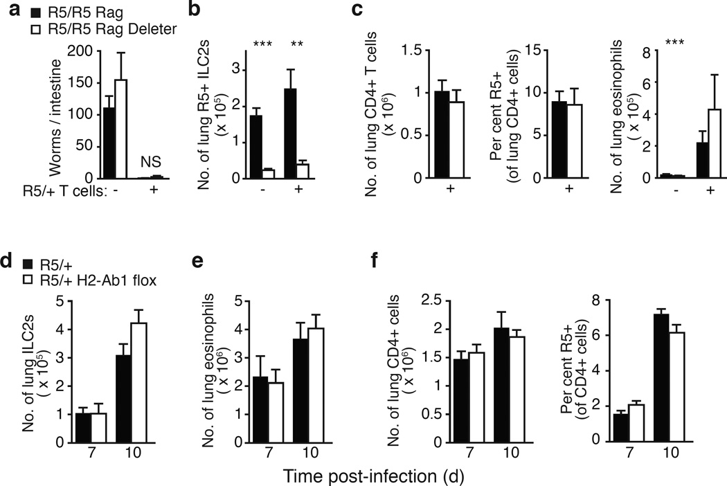
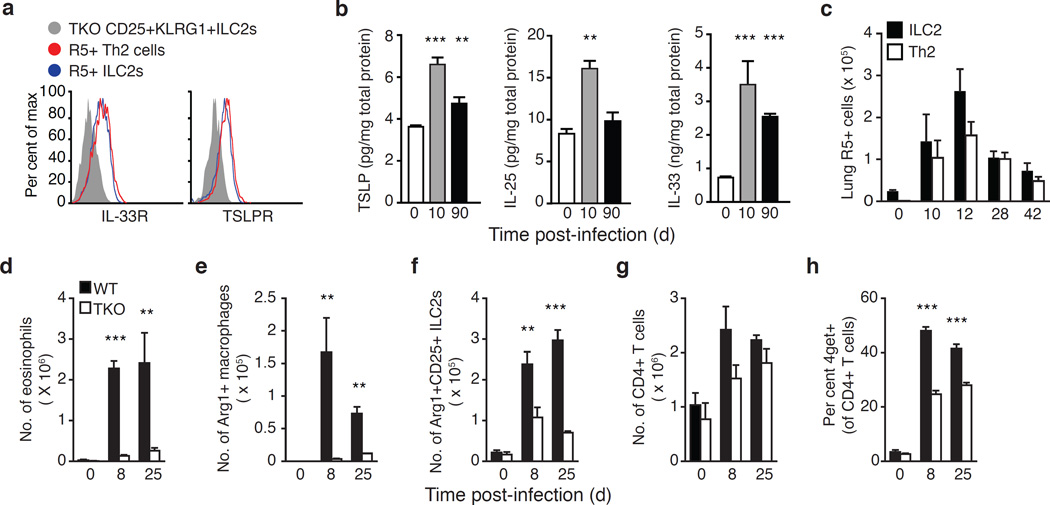
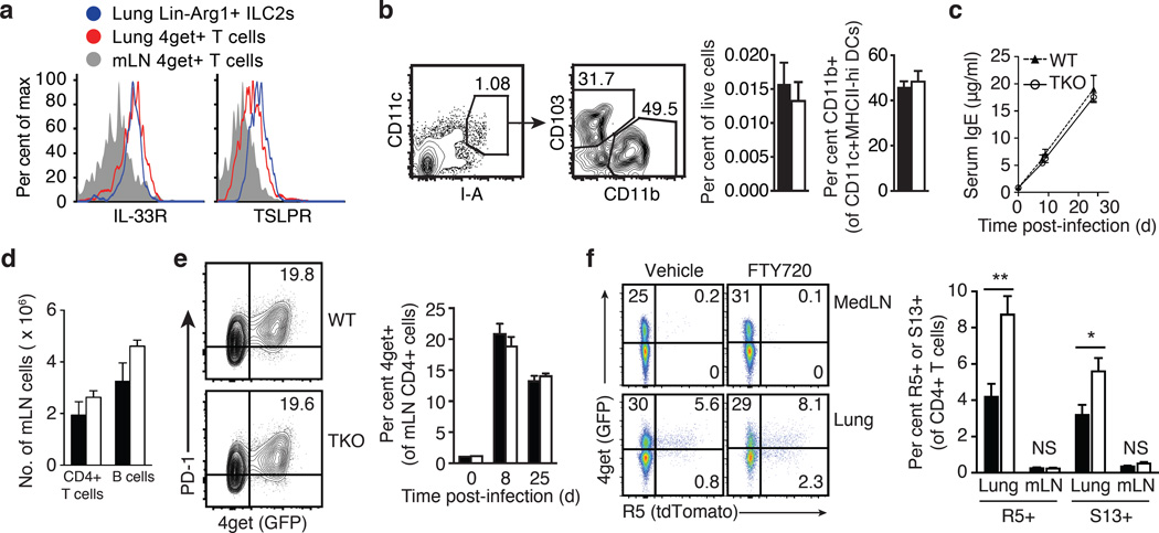
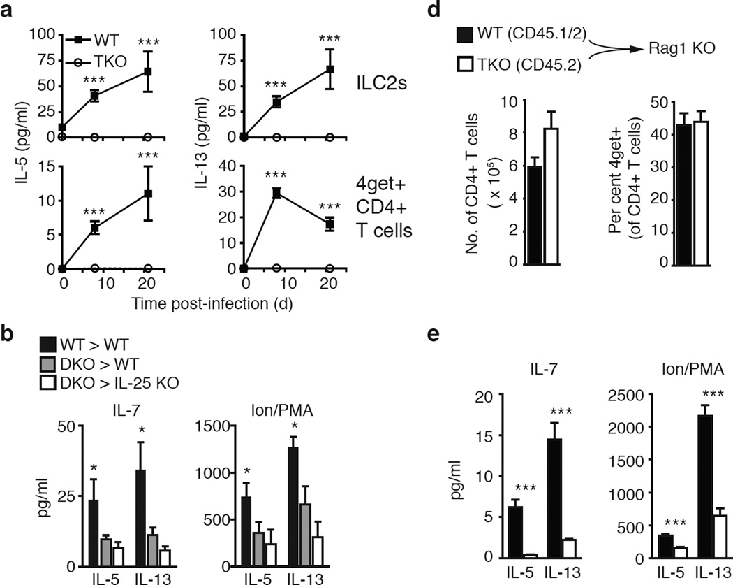
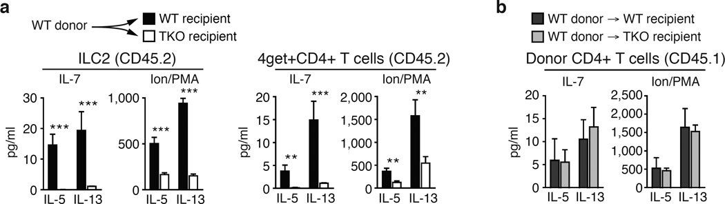
Group 2 innate lymphoid cells (ILC2s) and CD4+ type 2 helper T cells (TH2 cells) are defined by their similar effector cytokines, which together mediate the features of allergic immunity. We found that tissue ILC2s and TH2 cells differentiated independently but shared overlapping effector function programs that were mediated by exposure to the tissue-derived cytokines interleukin 25 (IL-25), IL-33 and thymic stromal lymphopoietin (TSLP). Loss of these three tissue signals did not affect lymph node priming, but abrogated the terminal differentiation of effector TH2 cells and adaptive lung inflammation in a T cell-intrinsic manner. Our findings suggest a mechanism by which diverse perturbations can activate type 2 immunity and reveal a shared local-tissue-elicited checkpoint that can be exploited to control both innate and adaptive allergic inflammation.


Figures

Fig. 1
Fig. 1
Cytokine reporters mark innate and adaptive tissue effectors. a, Flow cytometry of lung CD4+ T cells from uninfected or N. brasiliensis-infected R5/4get dual reporter mice. b, R5+ ILC2s and R5+ CD4+ T cells in lungs of mice on the indicated days of infection, presented as mean ± SEM. c, representative tracks pooled from ATAC-Seq reads aligning to Il5, Il13, Gata3, and Rora, shown with identical vertical scale. d, Representative flow cytometry of lung CD8CD19NK1.1+-CD90.2+ cells and schematic Venn diagram of secreted proteins, cell surface markers, and nuclear factors that were significantly enriched in ILC2s (blue), Th2 cells (red), or shared without detection of statistical difference (purple). Data are (a) representative of 3 independent experiments; (b) pooled from 2 independent experiments for a total of at least 3 mice per time point; (c) from 2 (lung ILC2s), 3 (lung T cells), and 4 (LN T cells) biological replicates; (d) collected from 2 independent infections prior to sequencing for a total of 6 ILC2 and 5 Th2 cell biological replicates.
Fig. 2
Fig. 2
IL-5-producing cells drive type 2 immunity in the lung, but not the draining lymph node. a, Flow cytometry of lung ILC2s from R5/+ or R5/S13 mice 10 days post infection (d.p.i.) with Nbb, worm clearance, c, lung LinKLRG1+ ILC2s, d, lung eosinophils, e, lung CD4+ T cells and percent R5+ Th2 effector cells f, percent CD4+ T cells and PD-1+CXCR5+ Tfh cells in the draining lymph nodes, and g, serum IgE in R5/R5 or R5/R5 Deleter mice on the indicated d.p.i. Data are (a) representative of 3 independent experiments, (b–e) pooled from 4 independent experiments for at least 5 mice per genotype, (f,g) pooled from 2 independent experiments for at least 5 mice per genotype. Lin, lineage; Med LN, mediastinal lymph node; Tfh, T follicular helper cells; *, p < 0.05; **, p < 0.01; ***, p < 0.001.
Fig 3
Fig 3
Activation of adaptive type 2 immunity despite ILC2 deficiency. Rag1-deficient mice (either R5/R5 or R5/R5 Deleter) received no cells or naïve R5/+ CD4+ T cells and were analyzed 10 days post infection (d.p.i.) for a, worm counts, b, R5+ ILC2s, and c, CD4+ T cells, percent of T cells expressing R5, and lung eosinophils. d, LinThy1+KLRG1+ ILC2s, e, eosinophils, and f, CD4+ T cells and R5+ Th2 effector cells in the lungs on the indicated d.p.i. Data are pooled from (a–c) 4 independent experiments for at least 5 mice per group or (d–e) 2 experiments for at least 3 mice per group. NS, not significant; *, p < 0.05; **, p < 0.01; ***, p < 0.001.
Fig. 4
Fig. 4
Multiple epithelial cytokines are induced in tissue during type 2 immunity. a, Histogram showing surface staining of IL-33 receptor and TSLP receptor on lung R5+ ILC2s (blue) and R5+ CD4+ T cells (red) 14 d.p.i. compared with lung ILC2s from a TKO mouse (gray). b, TSLP, IL-25, and IL-33 protein amounts in whole lung lysates from WT C57BL/6 mice at indicated days post Nb infection (d.p.i.). c, Number of R5+ ILC2s and R5+ CD4+ T cells in lungs of mice at the indicated d.p.i. d, Lung eosinophils, e, Arg1+ macrophages, and f, Arg1+CD25+ILC2s in BALB/c wild-type (WT) or Crlf2−/−Il25−/−Il1rl1−/− triple-deficient (TKO) mice on Arg1 (Yarg) / IL4 (4get) dual reporter background. g, CD4+ T cells in 4get/Arg1 BALB/c (WT) or TKO lungs at indicated d.p.i. h, 4get+ cells as a percent of total lung CD4+ T cells. Data in (a) representative of 5 mice of each genotype from 2 independent experiments and in (b–h) pooled from 2 independent experiments for a total of at least 3 mice per group, represented as mean ± SEM, **, p < 0.001; ***, p < 0.0001.
Fig. 5
Fig. 5
Tissue cytokines are not required for lymph node adaptive immunity. a, Histogram showing surface staining of IL-33 receptor and TSLP receptor on lung ILC2s (blue), lung 4get+ T cells (red), and mediastinal lymph node (mLN) 4get+ T cells (gray) 10 days post infection (d.p.i). b, Representative flow cytometry and quantification of mLN CD11c+MHCIIhi dendritic cells 3 d.p.i., previously gated on CD4CD8CD19 cells. c, Serum IgE concentrations at indicated d.p.i. in WT and TKO mice. d, Numbers of CD4+ T cells, B cells, and e, percent of CD4+ T cells expressing Tfh markers PD-1 and 4get in the mLN of indicated mice 8 d.p.i. f, Representative flow cytometry and quantification of the R5+ or S13+ percentage of CD4+ T cells in the lung and mLN of dual reporter mice 7 d.p.i that were treated with FTY720 or vehicle. Flow cytometric analysis in (a), (b), and (e) representative of 3 independent experiments, quantification in (b) and (f) representative of 2 independent experiments with at least 3 mice per group, and data in (c–e) pooled from 2 independent experiments for at least 3 mice per group, all presented as mean ± SEM, *, p < 0.05; **, p < 0.01; NS, no statistically significant difference.
Fig. 6
Fig. 6
Tissue cytokine licensing of Th2 cells is cell-intrinsic. a, IL-5 and IL-13 in supernatants of lung ILC2s or 4get+CD4+ T cells sorted at the indicated days post Nb infection (d.p.i.) and then cultured in IL-7 for 24 hours. b, IL-5 and IL-13 in supernatants of 24-hour IL-7 or Ion/PMA cultures of donor WT or IL-33R / TSLPR double-deficient (DKO) 4get+CD4+ T cells sorted from the lungs of WT or IL-25 KO recipients 10 d.p.i. c, Numbers of total donor-derived congenic WT and TKO CD4+ T cells and percentage 4get+ cells in the lungs of Rag1-deficient recipients and d, IL-5 and IL-13 in the supernatants of 24-hour IL-7 or Ion/PMA cultures of sorted lung 4get+CD4+ T cells 8 d.p.i. Data are presented as mean ± SEM, pooled from 2 independent experiments for at least 3 (a) and (b) or 6 (c) and (d) mice per group, represented as mean ± SEM. *, p < 0.05 in (b) refers to comparison between WT and similarly treated DKO cells; ***, p < 0.001.
Fig. 7
Fig. 7
Cell-intrinsic epithelial cytokine signaling is sufficient for terminal Th2 cell differentiation. IL-5 and IL-13 in supernatant of 24-hour IL-7 or Ion/PMA cultures of a, recipient CD45.2+ ILC2s or 4get+CD4+ T cells sorted from WT or TKO CD45.2+ recipients or b, donor WT CD45.1+CD4+ T cells sorted from lungs of WT or TKO recipients 8 days after Nb infection. Data are presented as mean ± SEM and represent at least 3 mice per group pooled from 2 independent experiments; **, p < 0.01; ***, p < 0.001, compared to similarly-treated TKO.

Comment in

Similar articles

Cited by

References

    1. Spits H, et al. Innate lymphoid cells--a proposal for uniform nomenclature. Nat Rev Immunol. 2013;13:145–149. - PubMed
    1. Patel N, et al. A2B adenosine receptor induces protective antihelminth type 2 immune responses. Cell Host Microbe. 2014;15:339–350. - PubMed
    1. Doherty TA, et al. Lung type 2 innate lymphoid cells express cysteinyl leukotriene receptor 1, which regulates Th2 cytokine production. J Allergy Clin Immunol. 2013;132:205–213. - PMC - PubMed
    1. Halim TY, et al. Group 2 innate lymphoid cells license dendritic cells to potentiate memory Th2 cell responses. Nat Immunol. 2015 - PMC - PubMed
    1. Oliphant CJ, et al. MHCII-mediated dialog between group 2 innate lymphoid cells and CD4(+) T cells potentiates type 2 immunity and promotes parasitic helminth expulsion. Immunity. 2014;41:283–295. - PMC - PubMed

METHODS-ONLY REFERENCES

    1. Reese TA, et al. Chitin induces accumulation in tissue of innate immune cells associated with allergy. Nature. 2007;447:92–96. - PMC - PubMed
    1. Van Dyken SJ, et al. Fungal chitin from asthma-associated home environments induces eosinophilic lung infiltration. J Immunol. 2011;187:2261–2267. - PMC - PubMed
    1. Li H, Durbin R. Fast and accurate short read alignment with Burrows-Wheeler transform. Bioinformatics. 2009;25:1754–1760. - PMC - PubMed
    1. Quinlan AR, Hall IM. BEDTools: a flexible suite of utilities for comparing genomic features. Bioinformatics. 2010;26:841–842. - PMC - PubMed
    1. Picard Tools. 2015 < http://broadinstitute.github.io/picard/>.

MeSH terms