. 2016 Oct 20;538(7625):378-382.
doi: 10.1038/nature19823. Epub 2016 Oct 12.

A renewed model of pancreatic cancer evolution based on genomic rearrangement patterns

Affiliations

A renewed model of pancreatic cancer evolution based on genomic rearrangement patterns

Faiyaz Notta et al. Nature. .

Erratum in

  • Erratum: A renewed model of pancreatic cancer evolution based on genomic rearrangement patterns.
    Notta F, Chan-Seng-Yue M, Lemire M, Li Y, Wilson GW, Connor AA, Denroche RE, Liang SB, Brown AM, Kim JC, Wang T, Simpson JT, Beck T, Borgida A, Buchner N, Chadwick D, Hafezi-Bakhtiari S, Dick JE, Heisler L, Hollingsworth MA, Ibrahimov E, Jang GH, Johns J, Jorgensen LG, Law C, Ludkovski O, Lungu I, Ng K, Pasternack D, Petersen GM, Shlush LI, Timms L, Tsao MS, Wilson JM, Yung CK, Zogopoulos G, Bartlett JM, Alexandrov LB, Real FX, Cleary SP, Roehrl MH, McPherson JD, Stein LD, Hudson TJ, Campbell PJ, Gallinger S. Notta F, et al. Nature. 2017 Feb 2;542(7639):124. doi: 10.1038/nature20164. Epub 2016 Nov 16. Nature. 2017. PMID: 27851734 No abstract available.

Abstract

Pancreatic cancer, a highly aggressive tumour type with uniformly poor prognosis, exemplifies the classically held view of stepwise cancer development. The current model of tumorigenesis, based on analyses of precursor lesions, termed pancreatic intraepithelial neoplasm (PanINs) lesions, makes two predictions: first, that pancreatic cancer develops through a particular sequence of genetic alterations (KRAS, followed by CDKN2A, then TP53 and SMAD4); and second, that the evolutionary trajectory of pancreatic cancer progression is gradual because each alteration is acquired independently. A shortcoming of this model is that clonally expanded precursor lesions do not always belong to the tumour lineage, indicating that the evolutionary trajectory of the tumour lineage and precursor lesions can be divergent. This prevailing model of tumorigenesis has contributed to the clinical notion that pancreatic cancer evolves slowly and presents at a late stage. However, the propensity for this disease to rapidly metastasize and the inability to improve patient outcomes, despite efforts aimed at early detection, suggest that pancreatic cancer progression is not gradual. Here, using newly developed informatics tools, we tracked changes in DNA copy number and their associated rearrangements in tumour-enriched genomes and found that pancreatic cancer tumorigenesis is neither gradual nor follows the accepted mutation order. Two-thirds of tumours harbour complex rearrangement patterns associated with mitotic errors, consistent with punctuated equilibrium as the principal evolutionary trajectory. In a subset of cases, the consequence of such errors is the simultaneous, rather than sequential, knockout of canonical preneoplastic genetic drivers that are likely to set-off invasive cancer growth. These findings challenge the current progression model of pancreatic cancer and provide insights into the mutational processes that give rise to these aggressive tumours.


Figures
Extended Data Figure 1
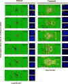
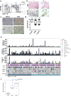
Extended Data Figure 1. Tumour enrichment and overview of somatic alterations in the cohort used in this study
(a) Flow cytometric profiles of EpCAM and CD45 from two representatives cases of pancreas ductal adenocarcinoma (PDA) (i,ii). On the right, post-sort analysis of EpCAM+ cells (Tu) and CD45+ lymphocytes (Ly) demonstrates the high level of purity obtained from flow sorting. (b) Immunohistochemical analysis of formalin-fixed tumours using the EpCAM clone for flow sorting in a (H&E – hematoxylin and eosin). Two independent cases are shown (i,ii). (c) Profiles of hematoxylin stained sections of PDA before and after LCM from two representative cases (i,ii). (d) Box whisker plots showing median tumour cellularity of flow-sorted (n=21), LCM (n=86) and the total cohort (n=107). Dashed line depicts cellularity of bulk tumours that have not undergone enrichment. (e) Overview of somatic alterations of the cohort used in the study. (f) X chromosome mutation ratio in diploid PC genomes showing hypermutation on this chromosome in females. Males were corrected for single copy of X chromosome by doubling the raw value. p values were derived from t-test. A more detailed description of these data is provided in Supplementary results.
Extended Data Figure 2
Extended Data Figure 2. CELLULOID validation
Copy number for common alterations (TP53, SMAD4 – shown by black arrow) was derived from ploidy estimates generated by CELLULOID. Six diploid and five polyploid tumours analyzed by FISH (shown on the right of each contour plot). In all cases, CN from CELLULOID ploidy estimates were confirmed. In Pcsi_0084 (diploid), CELLULOID predicted zero copies of SMAD4. The allelic ratio in this region was 50% (heterozygous) as only reads from normal cells spanned this region. In Ashpc_0027, both CELLULOID and FISH indicate that this tumour is polyploid. The CELLULOID plot demonstrates that there is a further subclonal amplification in TP53 from polyploid clone (copy state = 3.2 derived from one allele). FISH analysis shows tumour cells with 2 or 3 copies of TP53 supporting this is subclonal. Copy number by FISH for SMAD4 (top right of each plot) and TP53 (top right of each plot) is indicated in red.
Extended Data Figure 3
Extended Data Figure 3. Tumour ploidy and genomic instability in PC
(a) Tumour ploidy and sample cellularity estimates are interconnected: while the ploidy of a tumour can always be doubled and still provide CN segments at integer levels (albeit only at even values), the estimate of cellularity would have to be decreased. Indeed, in order to maintain an allelic ratio at a given value, the proportion of tumour cells has to be reduced to compensate the higher number of copies in them (from a cellularity value t to a value t/(2-t) in the case of a doubling of the ploidy). A test can thus be designed to verify that ploidy estimates have not been systematically over- or underestimated, simply by comparing the distribution of cellularity estimates stratified by ploidy. p value was derived using Kruskal-Wallis test. (b) Deviation from baseline ploidy in diploids (ploidy = 2), tetraploids (ploidy = 4) and hexaploids (ploidy = 6) indicates dramatic loss of genomic material in polyploids. (c) Box whisker plots of total CN alterations in polyploid and diploid tumours. (d) Mutational signatures of the 107 genomes used in this study. The signatures were derived using the trinucleotide mutation context as previously published. The proportion of individual signature operative in each tumour is shown in the bar plot. The overall classification of each case is indicated on the bottom. Signatures of polyploidy tumours is shown on the left versus diploids is shown on the right. (ND – not done – 1 polyploid and 4 diploid cases). Detailed analysis of mutational signatures in PDA is covered in another manuscript under review (Connor et al.) (e) Percentage of CN losses (left) and gains (right) that occurred before (yellow) or after (blue) genome duplication for each polyploid tumour. Box whisker plots depict median +/− 10–90 percentile. p values were derived using t-test.
Extended Data Figure 4
Extended Data Figure 4. Characterization of chromothripsis events in PC
(a) The distribution of chromothripsis events across the genome (single chromosome – white; multi-chromosome – black). **, p<0.001 (Monte Carlo Sampling, Supplementary methods). (b) The specific effects of chromothripsis on the CN of chr18 (top, n=22), chr12 (middle, n=15), and chr19 (bottom, n=5). Statistical differences in CN between the groups were performed using Wilcoxon test using 10kb bins that covered GATA6 (chr18), KRAS (chr12) and PAK4 (chr19) genes (description of PAK4 event is covered in supplementary results). CN profiles of polyploids were adjusted according to tumour ploidy to allow comparison against diploids (referred to as Normalized CN on the y-axis). Interquartile ranges for chromothripsis cases are indicated in pale red, and for non-chromothripsis cases in pale blue. (c) Two cases of chromothripsis resulting in the amplification of the mutant KRAS allele. In Pcsi_0290, the mutant allele was amplified as part of a multi-chromosomal with chr18 (top). In Pcsi_0356, the chromothripsis event was coopted with cycles of BFB to knockout the wildtype allele (bottom). The absolute CN of the locus encompassing KRAS and mutation is shown for each case. (d) Cumulative incidence of chromothripsis events in polyploid and diploid tumours (p=0.013, Fisher’s exact test).
Extended Data Figure 5
Extended Data Figure 5. Most CN alterations arise from individual chromothripsis events
(a) In Pcsi_0082, five distinct chromothripsis events on chr15 (top, ➀), chr18 (top, ➁), chr8 (top, ➂), chr13 (bottom, ➃), and chr16 (bottom, ➄) are displayed. CN steps on chr15 (➀), chr8 (➁) are 2 or greater indicating that these events occurred before polyploidization. Single CN steps on chr18 (➁), chr13 (➃) and chr16 (➄) indicate that these events were sustained after polyploidization. The single rearrangement between chr15 and chr18 appears to be independent from the chromothripsis on chr18. Pie graphs depict the proportion of CN alterations derived from each chromothripsis event. (b) Distribution of CN alterations due to chromothripsis for all cases where such an event was detected by Chrom-AL. (c) In Ashpc_0008, two multi-chromosomal chromothripsis events joining chr14, chr6, chr18 (top - ➀), and chr3, chr20 (bottom - ➁) are shown (discussed in supplementary results).
Extended Data Figure 6
Extended Data Figure 6. Characterization of chromothripsis and polyploidy in metastases
(a) CELLULOID plots illustrating polyploidy in metastases. In Pcsi_0380, the primary tumour was directly available for analysis. Similarly to Pcsi_0378, multiple metastases were polyploid suggesting primary tumour was also polyploid. The primary tumour was unavailable for sequencing in this case. (b) A case (Pcsi_0407) with discordant ploidy amongst different metastases. (c) Percent of diploid mutations from liver metastases that are shared (white) or unique (black) in comparison to the primary tumour or the lymph node metastasis. (d) Plots of chromothripsis events in metastases. pa – primary tumour; lv – liver; ln – lymph node.
Extended Data Figure 7
Extended Data Figure 7. Chromothripsis and polyploidy in Pcsi_0410 (accompany to Figure 2)
(a) CELLULOID (left) and chromothripsis plots (middle and right panels) of the different metastases from a patients with fulminant metastatic progression. (b) CN and LOH from chr8 (left) and chr6 (right) chromothripsis events indicate that these events were sustained before polyploidization.
Extended Data Figure 8
Extended Data Figure 8. Case of a simultaneous loss of CDKN2A and SMAD4 due to a chromothripsis event
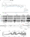
(a) Rearrangement and CN profile of a multi-chromosome chromothripsis event between chr9 and chr18 (Pcsi_0171). (b) Detailed view of the two inversions (one head-head –HH, the other tail-tail – TT) in the chromothripsis event that resulted in the concurrent loss of CDKN2A and SMAD4. (c) Schematic depiction of the temporal order of events derived from the rearrangement profile shown in A. (d) Summary of tumour evolution in Pcsi_0171. A more detailed description of Pcsi_0171 is provided in Supplementary results.
Extended Data Figure 9
Extended Data Figure 9. Reconstruction of the evolutionary events in a case where rearrangements did not span the classical PC drivers using single cell sequencing
(a) Fresh tumour specimen (Ashpc_0008) was dissociated and single tumour cells were deposited using flow sorting. 96 single cells were whole genome amplified using REPLI-g and paired end whole genome sequencing was performed using HiSeq 2500. Single cells were sequenced to a median whole genome depth of 3.9X (Supplementary Fig. 18). Only cells with enough whole genome coverage (n=70) were used in the analysis. This sequencing depth allowed us to track heterozygous SNPs across the whole genome in single cells. Using this methodology, we were able to follow LOH events across the whole genome in single cells with high concordance with bulk tumour (Supplementary Fig. 18). Hierarchical clustering based on LOH events across the whole genome was performed and found 4 independent cell clusters. (b) Specific LOH events on chr3, chr9, chr17 and chr18 are shown from representative single cells. The chromothripsis event on chr3 is shown in greater detail in Fig. 2a. Summary of the sequence of allelic losses is shown on the bottom. Supportive data that allelic losses precede mutational inactivation is shown in Supplementary Figure 13 and Supplementary Figure 14. (c) Representative plot of the shared chromosomal breakpoint on chr18 on the bulk (top), preneoplastic single cell (middle) and tumour single cell (bottom). (d) Classical model of pancreas tumour progression.
Extended Data Figure 10
Extended Data Figure 10. Theoretical model of PC tumour progression
The classical model of tumour evolution driven by a gradual pace (grey), and an alternate model driven by punctuated equilibrium (red). In gradualism, there is a period of latency between driver alterations that lead to tumour development, and multiple, independent transforming events are required for tumour development (top – grey line; bottom left). In punctuated equilibrium, tumour development can be divided into two major events: the cancer-initiating event and cancer-transforming event (top – red; bottom right). Under this model, most mutations (indicated with x) would accrue in an extended phase of preneoplastic tumour development. Transformation, likely due to a genomic instability from copy number changes (arrow heads) ensuing from cataclysmic event, would rapidly lead to invasive cancer and metastases. Classical drivers (KRAS, CDKN2A, TP53, SMAD4) from the PanIN progression model are overlaid onto these models. Theoretical PanIN stages are shown as P1–P3.
Figure 1
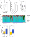
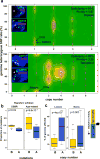
Figure 1. Mutational dynamics of polyploidization
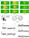
(a) CELLULOID profiles of a diploid (top – Ashpc_0008) and a tetraploid (bottom – Ashpc_0005) case that underwent tumour purification by flow cytometry followed by whole genome sequencing. Absolute copy number of of SMAD4 and TP53 allelic losses as predicted by CELLULOID is shown with black arrows on the contour plot. FISH validation of tumour copy number of SMAD4 and TP53 genes and corresponding centromeres (CEP17 and CEP18) are shown as an inset. (b) Proportion of mutations that occurred before (yellow) or after (blue) polyploidization event in the two predominant mutational signature subtypes of PC: DSBR (n=5; left) and Age-related (n=32; right) mutational signature. Due to increased instability in polyploids, mutations at a copy number of 4 in tetraploids were utilized in this analysis. (c) Fraction of the genome lost and gained either before (yellow) or after (blue) polyploidization. Legend is shown on the right of this plot. Box whisker plots represent 10–90% quartiles. p values were derived using t-test. A detailed description of these data is given in Supplementary results.
Figure 2
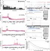
Figure 2. Characterization of genomic events in a patient with fulminant metastatic progression
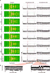
(a) Top: Progression timeline of patient Pcsi_0410. Bottom: Computerized tomography (CT) images of the abdomen at diagnosis (right) and one year later (left). No metastases are present in the liver at diagnosis. Within a year, the liver was decimated with metastases (right – white hashed line). At the rapid autopsy (RAP), 8 distinct metastases (see image in c) were harvested for sequencing. (b) Representative image of polyploidization (top) and chromothripsis (bottom) event from the adrenal gland metastasis. Analyses of all metastases are shown in Extended Data Figure 7. (c) FISH analysis c-MYC amplification in primary tumour and all metastases. Fibroblasts surrounding the tumour cells were used as a control (ctr - white arrows). Scattered nuclear staining in the primary tumour is consistent with presence of double minutes (dm – white arrow in pa). Homogenous staining areas (hsr – white arrow in ag) suggest reintegration into the genome. (d) Proportion of SVs common to all (black), shared by two or more (blue), or unique to each metastases is shown on the left. CN and SVs were used to reconstruct radial phylogenetic tree of metastatic progression (right). The primary tumour was surgically removed one year before autopsy and fresh frozen material was not available for WGS. It is possible that branch lengths of the phylogenetic tree would vary if the primary tumour were included in this analysis. Lines are to scale with CN based clustering dendrogram presented in Supplementary Figure 15, with the exception of germline origin (GL) that is half the length.
Figure 3
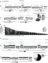
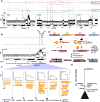
Figure 3. Simultaneous knockout of driver genes in PC evolution
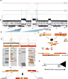
(a) Rearrangement and CN profile of a chromothripsis event in Ashpc_0005. The positions of CDKN2A, TP53, and SMAD4 genes are shown bottom of the plot. (b) A detailed view of chr9 with two distinct sets of rearrangements (window 1 and window 2), each responsible for the loss of one copy of CDKN2A. In window 2, three fold-back inversions (2 mapped and 1 unmapped*) highlighted with curved black arrows indicate three cycles of BFB. The final CN state of amplifications resulting from BFB cycles are shown on the plot (arrows). Zigzag symbol denotes DNA double strand break required to initiate a BFB cycle. (c) Schematic depiction of the three cycles of BFB that generated the final CN state of the amplifications shown in window 2 in b. (d) Temporal order of events derived from rearrangement profile for Ashpc_0005. The leftover TP53 and SMAD4 allele carry inactivating mutations (x). As both TP53 alleles carry the mutations (ploidy >1), this mutation was acquired prior to genome duplication. The relative timing of the SMAD4 mutation cannot be inferred because there is only one copy of this allele leftover and the mutation is fully clonal. (e) Summary of tumour evolution in Ashpc_0005.

Comment in

Similar articles

Cited by

References

    1. Hruban RH, Goggins M, Parsons J, Kern SE. Progression model for pancreatic cancer. Clinical cancer research: an official journal of the American Association for Cancer Research. 2000;6:2969–2972. - PubMed
    1. Moskaluk CA, Hruban RH, Kern SE. p16 and K-ras gene mutations in the intraductal precursors of human pancreatic adenocarcinoma. Cancer research. 1997;57:2140–2143. - PubMed
    1. Wilentz RE, et al. Inactivation of the p16 (INK4A) tumor-suppressor gene in pancreatic duct lesions: loss of intranuclear expression. Cancer research. 1998;58:4740–4744. - PubMed
    1. Wilentz RE, et al. Loss of expression of Dpc4 in pancreatic intraepithelial neoplasia: evidence that DPC4 inactivation occurs late in neoplastic progression. Cancer research. 2000;60:2002–2006. - PubMed
    1. Lüttges J, et al. Allelic loss is often the first hit in the biallelic inactivation of the p53 and DPC4 genes during pancreatic carcinogenesis. The American journal of pathology. 2001;158:1677–1683. - PMC - PubMed

MeSH terms