
Decreased somatic hypermutation induces an impaired peripheral B cell tolerance checkpoint

Tineke Cantaert et al. J Clin Invest. .

Abstract

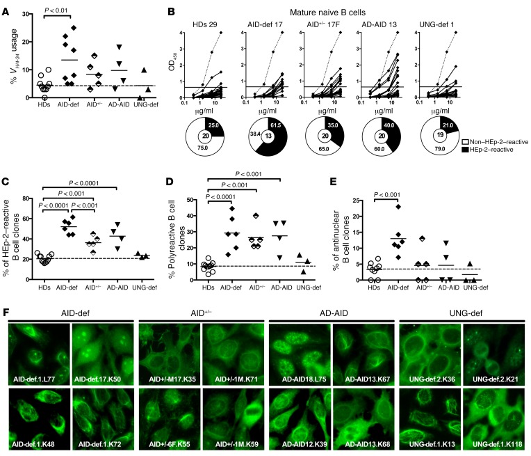
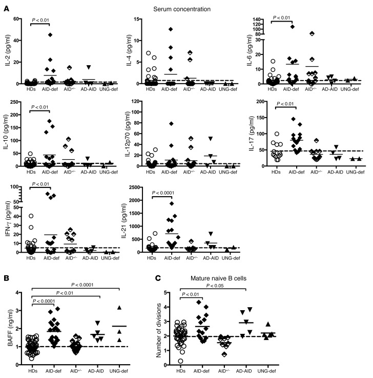
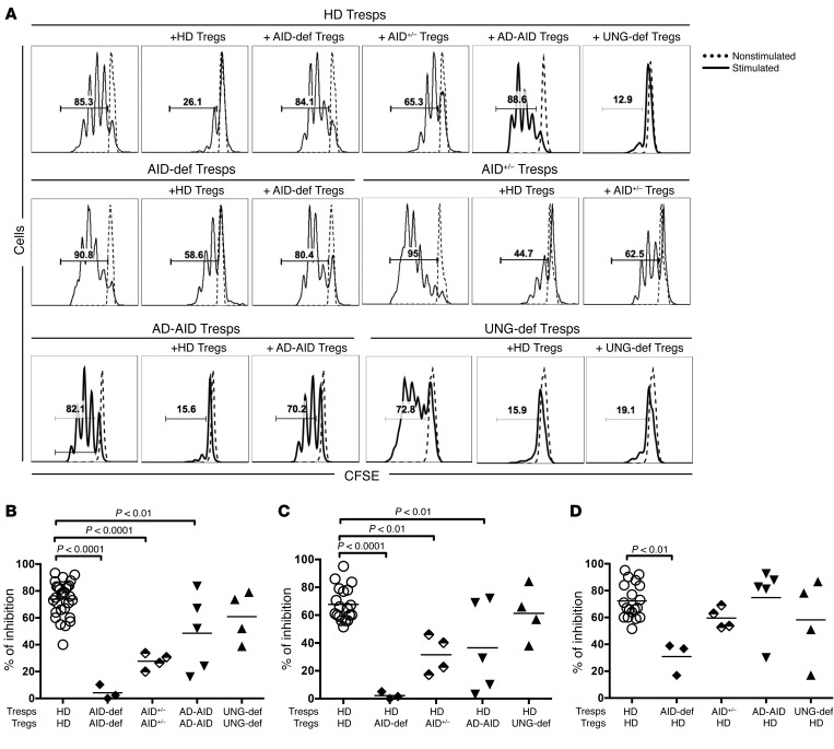
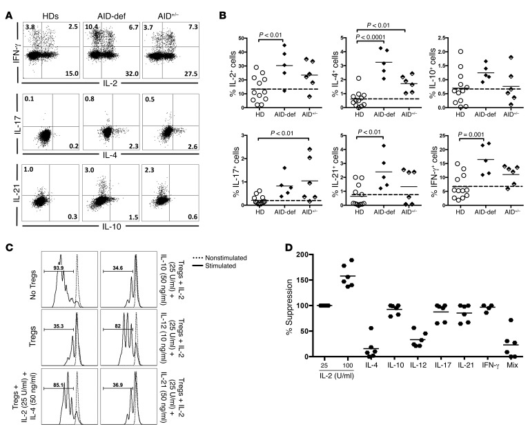
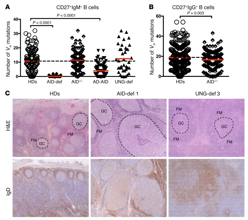
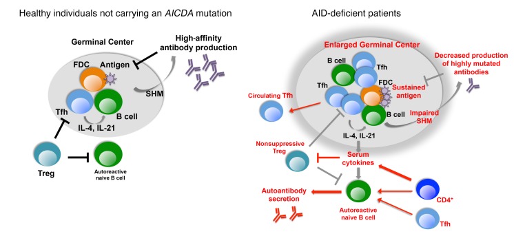
Patients with mutations in AICDA, which encodes activation-induced cytidine deaminase (AID), display an impaired peripheral B cell tolerance. AID mediates class-switch recombination (CSR) and somatic hypermutation (SHM) in B cells, but the mechanism by which AID prevents the accumulation of autoreactive B cells in blood is unclear. Here, we analyzed B cell tolerance in AID-deficient patients, patients with autosomal dominant AID mutations (AD-AID), asymptomatic AICDA heterozygotes (AID+/-), and patients with uracil N-glycosylase (UNG) deficiency, which impairs CSR but not SHM. The low frequency of autoreactive mature naive B cells in UNG-deficient patients resembled that of healthy subjects, revealing that impaired CSR does not interfere with the peripheral B cell tolerance checkpoint. In contrast, we observed decreased frequencies of SHM in memory B cells from AD-AID patients and AID+/- subjects, who were unable to prevent the accumulation of autoreactive mature naive B cells. In addition, the individuals with AICDA mutations, but not UNG-deficient patients, displayed Tregs with defective suppressive capacity that correlated with increases in circulating T follicular helper cells and enhanced cytokine production. We conclude that SHM, but not CSR, regulates peripheral B cell tolerance through the production of mutated antibodies that clear antigens and prevent sustained interleukin secretions that interfere with Treg function.


Figures

Figure 1
Figure 1. Defective peripheral tolerance checkpoint in patients with AICDA gene mutations.
(A) Increased frequency of VH/4-34 gene usage in AID-deficient (AID-def) patients (n = 8), asymptomatic healthy heterozygotes (AID+/–) (n = 5), and AD-AID patients (n = 4) compared with that of HDs (n = 11) or UNG-deficient (UNG-def) patients (n = 3). Bars indicate the mean ± SD; dashed line indicates the mean value for the HDs. (B) Antibodies from mature naive B cells were tested by ELISA for anti–HEp-2 cell reactivity. Dotted lines show ED38-positive control, and solid lines show binding for each cloned recombinant antibody. Horizontal lines define the cutoff OD405 for positive reactivity. For each individual, the frequency of autoreactive (black area) and nonautoreactive (white area) clones is summarized in pie charts, with the total number of clones tested indicated in the centers. Summaries of the frequencies of HEp-2–reactive (C), polyreactive (D), and antinuclear (E) mature naive B cells in HDs (n = 11) and AID-deficient (n = 6), AID+/– (n = 5), AD-AID (n = 4), and UNG-deficient (n = 3) patients. Bars indicate the mean, and dashed lines indicate the mean value for HDs as a comparison. (F) Diverse staining patterns of antibodies expressed by mature naive B cells obtained by IFA on HEp-2 cells. Original magnification, ×40. P values were determined by ANOVA with Dunnett’s correction for multiple comparisons.
Figure 2
Figure 2. Increased serum cytokine and BAFF concentrations and B cell proliferation in AID-deficient and AD-AID patients.
(A) Serum concentrations of various cytokines measured by Luminex or ELISA (pg/ml) in HDs (n = 36), AID-deficient patients (n = 14), asymptomatic healthy heterozygotes (AID+/–) (n = 12), AD-AID patients (n = 4), and UNG-deficient patients (n = 2). (B) Serum BAFF concentrations (pg/ml) in HDs (n = 34), AID-deficient patients (n = 17), asymptomatic healthy heterozygotes (AID+/–) (n = 8), and AD-AID (n = 5) and UNG-deficient (n = 3) patients were measured by ELISA and found to be elevated in all patients with hyper-immunoglobulin M (HIGM) syndrome. (C) Evaluation of the number of cell divisions undergone in vivo by KREC analysis of mature naive B cells from HDs (n = 37) and AID-deficient (n = 14), AID+/– (n = 8), AD-AID (n = 5), and UNG-deficient (n = 5) patients. An increased proliferative history was observed in both AID-deficient and AD-AID patients. Bars represent the mean, and dashed lines indicate the mean for HDs as a comparison. P values were determined by ANOVA with Dunnett’s correction for multiple comparisons.
Figure 3
Figure 3. Altered Treg phenotype is restricted to AID-deficient patients.
(A) Representative CD25 and FOXP3 staining on CD3+CD4+ cells. (B and C) Summary of the data shows lower frequencies of CD3+CD4+CD25+CD127loFOXP3+ Tregs but a higher proportion of Ki-67+ Tregs in AID-deficient patients (n = 13) compared with frequencies detected in HDs (n = 48) and AID+/– (n = 11), AD-AID (n = 5), and UNG-deficient (n = 5) patients. Bars represent the mean, and dashed lines indicate the mean for HDs as a comparison. P values were determined by ANOVA with Dunnett’s correction for multiple comparisons.
Figure 4
Figure 4. Peripheral B cell tolerance defects in patients carrying AICDA mutations correlate with impaired Treg function.
(A) Representative histograms of Treg-mediated suppression of autologous and heterologous CFSE-labeled Tresps on day 3.5 from 1 AID-deficient patient, 1 asymptomatic healthy carrier, 1 AD-AID patient, and 1 UNG-deficient patient compared with those from a HD. Dashed line shows nonstimulated Tresps. (B) Autologous suppressive capacity of Tregs from HDs (n = 19), AID-deficient patients (n = 3), AID+/– subjects (n = 4), AD-AID patients (n = 5), and UNG-deficient patients (n = 4). Bars indicate the mean, whereas C and D display the heterologous suppressive capacity of mixed Treg/Tresp cocultures. P values were determined by ANOVA with Dunnett’s correction for multiple comparisons.
Figure 5
Figure 5. Increased cytokine production by Tresps interferes with proper Treg suppression in all individuals carrying AICDA mutations.
(A) Representative dot plots of IL-2+, IL-4+, IL-10+, IL-17+, IL-21+, and IFN-γ+ CD4+ T cells directly ex vivo after 4 hours of PMA/ionomycin stimulation of CD4+ T cells from HDs (n = 12), AID-deficient patients (n = 5), and AID+/– patients (n = 6). Data are summarized in B, where bars indicate the mean and dashed lines the mean for HDs. P values in B were determined by ANOVA with Dunnett’s correction for multiple comparisons. (C) Representative histograms of Tregs preincubated 4 days with or without the addition of IL-2, IL-4, IL-10, IL-12, or IL-21 prior to coculture with freshly isolated autologous, CFSE-labeled Tresps. Dashed lines show nonstimulated Tresps. Data are summarized in D (n = 6).
Figure 6
Figure 6. B cell tolerance is further breached in AID-deficient patients and correlates with the presence of circulating Tfh cells.
(A) IgM autoantibodies and ANAs with diverse staining patterns were only present in SLE and AID-deficient patients. Serum dilution: 1:80. (B) Representative dot plots of PD-1 and CXCR5 staining on CD3+CD4+ cells and histograms of ICOS expression on CD3+CD4+CXCR5PD-1 T cells (dashed line) compared with CD3+CD4+CXCR5+PD-1+ T cells (solid line) in all groups. (C) Summary of data comparing the frequency of circulating CD3+CD4+CXCR5+PD-1+ cells and the mean fluorescence intensity (MFI) of ICOS in HDs (n = 32) versus AID-deficient patients (n = 11), AID+/– subjects (n = 10), AD-AID patients (n = 5), and UNG-deficient patients (n = 5). Bars indicate the mean; dashed line indicates the mean for HDs as a comparison. (D) The frequency of circulating Tfh cells inversely correlated with the frequency of circulating Tregs in all subjects. Line was obtained by linear regression analysis. P values were determined by ANOVA with Dunnett’s correction for multiple comparisons.
Figure 7
Figure 7. Decreased SHM frequencies in memory B cells from subjects with AICDA gene mutation(s).
(A) Decreased SHM in IgM+ memory B cells from AD-AID patients. The number of mutations evaluated in VH sequences derived from single CD27+IgM+ B cells from HDs (n = 16), AID-deficient patients (n = 5), AID+/– subjects (n = 4), AD-AID patients (n = 4), and 1 UNG-deficient patient is shown. P values were determined by ANOVA with Dunnett’s correction for multiple comparisons. (B) Decreased SHM in IgG+ memory B cells from AID+/– subjects. The number of mutations in VH sequences from single CD27+IgG+ B cells from HDs (n = 15) and AID+/– healthy subjects (n = 7) is shown. Each sequence is depicted by a symbol, and bars represent the mean. P values were determined by an unpaired, 2-tailed Student’s t test. (C) Left: Control reactive cervical lymph node with lymphoid follicles composed of normal-sized GCs and a well-defined follicular mantle (FM). Middle: cervical lymph node biopsies from AID-deficient patient 1 showing lymphoid follicular hyperplasia with giant GCs and a characteristic reduced FM. Right: cervical lymph node biopsy from UNG-deficient patient 3 with lymphoid follicle hyperplasia with ill-defined GC separation from the normal-sized mantle zone. Original magnification, ×50.
Figure 8
Figure 8. Disruption of the peripheral B cell tolerance checkpoint by untamed GC reactions.
Decreased SHM processes result in a reduced efficiency at generating highly mutated specific antibodies, potentially leading to enhanced GC reactions, increased Tfh cell production, and cytokine secretion. These cytokines interfere with Treg suppressive function and potentially result in a defective peripheral B cell tolerance checkpoint. This impaired selection step allows the accumulation of large numbers of autoreactive B cells in the periphery that could be activated by Tfh cells and systemic cytokines that support not only autoantibody secretion but potentially the development of autoimmune manifestations.

Similar articles

Cited by

References

    1. Wardemann H, Yurasov S, Schaefer A, Young JW, Meffre E, Nussenzweig MC. Predominant autoantibody production by early human B cell precursors. Science. 2003;301(5638):1374–1377. doi: 10.1126/science.1086907. - DOI - PubMed
    1. Kinnunen T, et al. Accumulation of peripheral autoreactive B cells in the absence of functional human regulatory T cells. Blood. 2013;121(9):1595–1603. doi: 10.1182/blood-2012-09-457465. - DOI - PMC - PubMed
    1. Imai K, et al. Human uracil-DNA glycosylase deficiency associated with profoundly impaired immunoglobulin class-switch recombination. Nat Immunol. 2003;4(10):1023–1028. doi: 10.1038/ni974. - DOI - PubMed
    1. Lee WI, Torgerson TR, Schumacher MJ, Yel L, Zhu Q, Ochs HD. Molecular analysis of a large cohort of patients with the hyper immunoglobulin M (IgM) syndrome. Blood. 2005;105(5):1881–1890. doi: 10.1182/blood-2003-12-4420. - DOI - PubMed
    1. Muramatsu M, Kinoshita K, Fagarasan S, Yamada S, Shinkai Y, Honjo T. Class switch recombination and hypermutation require activation-induced cytidine deaminase (AID), a potential RNA editing enzyme. Cell. 2000;102(5):553–563. doi: 10.1016/S0092-8674(00)00078-7. - DOI - PubMed

Publication types

MeSH terms

Substances