

Increased soluble and membrane-bound PD-L1 contributes to immune regulation and disease progression in patients with tuberculous pleural effusion
- PMID: 27698705
- PMCID: PMC5038224
- DOI: 10.3892/etm.2016.3611
Increased soluble and membrane-bound PD-L1 contributes to immune regulation and disease progression in patients with tuberculous pleural effusion
Abstract
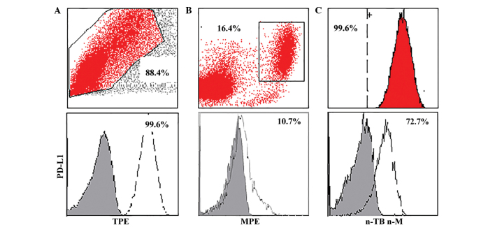
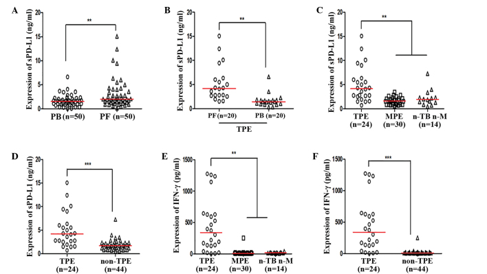
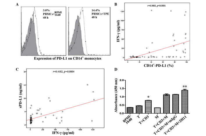
Soluble and membrane-bound programmed death ligand-1 (sPD-L1 and mPD-L1, respectively) have been demonstrated to participate in the immune suppression of non-small cell lung cancer. However, the contribution of sPD-L1 and mPD-L1 to immune regulation and disease progression in patients with pleural effusions remains unknown. The present study evaluated the levels of sPD-L1 and membrane-bound PD-1/PD-L1 in the peripheral blood and pleural effusions of patients with tuberculous pleural effusion (TPE), malignant pleural effusion (MPE) and non-tuberculous non-malignant pleural effusion (n-TB n-M). Furthermore, selected T lymphocytes and cluster of differentiation (CD)14+ monocytes were co-cultured to investigate the potential effect of the PD-1/PD-L1 pathway in TPE. Levels of sPD-L1 and PD-L1 on CD14+ monocytes were increased in the TPE group, as compared with the MPE and n-TB n-M groups. Furthermore, sPD-L1 levels and the expression levels of PD-L1 on CD14+ monocytes were demonstrated to be positively correlated with interferon (IFN)-γ concentration in pleural effusions. Therefore, IFN-γ may increase the expression of PD-L1 on CD14+ monocytes in vitro. Cell counting kit-8 analysis demonstrated that anti-PD-L1 antibody was able to partially reverse the proliferation of T lymphocytes in the co-culture system. The results of the present study indicated that sPD-L1 or mPD-L1 are associated with the immune regulation and disease progression of TPE, and may serve as possible biomarkers of TPE. Furthermore, sPD-L1 and the PD-1/PD-L1 pathway of TPE may be associated with the Th1 immune response; therefore, an anti-PD-1/PD-L1 pathway suggests a potential immune therapy strategy for the treatment of TPE.
Keywords: antibody; interferon-γ; programmed death ligand-1; soluble programmed death ligand-1; tuberculous pleural effusion.
Figures
Similar articles
-
[Level of soluble programmed death ligand 1 in pleural effusion and peripheral blood of patients with tuberculous pleural effusion and its clinical implications].Zhonghua Yi Xue Za Zhi. 2014 May 27;94(20):1543-6. Zhonghua Yi Xue Za Zhi. 2014. PMID: 25146741 Chinese.
-
Monocytes subtypes from pleural effusion reveal biomarker candidates for the diagnosis of tuberculosis and malignancy.J Clin Lab Anal. 2022 Aug;36(8):e24579. doi: 10.1002/jcla.24579. Epub 2022 Jul 12. J Clin Lab Anal. 2022. PMID: 35819097 Free PMC article.
-
Characterization of soluble PD-L1 in pleural effusions of mesothelioma patients: potential implications in the immune response and prognosis.J Cancer Res Clin Oncol. 2021 Feb;147(2):459-468. doi: 10.1007/s00432-020-03457-7. Epub 2020 Nov 20. J Cancer Res Clin Oncol. 2021. PMID: 33216211
-
Soluble Programmed Death Ligand-1 (sPD-L1): A Pool of Circulating Proteins Implicated in Health and Diseases.Cancers (Basel). 2021 Jun 17;13(12):3034. doi: 10.3390/cancers13123034. Cancers (Basel). 2021. PMID: 34204509 Free PMC article. Review.
-
Pleural manometry: techniques, applications, and pitfalls.J Thorac Dis. 2020 May;12(5):2759-2770. doi: 10.21037/jtd.2020.04.04. J Thorac Dis. 2020. PMID: 32642184 Free PMC article. Review.
Cited by
-
The clinical implication of soluble PD-L1 (sPD-L1) in patients with breast cancer and its biological function in regulating the function of T lymphocyte.Cancer Immunol Immunother. 2021 Oct;70(10):2893-2909. doi: 10.1007/s00262-021-02898-4. Epub 2021 Mar 10. Cancer Immunol Immunother. 2021. PMID: 33688997 Free PMC article.
-
PDL1 expression on monocytes is associated with plasma cytokines in Tuberculosis and HIV.PLoS One. 2021 Oct 1;16(10):e0258122. doi: 10.1371/journal.pone.0258122. eCollection 2021. PLoS One. 2021. PMID: 34597347 Free PMC article.
-
Diagnostic Usefulness of Cytokine and Chemokine Levels in the Cerebrospinal Fluid of Patients with Suspected Tuberculous Meningitis.Am J Trop Med Hyg. 2019 Aug;101(2):343-349. doi: 10.4269/ajtmh.18-0947. Am J Trop Med Hyg. 2019. PMID: 31264559 Free PMC article.
-
PD-L1 Expression in Monocytes Correlates with Bacterial Burden and Treatment Outcomes in Active Pulmonary Tuberculosis.Int J Mol Sci. 2022 Jan 30;23(3):1619. doi: 10.3390/ijms23031619. Int J Mol Sci. 2022. PMID: 35163542 Free PMC article.
-
Plasma immune profiling combined with machine learning contributes to diagnosis and prognosis of active pulmonary tuberculosis.Emerg Microbes Infect. 2024 Dec;13(1):2370399. doi: 10.1080/22221751.2024.2370399. Epub 2024 Jul 4. Emerg Microbes Infect. 2024. PMID: 38888093 Free PMC article.
References
-
- www.who.int/iris/bitstream/10665/191102/1/9789241565059_eng.pdf?ua=1 WHO global tuberculosis report. 2015
-
- Khan FY, Hamza M, Omran AH, Saleh M, Lingawi M, Alnaqdy A, Rahman MO, Ahmedullah HS, Hamza A, Ani AA, et al. Diagnostic value of pleural fluid interferon-gamma and adenosine deaminase in patients with pleural tuberculosis in Qatar. Int J Gen Med. 2013;6:13–18. doi: 10.2147/IJGM.S39345. - DOI - PMC - PubMed
LinkOut - more resources
Full Text Sources
Other Literature Sources
Research Materials