. 2016 Sep 23:7:12859.
doi: 10.1038/ncomms12859.

An imbalance between specialized pro-resolving lipid mediators and pro-inflammatory leukotrienes promotes instability of atherosclerotic plaques

Affiliations

An imbalance between specialized pro-resolving lipid mediators and pro-inflammatory leukotrienes promotes instability of atherosclerotic plaques

Gabrielle Fredman et al. Nat Commun. .

Abstract

Chronic unresolved inflammation plays a causal role in the development of advanced atherosclerosis, but the mechanisms that prevent resolution in atherosclerosis remain unclear. Here, we use targeted mass spectrometry to identify specialized pro-resolving lipid mediators (SPM) in histologically-defined stable and vulnerable regions of human carotid atherosclerotic plaques. The levels of SPMs, particularly resolvin D1 (RvD1), and the ratio of SPMs to pro-inflammatory leukotriene B4 (LTB4), are significantly decreased in the vulnerable regions. SPMs are also decreased in advanced plaques of fat-fed Ldlr-/- mice. Administration of RvD1 to these mice during plaque progression restores the RvD1:LTB4 ratio to that of less advanced lesions and promotes plaque stability, including decreased lesional oxidative stress and necrosis, improved lesional efferocytosis, and thicker fibrous caps. These findings provide molecular support for the concept that defective inflammation resolution contributes to the formation of clinically dangerous plaques and offer a mechanistic rationale for SPM therapy to promote plaque stability.


Figures
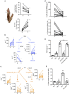
Figure 1
Figure 1. Human vulnerable atherosclerotic plaques have decreased 5-LOX SPM levels and a lower SPM:LT ratio than stable lesions.
(a) Flash-frozen human plaques were separated into vulnerable (V) and stable (S) regions (left panel), which were quantified for fibrous cap thickness:lesion area ratio (top right panel) and percent plaque necrosis (bottom right panel). (t-test, **P<0.01, n=8 donors). (b) Quantification of key DHA-derived lipid mediators, with their respective biosynthetic pathways indicated (LOX, lipoxygenase). (c) Quantification of key AA-derived lipid mediators. (d) Comparison of 5-LOX-derived SPMs (RvD1 and LXA4) and the SPM:LT ratio in vulnerable versus stable regions. For (bd), t-test, *P<0.05, n=15 donors. (e) Quantification of RvD1 by ELISA in human macrophages that were incubated for 5 h with vehicle control, or 35 μM of 7KC, followed by Veh or 10 μM DHA, for an additional 40 min, and then the media were assayed for RvD1 with or without a 1 h pre-incubation with 10 μM NAC. Data are shown as mean±s.e.m. (n=4 separate donors). Statistical analysis was conducted using one-way ANOVA with the Kruskal–Wallis test and Dunn's multiple comparison post-hoc analysis, *P<0.05 of n=4 separate donors. (f) Quantification nuclear:non-nuclear 5-LOX ratio by confocal microscopy in macrophages incubated as in e, with two additional groups, NAC alone and 7KC+NAC. Images were acquired on a Leica confocal microscope, and nuclear:non-nuclear 5-LOX MFI was quantified using Image J. Data are shown as mean±s.e.m. (n=4 separate donors). Statistical analysis was conducted using one-way ANOVA with the Kruskal–Wallis test with the Dunn's multiple comparison post-hoc analysis, *P<0.05.
Figure 2
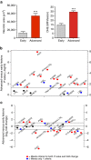
Figure 2. Advanced aortic root plaques of WD-fed Ldlr−/− mice exhibit an imbalance in 5-LOX-derived SPMs and LTs compared with earlier stage lesions.
(a) Aortic root lesions from Ldlr−/− mice fed the WD for 8 weeks (Early) or 17 weeks (Advanced) were quantified for necrotic area and mean fluorescence intensity after DHE staining (data are shown as mean±s.e.m., t-test, ***P<0.001, n=7 for early lesions and n=11 for advanced plaques). (b,c) Analysis of lipid mediators in advanced versus early atherosclerotic plaques. Red dots indicate mediators that both reached statistical difference by Student's t-test (−log10 of the P-value) and changed by at least twofold (log2); blue dots indicate mediators that met only one of these criteria; and black dots represent lipid mediators that met neither of these criteria. For b and c, n=8 for early lesions and n=11 for advanced lesions.
Figure 3
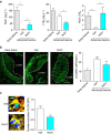
Figure 3. Administration of RvD1 to WD-fed Ldlr−/− mice with established atherosclerosis restores lipid medicator balance.
(a) Quantification of lesional RvD1 and LTB4 levels (left and middle panels) and RvD1:LTB4 ratio (right panel) in aortic lesions of 17-week WD-fed Ldlr−/− mice that were administered vehicle control (Veh) or 100 ng RvD1/mouse (3 × /week i.p.) during weeks 12–17; the ratio data in 3a right panel are also shown for mice fed the WD for 8 weeks (Early lesions). For a, data are shown as mean±s.e.m., one-way ANOVA with Fisher's least significant difference post-hoc analysis, *P<0.05, n=8 for early lesions, n=11 for advanced lesions/Veh group, and n=10 for advanced lesions/RvD1 group. (b) Representative images and quantification of lesional oxCaMKII by immunofluorescence microscopic analysis. MFI data are shown as mean±s.e.m., one-way ANOVA with Tukey's multiple comparison test, *P<0.05 and ***P<0.001 early lesions versus other groups. ^P<0.05 Veh versus RvD1 groups, n=8 for early lesions, n=11 for advanced lesions/Veh group, and n=10 for advanced lesions/RvD1 group. Scale bar, 100 μm. (c) Representative confocal immunofluorescence images and quantification of 5-LOX localization in aortic lesional macrophages of 17-week WD-fed Ldlr−/− mice given vehicle control (Veh) or RvD1 during weeks 12–17. Macrophage F4/80 is red, 5-LOX is green, and Hoechst (nuclei) is blue. Arrows indicate nuclear region. In these images, nuclear 5-LOX appears as blotchy light blue staining within the dark blue Hoechst-stained nuclei. The quantitative data are shown as mean±s.e.m., t-test, *P<0.05, n=11 for Veh group and n=10 for RvD1 group. Scale bar, 5 μm.
Figure 4
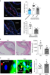
Figure 4. Administration of RvD1 to mice with established atherosclerosis suppresses lesional ROS and necrosis and enhances efferocytosis.
(a) Representative images of DHE and quantification of immunoreactive NOX2 and DHE in aortic root lesions of 8-week and 17-week WD-fed Ldlr−/− mice given vehicle control (Veh) or RvD1 for weeks 12–17 (Fig. 3). Scale bar, 100 μm. Data are shown as mean±s.e.m., one-way ANOVA with Tukey's multiple comparison test, ***P<0.001 early lesions versus other groups. ^^P<0.01 Veh versus RvD1 groups, n=8 for early lesions, n=11 for advanced lesions, n=10 for advanced lesions/RvD1 group. (b) Representative images and quantification of lesional necrosis in the two cohorts of mice. Scale bar, 100 μm. (c) Representative images and quantification of lesional efferocytosis, quantified as the ratio of TUNEL+ apoptotic cells (red) associated with lesional macrophages (green) versus apoptotic cells not associated with macrophages (‘free'). Yellow indicates red/green overlap, and blue indicates DAPI-stained nuclei. Scale bar, 5 μm. Data are shown as mean±s.e.m., t-test, *P<0.05, **P<0.01, n=11 for Veh group and n=10 for RvD1 group.
Figure 5
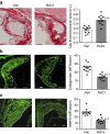
Figure 5. Administration of RvD1 to WD-fed Ldlr−/− mice with established atherosclerosis enhances fibrous caps and decreases collagenase and MMP9.
(a) Representative images and quantification of collagen of 17-week WD-fed Ldlr−/− mice given vehicle control (Veh) or RvD1 for weeks 12–17 (Fig. 3). Scale bar, 100 μm. (b,c) Representative images and quantification of collagenase and MMP9. Scale bar, 100 μm. Data are shown as mean±s.e.m., t-test, **P<0.01 and ***P<0.001, n=11 for Veh group and n=10 for RvD1 group.

Similar articles

Cited by

References

    1. Merched A. J., Ko K., Gotlinger K. H., Serhan C. N. & Chan L. Atherosclerosis: evidence for impairment of resolution of vascular inflammation governed by specific lipid mediators. FASEB J. 22, 3595–3606 (2008). - PMC - PubMed
    1. Tabas I. Macrophage death and defective inflammation resolution in atherosclerosis. Nat. Rev. Immunol. 10, 36–46 (2010). - PMC - PubMed
    1. Libby P., Tabas I., Fredman G. & Fisher E. A. Inflammation and its resolution as determinants of acute coronary syndromes. Circ. Res. 114, 1867–1879 (2014). - PMC - PubMed
    1. Viola J. & Soehnlein O. Atherosclerosis—a matter of unresolved inflammation. Semin. Immunol. 27, 184–193 (2015). - PubMed
    1. Serhan C. N. Pro-resolving lipid mediators are leads for resolution physiology. Nature 510, 92–101 (2014). - PMC - PubMed