Review
. 2016 Oct;96(4):1567-91.
doi: 10.1152/physrev.00004.2016.

Idiopathic Pulmonary Fibrosis: A Genetic Disease That Involves Mucociliary Dysfunction of the Peripheral Airways

Affiliations
Review

Idiopathic Pulmonary Fibrosis: A Genetic Disease That Involves Mucociliary Dysfunction of the Peripheral Airways

Christopher M Evans et al. Physiol Rev. 2016 Oct.

Abstract

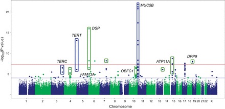
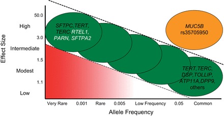
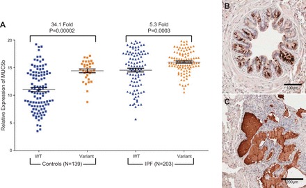
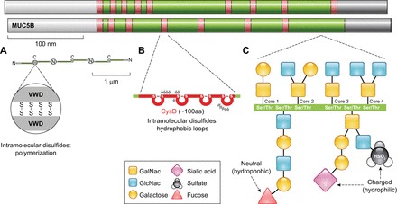
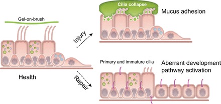
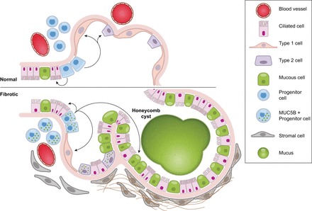
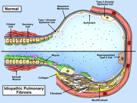
Idiopathic pulmonary fibrosis (IPF) is an incurable complex genetic disorder that is associated with sequence changes in 7 genes (MUC5B, TERT, TERC, RTEL1, PARN, SFTPC, and SFTPA2) and with variants in at least 11 novel loci. We have previously found that 1) a common gain-of-function promoter variant in MUC5B rs35705950 is the strongest risk factor (genetic and otherwise), accounting for 30-35% of the risk of developing IPF, a disease that was previously considered idiopathic; 2) the MUC5B promoter variant can potentially be used to identify individuals with preclinical pulmonary fibrosis and is predictive of radiologic progression of preclinical pulmonary fibrosis; and 3) MUC5B may be involved in the pathogenesis of pulmonary fibrosis with MUC5B message and protein expressed in bronchiolo-alveolar epithelia of IPF and the characteristic IPF honeycomb cysts. Based on these considerations, we hypothesize that excessive production of MUC5B either enhances injury due to reduced mucociliary clearance or impedes repair consequent to disruption of normal regenerative mechanisms in the distal lung. In aggregate, these novel considerations should have broad impact, resulting in specific etiologic targets, early detection of disease, and novel biologic pathways for use in the design of future intervention, prevention, and mechanistic studies of IPF.


Figures

FIGURE 1.
FIGURE 1.
Traditional and hypothesized relationship between fibroblastic foci and honeycomb cysts. The traditional thinking is that honeycomb cysts are a consequence of extensive fibroproliferation throughout the lung parenchymal and represent a pathophysiological consequence of persistent fibroproliferation. Based on our findings and observations made by other investigators, we have hypothesized persistent bronchiolo-alveolar injury results in a failure to repair the airway epithelia and that independent of fibroproliferation, this inability to regenerate the distal airway epithelia results in honeycomb formation. We further hypothesize that overexpression of MUC5B by airway progenitor cells inhibits normal repair and accelerates the development of honeycomb cysts.
FIGURE 2.
FIGURE 2.
Kaplan-Meier survival curves for patients with IPF by MUC5B promoter variant rs35705950 genotype for the INSPIRE cohort (A) and the Chicago cohort (B). In the INSPIRE cohort, the hazard ratios controlled for age, gender, FVC, DLCO, and MMP7. In the Chicago cohort, the hazard ratios controlled for age, gender, FVC, and DLCO. [Adapted from Peljto et al. (158), with permission from American Medical Association.]
FIGURE 3.
FIGURE 3.
GWAS results at 439,828 SNPs with 1,616 cases and 4,683 controls under additive model. SNPs above red line were genome-wide significant at P < 5 × 10⁻⁸. These SNPs and SNPs between red and blue lines, corresponding to 5 × 10⁻⁸ < P value < 0.0001 were followed up in 876 cases and 1,890 controls. [Adapted from Fingerlin et al. (47), with permission from Nature Publishing Group.]
FIGURE 4.
FIGURE 4.
The genetic basis of familial and sporadic idiopathic interstitial pneumonia is similar. A similar risk is conferred by common variants at each of the major IPF loci for both familial and sporadic forms of IPF. [Data derived from recent GWAS in Fingerlin et al. (47) and previously presented in Mathai et al. (128), with permission from Wolters Kluwer Health, Inc.]
FIGURE 5.
FIGURE 5.
Relationship between allele frequency and penetrance of the risk allele, with examples of genes with risk alleles for IPF. Genes in black font have variants reported for both familial and sporadic IPF, and genes in white font have variants reported only for familial IPF. [Adapted from Figure 1 in Antonarakis et al. (2), with permission from Nature Publishing Group.]
FIGURE 6.
FIGURE 6.
A: MUC5B expression in IPF (n = 203) and unaffected subjects (n = 139) stratified by MUC5B promoter variant (rs35705950) genotype. MUC5B is produced in the bronchiolo-alveolar epithelia (B) and honeycomb cysts (C) of patients with IPF. [Adapted from Seibold et al. (178).]
FIGURE 7.
FIGURE 7.
Polymeric mucin structures. MUC5AC and MUC5B apoprotein backbones are comprised by NH2-terminal (white) and COOH-terminal (gray) cysteine-rich domains that are conserved in polymeric mucins and von Willebrand factor. The central portions of MUC5AC and MUC5B are comprised of a mucin glycosylation domain (green) that is rich in serines and threonines that are sites of O-linked glycan attachments. The glycosylation domain is interrupted by additional cysteine-rich CysD domains (red). A: the COOH-terminal von Willebrand-like domain (VWD) has a cysteine-knot structure that is the site of disulfide dimerization in the endoplasmic reticulum; each dimer is ∼1 μm in length. In the Golgi, dimers further assemble at their NH2 termini via covalent disulfide bonds and other noncovalent interactions to form extensive polymeric structures that are 10s to 100s of microns long. B: in cysteine-rich CysD domains, intramolecular disulfide bonds also assemble. CysDs contain hydrophobic amino acids and form loops that may promote mucin mesh network alignment. C: in the mucin glycosylation domain, glycan linkages occur on the hydroxylated ends of serine and threonine residues through the initial attachment of N-acetylgalactosamine (GalNac). This initial O-glycosylation step is followed by galactose and/or N-acetylglucosamine (GlcNac) attachments in specific core linkages. These can be elaborated with additional galactose and GlcNac moieties and be modified further by the attachment of fucose, sialic acid, and sulfate end groups. These terminal groups carry charges and structural characteristics that mediate mucin biophysical properties and interactions with host-derived and foreign materials.
FIGURE 8.
FIGURE 8.
Regulation of motile and nonmotile cilia in the lungs. Under conditions of health, multiciliated cells are present in conducting airways. These specialized epithelial cells maintain the normal structure and function of apically localized motile cilia clusters through a balance of electrolyte and osmolyte homeostasis. In addition, membrane tethered mucins present along cilia adsorb water and maintain a stable “grafted brush” structure that supports an overlying gel. Normal repair programs following injury activate progenitor basal-like cells to differentiate fully to secretory and ciliated cells. Primary nonmotile cilia precede motile cilia in this normal repair process. Under conditions where mucins are overproduced or hypersecreted (such as the MUC5B promoter variant rs35705950), or when electrolyte homeostasis is disrupted (e.g., through excessive Na+ absorption), cilia collapse and aggregates of mucus adhere to airway surfaces, potentially worsening injury. Under injurious conditions, resulting from excess mucus or other forms of lung injury, aberrant repair could lead to abnormal activation of progenitor basal-like cells, resulting in partially differentiated ciliated cells that retain primary cilia, have poorly developed apical motile cilia, and developmental programs that are aberrantly activated.
FIGURE 9.
FIGURE 9.
Indicates that the “at-risk” population and the population with preclinical pulmonary fibrosis is large (19% with the MUC5B promoter variant and 1.8% of individuals = 50 years of age, respectively), IPF is diagnosed in a small population with established end-stage disease (111, 163), and preclinical pulmonary fibrosis can be identified using the MUC5B variant rs35705950. In addition, recent findings (3) indicate that preclinical pulmonary fibrosis (detected via chest CT scan) is associated with a poor prognosis, suggesting that preclinical pulmonary fibrosis may be a precursor of IPF.
FIGURE 10.
FIGURE 10.
Gene expression profiling identifies two subtypes of IPF. mRNA profiles from 119 IPF lungs were subject to hierarchical clustering based on the expression of 472 transcripts that are differentially expressed at 5% FDR and with greater than 2-fold change in IPF compared with control lung. The most prominent feature of the heatmap is the group of 51 subjects (43%; subject group II) with relatively high expression compared with 68 subjects (57%; subject group I) of a large set of transcripts (transcript clusters A and B) and low expression of another set of transcripts (transcript cluster C). Transcript cluster B contains 80 unique transcripts upregulated in group II compared with group I IPF that include a number of genes that have been previously shown to be upregulated in IPF/UIP, namely, osteopontin, MMP1, MMP7, PLUNC, MUC5B, collagen COL17A1, and keratins 5, 6C, 15, and 17. Cluster C contains 71 unique transcripts that are downregulated in group II compared with group I IPF with a few previously implicated in IPF [advanced glycosylation end product-specific receptor (AGER)] or other chronic lung diseases [hedgehog interacting protein (HHIP)] and many novel IPF candidate genes. Functional enrichment analysis, using Fisher exact test, of the 121 unique transcripts in cluster A showed it to be strongly enriched in transcripts associated with the cilium genes (Benjamini corrected P value 3.7 × 10⁻¹¹) and their structural components (axoneme, 3.9 × 10⁻¹¹; dynein, 9.4 × 10⁻⁷). This cluster also contains a number of genes with unknown function. Decreased expression is represented by blue color while red indicates increased expression. [Adapted from Yang et al. (232), with permission from BMJ Publishing Group Ltd.]
FIGURE 11.
FIGURE 11.
Model of stem cells repopulating bronchioles and alveoli under normal physiological conditions and when challenged with increased expression of MUC5B. We hypothesize that excessive production of MUC5B by stem cells that attempt to regenerate injured bronchiolar and alveolar epithelium disrupt normal developmental pathways and highjack the normal reparative mechanisms in the distal lung, resulting in chronic fibroproliferation and honeycomb cyst formation.
FIGURE 12.
FIGURE 12.
Model of recurrent inflammation/injury/repair at the bronchoalveolar junction that is initiated and exacerbated by overexpression of MUC5B, retention of inhaled particles, and enhanced lung injury. Top panel is the normal bronchoalveolar region, and bottom panel represents a bronchoalveolar region affected by IPF. We hypothesize that IPF is a mucociliary disease that is caused by recurrent injury/inflammation/repair at the bronchoalveolar junction that is initiated and exacerbated by overexpression of MUC5B leading to reduced ciliary function, retention of particles, and enhanced injury.

Similar articles

Cited by

References

    1. Adelman AG, Chertkow G, Hayton RC. Familial fibrocystic pulmonary dysplasia: a detailed family study. Can Med Ass J 95: 603–610, 1966. - PMC - PubMed
    1. Antonarakis SE, Chakravarti A, Cohen JC, Hardy J. Mendelian disorders and multifactorial traits: the big divide or one for all? Nat Rev Genet 11: 380–384, 2010. - PubMed
    1. Araki T, Putman RK, Hatabu H, Gao W, Dupuis J, Latourelle JC, Nishino M, Zazueta OE, Kurugol S, Ross JC, Estepar RS, Schwartz DA, Rosas IO, Washko GR, O'Connor GT, Hunninghake GM. Development and progression of interstitial lung abnormalities in the Framingham Heart Study. Am J Respir Crit Care Med. In press. - PMC - PubMed
    1. Armanios MY, Chen JJ, Cogan JD, Alder JK, Ingersoll RG, Markin C, Lawson WE, Xie M, Vulto I, Phillips JA 3rd, Lansdorp PM, Greider CW, Loyd JE. Telomerase mutations in families with idiopathic pulmonary fibrosis. N Engl J Med 356: 1317–1326, 2007. - PubMed
    1. Baek HA, Kim do S, Park HS, Jang KY, Kang MJ, Lee DG, Moon WS, Chae HJ, Chung MJ. Involvement of endoplasmic reticulum stress in myofibroblastic differentiation of lung fibroblasts. Am J Respir Cell Mol Biol 46: 731–739, 2012. - PubMed

Publication types

MeSH terms