

Aberrant expression of KPNA2 is associated with a poor prognosis and contributes to OCT4 nuclear transportation in bladder cancer
- PMID: 27611951
- PMCID: PMC5341943
- DOI: 10.18632/oncotarget.11889
Aberrant expression of KPNA2 is associated with a poor prognosis and contributes to OCT4 nuclear transportation in bladder cancer
Abstract
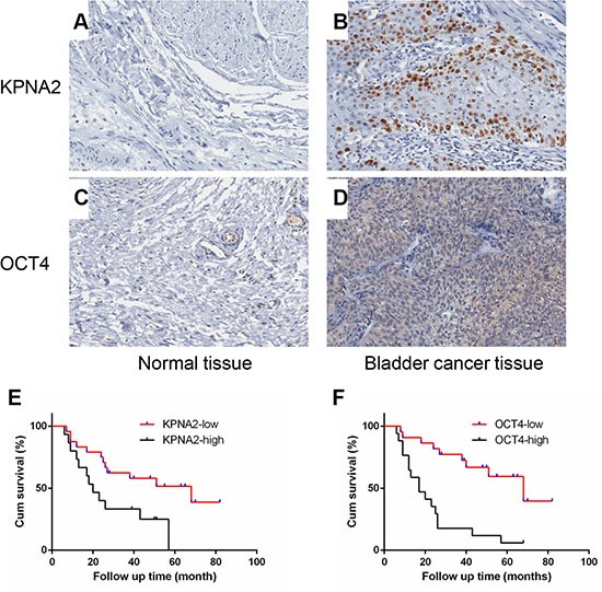
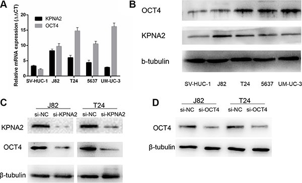
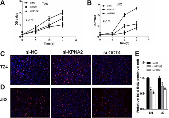
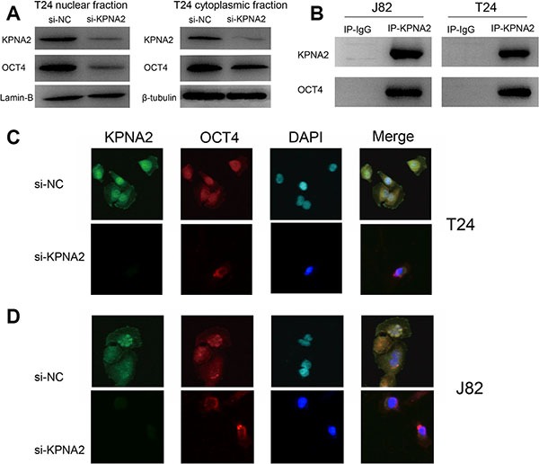
Recent studies show that Karyopherin alpha 2 (KPNA2) is up-regulated in quite a number of cancers and associated with poor prognosis. Here, we found that expression levels of KPNA2 and OCT4 are up-regulated in bladder cancer tissues and significantly associated with primary tumor stage and bladder cancer patients' poorer prognosis. Our data also showed decreased cell proliferation and migration rates of bladder cancer cell lines when the expression of KPNA2 and OCT4 was silenced. Meanwhile, cell apoptosis rate was increased. Furthermore, Co-IP and immunofluorescence assay showed the KPNA2 interacts with OCT4 and inhibits OCT4 nuclear transportation when KPNA2 was silenced. Thus, we confirmed that up-regulated KPNA2 and OCT4 expression is a common feature of bladder cancer that is correlated with increased aggressive tumor behavior. Also, we propose that KPNA2 regulates the process of OCT4 nuclear transportation in bladder cancer.
Keywords: KPNA2; OCT4; bladder cancer; nucleo-cytoplasmic transport; prognostic.
Conflict of interest statement
None.
Figures

Similar articles
-
Downregulation of KPNA2 in non-small-cell lung cancer is associated with Oct4 expression.J Transl Med. 2013 Sep 26;11:232. doi: 10.1186/1479-5876-11-232. J Transl Med. 2013. PMID: 24070213 Free PMC article.
-
Karyopherin alpha 2 is a novel prognostic marker and a potential therapeutic target for colon cancer.J Exp Clin Cancer Res. 2015 Dec 1;34:145. doi: 10.1186/s13046-015-0261-3. J Exp Clin Cancer Res. 2015. PMID: 26626145 Free PMC article.
-
MiR-26b/KPNA2 axis inhibits epithelial ovarian carcinoma proliferation and metastasis through downregulating OCT4.Oncotarget. 2015 Sep 15;6(27):23793-806. doi: 10.18632/oncotarget.4363. Oncotarget. 2015. PMID: 26204489 Free PMC article.
-
The emerging roles of KPNA2 in cancer.Life Sci. 2020 Jan 15;241:117140. doi: 10.1016/j.lfs.2019.117140. Epub 2019 Dec 6. Life Sci. 2020. PMID: 31812670 Review.
-
Novel roles of karyopherin subunit alpha 2 in hepatocellular carcinoma.Biomed Pharmacother. 2023 Jul;163:114792. doi: 10.1016/j.biopha.2023.114792. Epub 2023 Apr 28. Biomed Pharmacother. 2023. PMID: 37121148 Review.
Cited by
-
Polygenic risk modeling of tumor stage and survival in bladder cancer.BioData Min. 2022 Sep 30;15(1):23. doi: 10.1186/s13040-022-00306-w. BioData Min. 2022. PMID: 36175974 Free PMC article.
-
Low Expression of Pseudogene POU5F1B Affects Diagnosis and Prognosis in Acute Myeloid Leukemia (AML).Med Sci Monit. 2019 Jul 4;25:4952-4959. doi: 10.12659/MSM.914352. Med Sci Monit. 2019. PMID: 31271156 Free PMC article.
-
Identification of Key Biomarkers in Bladder Cancer: Evidence from a Bioinformatics Analysis.Diagnostics (Basel). 2020 Jan 24;10(2):66. doi: 10.3390/diagnostics10020066. Diagnostics (Basel). 2020. PMID: 31991631 Free PMC article.
-
Increased Nuclear Transporter KPNA2 Contributes to Tumor Immune Evasion by Enhancing PD-L1 Expression in PDAC.J Immunol Res. 2021 Mar 1;2021:6694392. doi: 10.1155/2021/6694392. eCollection 2021. J Immunol Res. 2021. PMID: 33728352 Free PMC article.
-
Prognostic value of octamer binding transcription factor 4 for patients with solid tumors: A meta-analysis.Medicine (Baltimore). 2020 Oct 16;99(42):e22804. doi: 10.1097/MD.0000000000022804. Medicine (Baltimore). 2020. PMID: 33080755 Free PMC article.
References
-
- Chen W, Zheng R, Baade PD, Zhang S, Zeng H, Bray F, Jemal A, Yu XQ, He J. Cancer statistics in China, 2015. CA Cancer J Clin. 2016;66:115–132. - PubMed
-
- Jacobs BL, Lee CT, Montie JE. Bladder cancer in 2010: how far have we come? CA Cancer J Clin. 2010;60:244–272. - PubMed
-
- Hurst CD, Knowles MA. Molecular subtyping of invasive bladder cancer: time to divide and rule? Cancer Cell. 2014;25:135–136. - PubMed
-
- Dal Moro F, Valotto C, Guttilla A, Zattoni F. Urinary markers in the everyday diagnosis of bladder cancer. Urologia. 2013;80:265–275. - PubMed
-
- Al Hussain TO, Akhtar M. Molecular basis of urinary bladder cancer. Adv Anat Pathol. 2013;20:53–60. - PubMed
MeSH terms
Substances
LinkOut - more resources
Full Text Sources
Other Literature Sources
Medical
Miscellaneous
