. 2016 Aug 19:7:12531.
doi: 10.1038/ncomms12531.

β-arrestin-2 regulates NMDA receptor function in spinal lamina II neurons and duration of persistent pain

Affiliations

β-arrestin-2 regulates NMDA receptor function in spinal lamina II neurons and duration of persistent pain

Gang Chen et al. Nat Commun. .

Abstract

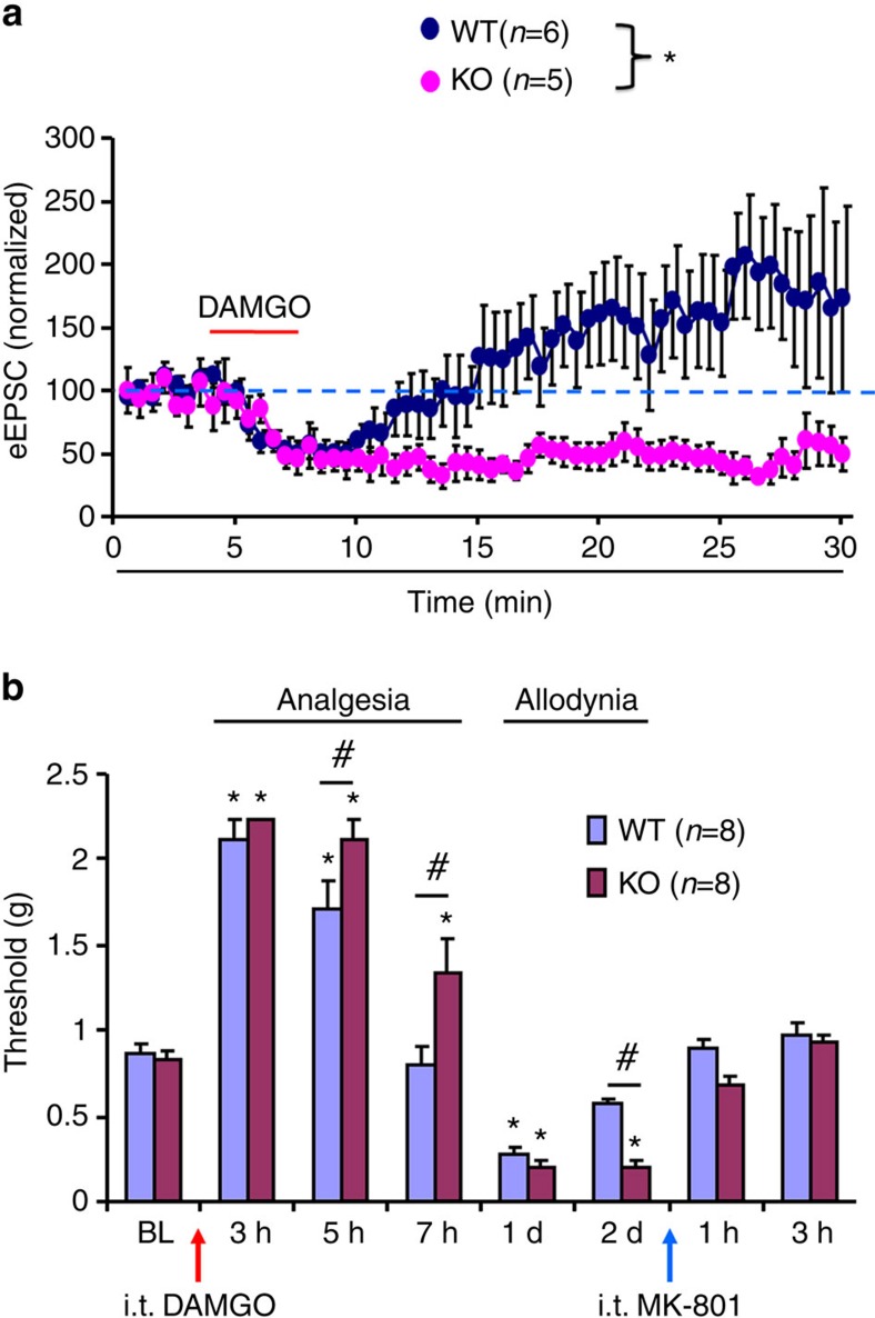
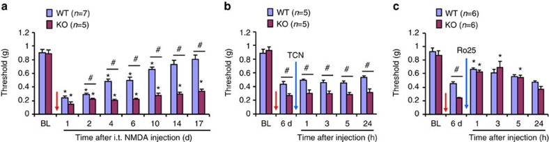
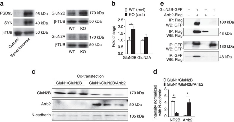
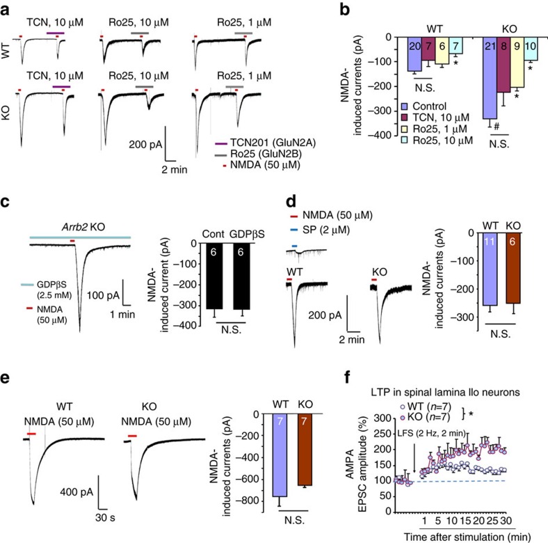
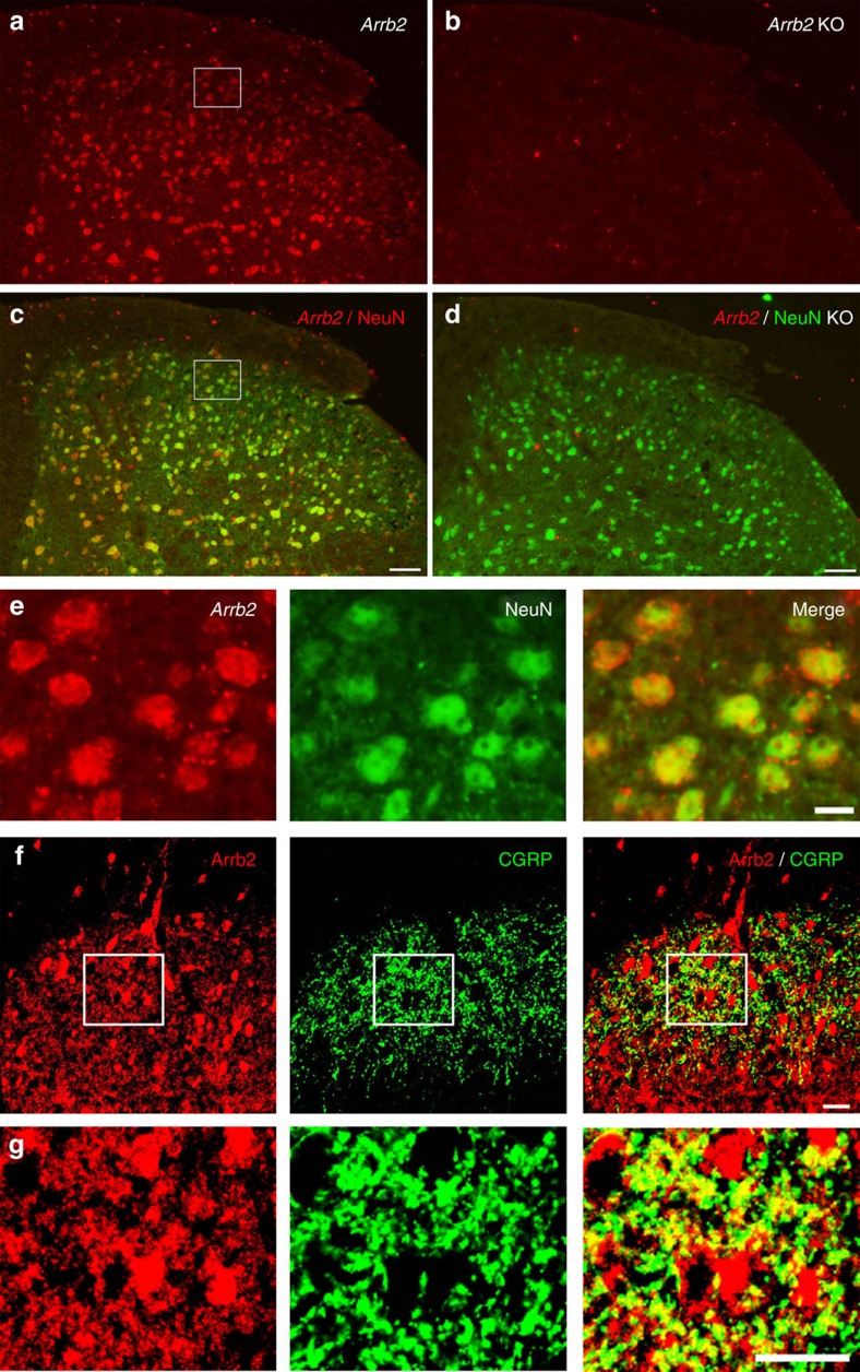
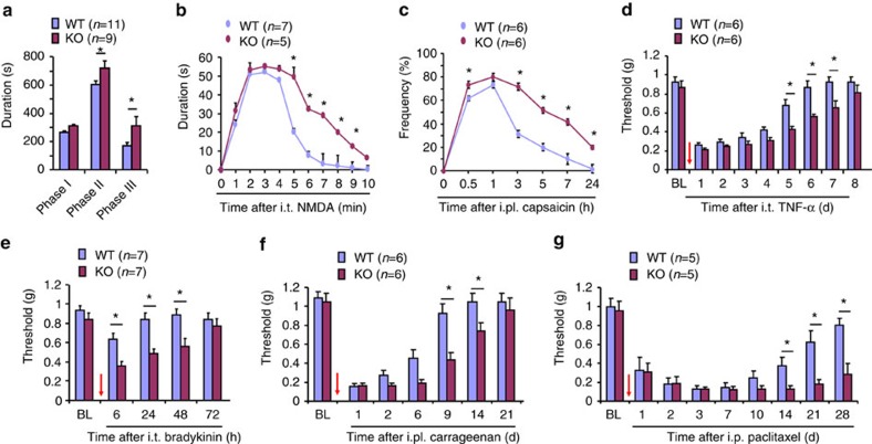
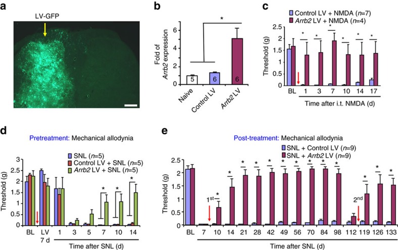
Mechanisms of acute pain transition to chronic pain are not fully understood. Here we demonstrate an active role of β-arrestin 2 (Arrb2) in regulating spinal cord NMDA receptor (NMDAR) function and the duration of pain. Intrathecal injection of the mu-opioid receptor agonist [D-Ala(2), NMe-Phe(4), Gly-ol(5)]-enkephalin produces paradoxical behavioural responses: early-phase analgesia and late-phase mechanical allodynia which requires NMDAR; both phases are prolonged in Arrb2 knockout (KO) mice. Spinal administration of NMDA induces GluN2B-dependent mechanical allodynia, which is prolonged in Arrb2-KO mice and conditional KO mice lacking Arrb2 in presynaptic terminals expressing Nav1.8. Loss of Arrb2 also results in prolongation of inflammatory pain and neuropathic pain and enhancement of GluN2B-mediated NMDA currents in spinal lamina IIo not lamina I neurons. Finally, spinal over-expression of Arrb2 reverses chronic neuropathic pain after nerve injury. Thus, spinal Arrb2 may serve as an intracellular gate for acute to chronic pain transition via desensitization of NMDAR.


Conflict of interest statement

The authors declare no competing financial interests.

Figures

Figure 1
Figure 1. Arrb2 regulates sLTP and DAMGO-induced analgesia and allodynia.
(a) Normalized amplitudes of dorsal root stimulation evoked EPSCs (eEPSCs) in lamina IIo neurons before, during and after DAMGO (500 nM) perfusion in spinal cord slices in wild-type (WT) and Arrb2-knockout (KO) mice. In WT mice, DAMGO produces initial suppression of EPSCs followed by long-term potentiation (LTP) of eEPSCs during withdrawal phase. Spinal LTP (sLTP) is abolished in KO mice, only showing prolonged suppression of eEPSCs. *P<0.05, Two-Way ANOVA (WT. versus KO), n=5, 6 neurons per group. (b) von Frey test shows acute analgesia and late-onset mechanical allodynia following i.t. DAMGO (1 μg) in WT and KO mice and the reversal of allodynia by MK-801 (i.t., 10 nmol). *P<0.05, versus baseline (BL), #P<0.05 (WT versus KO). Two-Way ANOVA, n=8 mice per group. All the data are mean±s.e.m. d, day.
Figure 2
Figure 2. Arrb2 deficiency causes prolongation of NMDA-induced allodynia.
(a) Prolongation of NMDA (1 nmol, i.t.)-induced mechanical allodynia in Arrb2 KO mice. *P<0.05, versus baseline (BL), #P<0.05 (KO versus WT), Two-Way ANOVA, n=5, 7 mice per group. (b) GluN2A antagonist TCN-201 (10 nmol, i.t.) fails to reduce the NMDA-induced allodynia. (c) GluN2B antagonist Ro25-6091 (10 nmol, i.t.) reduces the NMDA-induced allodynia in WT and KO mice. *P<0.05, versus pre-NMDA injection baseline (6 d), One-Way ANOVA, n=6 mice per group. #P<0.05 (WT versus KO), Two-Way ANOVA, n=6 mice per group. BL, baseline. The antagonists were given 6 d after i.t. NMDA injection. Note that the difference between WT and KO mice is abolished by the GluN2B inhibition. All the data are mean±s.e.m. d, day.
Figure 3
Figure 3. Arrb2 deficiency increases synaptic GluN2B in SDH neurons.
(a,b) Expression of GluN2A and GluN2B in synaptosome-containing P2 fraction of spinal cord dorsal horn (SDH) of WT and Arrb2-KO mice. (a) Enrichment of synaptic proteins PSD-95 and synaptophysin (SYN) in synaptosome-contacting P2 fraction but not cytosol fraction. (b) Quantification of GluN2A and GluN2B expression. *P<0.05, Student t-test, n=4 mice per group. (c) Surface expression of GluN2B and Arrb2 in HeLa cells transfected with GluN1/GluN2B and GluN1/GluN2B/Arrb2. (d) Relative expression levels of GluN2B and Arrb2, normalized with N-cadherin, a positive control for surface expression. *P<0.05, Student’s t-test, n=3 cultures per group. (e) Pull down assay showing Co-IP of Arrb2 with GluN2B in HeLa cells. All the data are mean±s.e.m. Gel images have been cropped for presentation. Full size images are presented in Supplementary Fig. 8a.
Figure 4
Figure 4. Arrb2 deficiency enhances GluN2B currents in spinal lamina IIo neurons.
(a) Representative traces of inward currents in WT and KO mice, induced by NMDA (50 μM) via bath application. Note a remarkable potentiation of the current in KO mice. Also note different effects of TCN-201 (GluN2A antagonist) and Ro25-6091 (GluN2B antagonist). (b) Amplitude of NMDA-induced currents from (a). *P<0.05, versus corresponding control, #P<0.05 (WT versus KO), One-Way ANOVA, n=6–21 neurons per group (shown in each column). N.S., not significant. (c) Representative trace of NMDA current in KO mice in the presence of G-protein inhibitor GDPβS (2.5 mM) via intracellular delivery through the recording pipette. Right, amplitude of NMDA-induced currents. N.S., no significance, Student’s t-test, n=6 neurons per group. (d,e) NMDA currents in spinal lamina I neurons and hippocampal CA1 neurons are comparable in WT and Arrb2-KO mice. (d) Traces of NMDA (50 μM)-induced currents in lamina I neurons of spinal slices. The projection neurons respond to substance P (2 μM). Right, amplitude of NMDA-induced currents. N.S., no significance, n=6 and 11 neurons per group. (e) Traces of NMDA (50 μM)-induced currents in hippocampal CA1 neurons from WT and KO mice. Right, amplitude of NMDA currents in hippocampal CA1 neurons. N.S., no significance, Student’s t-test, n=7 neurons per group. (f) Spinal LTP of C-fibre evoked EPSCs (eEPSCs) in lamina IIo neurons of spinal cord slices in WT and KO mice following low frequency dorsal root stimulation (LFS, 2 Hz). *P<0.05, WT versus KO, Two-way ANOVA, n=7 neurons per group. All data are expressed as mean±s.e.m.
Figure 5
Figure 5. Expression of Arrb2 mRNA and Arrb2 protein in SDH.
(a-d) Double staining of in situ hybridization and immunohistochemistry showing colocalization of Arrb2 mRNA and NeuN immunoreactivity in SDH neurons. (a,c) Colocalization of Arrb2 mRNA and NeuN immunoreactivity in SDH neurons of WT mice. (b,d) Loss of Arrb2 mRNA labelling in Arrb2 KO mice. Scale, 50 μm. (e) Enlarged images from the boxes in (a) and (c) in the superficial SDH. Note that Arrb2 mRNA is highly co-localized with NeuN in SDH neurons. Scale, 10 μm. (f) Double staining of Arrb2 and CGRP in axonal terminals in the superficial SDH. Scale, 20 μm. (g) High magnification images of boxes in f showing co-localization of Arrb2 and CGRP in axonal terminals but not in cell bodies. Scale, 20 μm.
Figure 6
Figure 6. Arrb2 deficiency causes prolongation of inflammatory and neuropathic pain.
(a-g) Inflammatory pain (a-f) and neuropathic pain (g) in wildtype (WT) and Arrb2-knockout (KO) mice. (a) Acute inflammatory pain in Phase-I (0–10 min), Phase-II (10–45 min) and Phase-III (45–90 min) following intraplantar (i.pl.) formalin. (b) Spontaneous pain after i.t. NMDA (1 nmol). (c) 2nd mechanical allodynia following i.pl. capsaicin (5 μg), measured by frequency response to a 0.16 g filament. (d,e) Mechanical allodynia following i.t. TNF-α (20 ng, d) and i.t. bradykinin (1 μg, e). (f) Persistent inflammatory pain (mechanical allodynia) following i.pl. carrageenan (1.5%). (g) Neuropathic pain (mechanical allodynia) after intraperitoneal paclitaxel (6 mg/kg). *P<0.05 (WT versus KO), n=5–11 mice per group, Two-Way ANOVA followed by post-hoc Bonferroni test. Arrows (d-g) indicate drug injections. All data are expressed as mean±s.e.m.
Figure 7
Figure 7. Spinal cord presynaptic Arrb2 deficiency enhances NMDAR function.
(a) Single-cell PCR shows the expression of Arrb1, Arrb2, Nav1.8 and Gapdh mRNA in 5 small-sized DRG neurons of WT and Arrb2 CKO mice with Arrb2 deletion in Nav1.8-expressing sensory neurons. Note that Arrb2 but not Arrb1 is absent in small-sized DRG neurons of CKO mice. M, molecular weight; N, negative control. Gapdh is used as positive control. Gel images have been cropped for presentation. Full size images are presented in Supplementary Fig. 8b. (b) Traces of NMDAR-induced eEPSCs in WT, KO, CKO and their littermate control (LC) mice following dorsal root stimulation. (c) Amplitude of synaptic currents (dorsal root stimulation-evoked and NMDAR-mediated eEPSCs) of WT and KO mice as well as in CKO mice and LC mice. *P<0.05, Two-Way ANOVA, n=7–10 neurons per group. Note that KO mice show larger currents than CKO mice. (d) Spontaneous pain (0–10 min) after i.t. NMDA (1 nmol) injection. *P<0.05 versus KO and CKO mice, One-way ANOVA. Note that spontaneous pain is potentiated in KO but not CKO mice. (e) Mechanical allodynia after i.t. NMDA (1 nmol) injection. $P<0.05, Two-Way ANOVA; *P<0.05, #P<0.05 (KO versus CKO), Two-Way ANOVA followed by post-hoc Bonferroni test, n=5–7 mice per group. Note that allodynia is prolonged in both KO and CKO mice. All data are expressed as mean±s.e.m.
Figure 8
Figure 8. Spinal over-expression of Arrb2 prevents and reverses neuropathic pain.
(a) Unilateral SDH microinjections of Arrb2-lentivirus (LV) target ipsilateral SDH. Yellow arrow indicates the injection site. Scale, 200 μm. (b) Arrb2 mRNA expression in SDH 2 weeks after the LV injections. *P<0.05, versus naïve control and Control LV, n=5–6 mice per group. Following laminectomy, LV injections (2 × 0.4 μl≈105 TU) were made into the L5-SDH via a glass pipette. Scale, 200 μm. (c,d) Prevention of mechanical allodynia after i.t. NMDA (1 nmol, c) and spinal nerve ligation (SNL, d) by intra-spinal pretreatment of Arrb2-LV, given 7 d before the NMDA injection or SNL. *P<0.05, Two-Way ANOVA. n=4–7 mice per group. Arrows indicate the injections. (e) Reversal of spinal nerve ligation (SNL)-induced mechanical allodynia by intra-spinal post-treatment of Arrb2-LV, given one week after SNL. Note that a second post-treatment (indicated by the red arrow) of LV on day 112 is still effective in reversing mechanical allodynia. *P<0.05, Two-Way ANOVA followed by post-hoc Bonferroni test. n=9 mice per group. Arrows indicate the injections. All data are expressed as mean±s.e.m.

Similar articles

Cited by

References

    1. Basbaum A. I., Bautista D. M., Scherrer G. & Julius D. Cellular and molecular mechanisms of pain. Cell 139, 267–284 (2009). - PMC - PubMed
    1. Hucho T. & Levine J. D. Signaling pathways in sensitization: toward a nociceptor cell biology. Neuron 55, 365–376 (2007). - PubMed
    1. Woolf C. J. & Salter M. W. Neuronal plasticity: increasing the gain in pain. Science 288, 1765–1769 (2000). - PubMed
    1. Ji R. R., Kohno T., Moore K. A. & Woolf C. J. Central sensitization and LTP: do pain and memory share similar mechanisms? Trends Neurosci. 26, 696–705 (2003). - PubMed
    1. Kuner R. Central mechanisms of pathological pain. Nat. Med. 16, 1258–1266 (2010). - PubMed

Publication types

MeSH terms