. 2016 Aug 15;214(4):445-58.
doi: 10.1083/jcb.201604061. Epub 2016 Aug 8.

VPS35 binds farnesylated N-Ras in the cytosol to regulate N-Ras trafficking

Affiliations

VPS35 binds farnesylated N-Ras in the cytosol to regulate N-Ras trafficking

Mo Zhou et al. J Cell Biol. .

Abstract

Ras guanosine triphosphatases (GTPases) regulate signaling pathways only when associated with cellular membranes through their C-terminal prenylated regions. Ras proteins move between membrane compartments in part via diffusion-limited, fluid phase transfer through the cytosol, suggesting that chaperones sequester the polyisoprene lipid from the aqueous environment. In this study, we analyze the nature of the pool of endogenous Ras proteins found in the cytosol. The majority of the pool consists of farnesylated, but not palmitoylated, N-Ras that is associated with a high molecular weight (HMW) complex. Affinity purification and mass spectrographic identification revealed that among the proteins found in the HMW fraction is VPS35, a latent cytosolic component of the retromer coat. VPS35 bound to N-Ras in a farnesyl-dependent, but neither palmitoyl- nor guanosine triphosphate (GTP)-dependent, fashion. Silencing VPS35 increased N-Ras's association with cytoplasmic vesicles, diminished GTP loading of Ras, and inhibited mitogen-activated protein kinase signaling and growth of N-Ras-dependent melanoma cells.


Figures

Figure 1.
Figure 1.
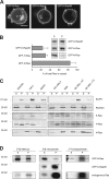
Prenylated N-Ras is largely cytosolic. (A) Jurkat T cells were transfected with GFP-tagged N-, H-, or K-Ras4B and imaged alive with a laser-scanning confocal microscope. N, nucleus. Arrowheads indicate Golgi, and the asterisk indicates cytosol. Bar, 10 µm. (B) S350 (S) and P350 (P) fractions were prepared from MDCK cells expressing the indicated GFP-tagged Ras protein that were metabolically labeled with [35S]methionine/cysteine for 17 h. GFP-tagged Ras proteins were immunoprecipitated from detergent extracts of the two fractions. Protein levels were analyzed by phosphorimager and the cytosolic fractions plotted as percentages of total Ras; mean ± SEM, n = 3. A representative radiograph is shown as an inset. (C) The indicated cell types were fractionated as in B. Cell equivalents from each fraction were loaded and analyzed for the indicated proteins by immunoblot. Epidermal growth factor receptor (EGFR) and RhoGDI serve as markers for membrane and cytosol, respectively. Representative data are shown (n = 3). (D) S350 and P350 fractions were generated from MDCK cells stably expressing GFP–N-Ras or GFP–K-Ras4B at endogenous levels that were metabolically labeled with [35S]methionine/cysteine, [3H]melavonate, or [125I]iodopalmitate, and immunoprecipitated Ras was analyzed by fluorimetry (3H) or phosphorimager (35S and 125I).
Figure 2.
Figure 2.
Prenylated N-Ras forms HMW complexes. (A) Cytosol (S350) from MDCK cells (±simvastatin) or rat liver homogenate or bacterially expressed H-Ras were subjected to Superdex 75 size-exclusion chromatography, and endogenous protein levels in each fraction were analyzed by immunoblot. (B) Protein levels of endogenous Ras isoforms in HMW and LMW fractions from A were analyzed by immunoblot. (C) Superdex 200 chromatography of S350 from MDCK cells stably expressing GFP–N-Ras. In A and C, peak elution of various molecular weight standards are indicated at the bottom.
Figure 3.
Figure 3.
VPS35 interacts with N-Ras in a prenyl-dependent fashion. (A and B) HEK293 cells were transiently transfected with VPS35 and/or N-Ras (wild type or C186S; A); N-Ras and either VPS35, VPS26A, or VPS29 (B); or all three. Whole-cell lysates (input) were immunoprecipitated with an anti-FLAG antibody (FLAG immunoprecipitation [IP]), and samples were analyzed by immunoblot. Data shown are representative of four independent experiments. (C) The cytosol (S350) of MDCK cells was subjected to Superdex 75 chromatography, and the indicated endogenous protein levels were analyzed by immunoblot. Data shown are representative of at least three independent experiments.
Figure 4.
Figure 4.
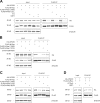
The VPS35–N-Ras interaction is dependent on prenylation, but not palmitoylation or GTP binding. (AD) HEK293 cells were transiently transfected with plasmids directing expression of the indicated proteins and analyzed by immunoprecipitation and immunoblot as in Fig. 3. Before lysis, cells were treated with farnesyltransferase inhibitor (FTI) and/or geranylgeranyltransferase inhibitor (GGTI; A) or 2-bromopalmitate (B). (D) Lysates were treated with GTP-γS or GDP before immunoprecipitation (IP). Data shown are representative of at least three independent experiments.
Figure 5.
Figure 5.
Retromer-deficient VPS35 mutants associate with N-Ras. HEK293 cells were transiently transfected with plasmids directing expression of the indicated proteins. Whole-cell lysates (input) were immunoprecipitated by anti-HA antibody (HA immunoprecipitation [IP]; A) or anti-FLAG antibody (FLAG immunoprecipitation; B), and samples were analyzed by immunoblot. Data shown are representative of at least three independent experiments.
Figure 6.
Figure 6.
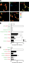
VPS35 associates with prenylated N-Ras, but not H- or K-Ras4B in intact cells. BHK cells were transiently transfected with the indicated constructs. GFP-tagged proteins are indicated in green, and mRFP-tagged proteins in red. The next day, cells were serum starved for 2 h before fixation, and FRET between the two tagged proteins was measured by FLIM. (A) Images of GFP lifetime represented in pseudocolor. Bar, 30 µm. (B and C) FRET efficiency values relative to the value of a GFP-mRFP fusion protein. Values plotted are mean ± SEM, n = 3 (>30 cells examined for each condition). ****, P < 0.0001; ns, not significant (compared with GFP-VPS35 plus mRFP–N-Ras). WT, wild type.
Figure 7.
Figure 7.
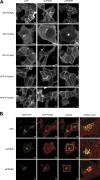
Silencing VPS35 promotes a vesicular distribution of GFP–N-Ras. (A and B) U2OS cells were transfected with untargeted siRNA (NT) or a validated pool targeting VPS35 or PDE6D. After 4 d, the cells were transfected with plasmids directing expression of the indicated fluorescent proteins and imaged live the next day. Representative images of U2OS cells expressing one of the four Ras isoforms (A) or coexpressing both N-Ras and β-1,4-galactosyltransferase (GalT; B). Bars, 10 µm.
Figure 8.
Figure 8.
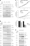
VPS35 is required for GTP loading of Ras and proliferation of N-Ras mutant melanoma cells. (A and B) HEK293 cells were transfected with untargeted siRNA (NT) or a validated pool targeting VPS35 or VPS26A. After 3 d, the cells were serum starved overnight and stimulated with EGF for the indicated time. The indicated proteins in cell lysates were quantified by immunoblot, and GTP loading was analyzed by GST-RBD pull down. Data shown are representative of four independent experiments. (C) Proliferation as measured by an MTS assay of SK-MEL-28 and -173 melanoma cell lines (BRAF or NRAS mutant, respectively) stably transfected with a tetracycline-inducible shRNA targeting NRAS or VPS35. shRNA was induced on the day of plating. Data shown are relative MTS values from day 1 to day 7 (top two panels) or at day 7 (bottom) after plating (mean ± SEM, n = 4; *, P < 0.05; **, P < 0.01; ***, P < 0.001; ns, not significant compared with untargeted siRNA). (D) Immunoblots show MEK, Erk, and Akt phosphorylation in the melanoma cell lines used in C.

Similar articles

Cited by

References

    1. Apolloni A., Prior I.A., Lindsay M., Parton R.G., and Hancock J.F.. 2000. H-ras but not K-ras traffics to the plasma membrane through the exocytic pathway. Mol. Cell. Biol. 20:2475–2487. 10.1128/MCB.20.7.2475-2487.2000 - DOI - PMC - PubMed
    1. Berg T.J., Gastonguay A.J., Lorimer E.L., Kuhnmuench J.R., Li R., Fields A.P., and Williams C.L.. 2010. Splice variants of SmgGDS control small GTPase prenylation and membrane localization. J. Biol. Chem. 285:35255–35266. 10.1074/jbc.M110.129916 - DOI - PMC - PubMed
    1. Casey P.J., Solski P.A., Der C.J., and Buss J.E.. 1989. p21ras is modified by a farnesyl isoprenoid. Proc. Natl. Acad. Sci. USA. 86:8323–8327. 10.1073/pnas.86.21.8323 - DOI - PMC - PubMed
    1. Chandra A., Grecco H.E., Pisupati V., Perera D., Cassidy L., Skoulidis F., Ismail S.A., Hedberg C., Hanzal-Bayer M., Venkitaraman A.R., et al. . 2011. The GDI-like solubilizing factor PDEδ sustains the spatial organization and signalling of Ras family proteins. Nat. Cell Biol. 14:148–158. 10.1038/ncb2394 - DOI - PubMed
    1. Choy E., Chiu V.K., Silletti J., Feoktistov M., Morimoto T., Michaelson D., Ivanov I.E., and Philips M.R.. 1999. Endomembrane trafficking of ras: the CAAX motif targets proteins to the ER and Golgi. Cell. 98:69–80. 10.1016/S0092-8674(00)80607-8 - DOI - PubMed

Publication types

MeSH terms