. 2016 Aug 8:6:31221.
doi: 10.1038/srep31221.

Marked Sexual Dimorphism in the Role of the Ryanodine Receptor in a Model of Pain Chronification in the Rat

Affiliations

Marked Sexual Dimorphism in the Role of the Ryanodine Receptor in a Model of Pain Chronification in the Rat

Luiz F Ferrari et al. Sci Rep. .

Abstract

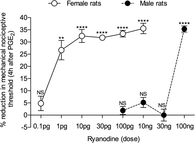
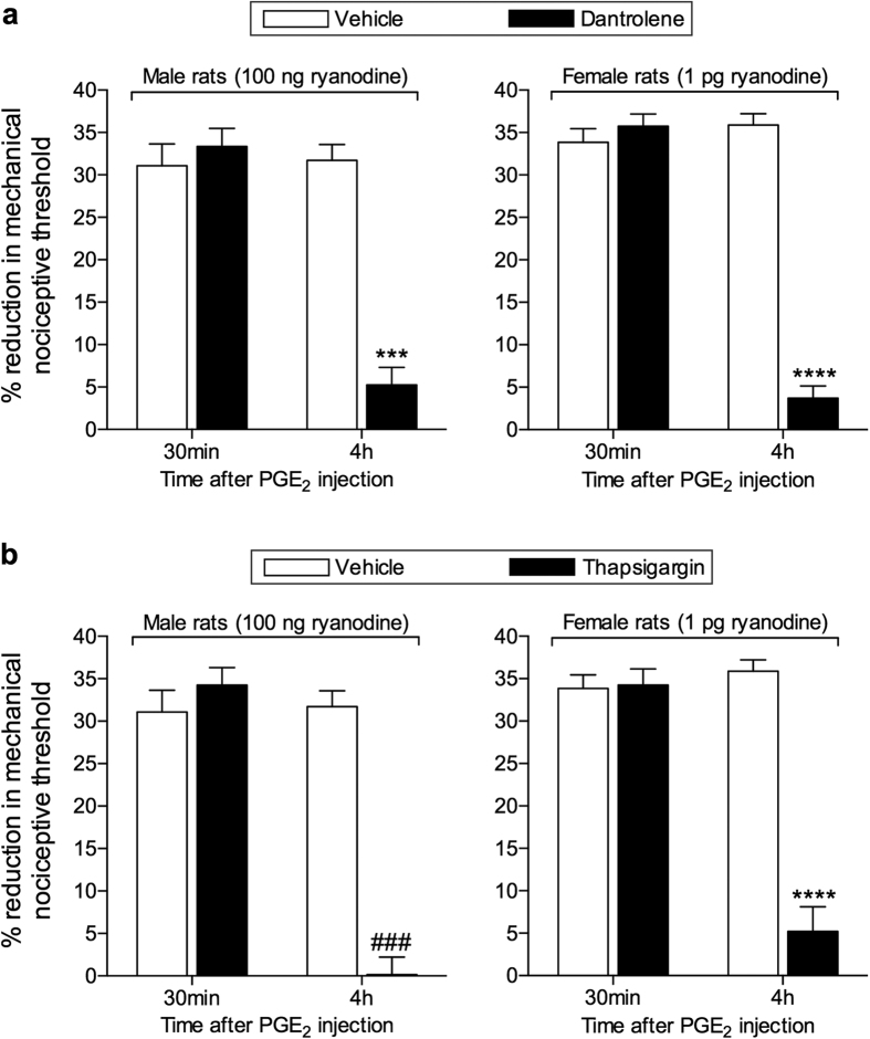
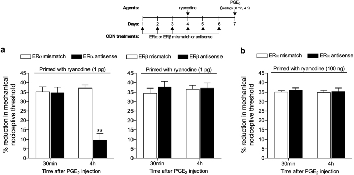
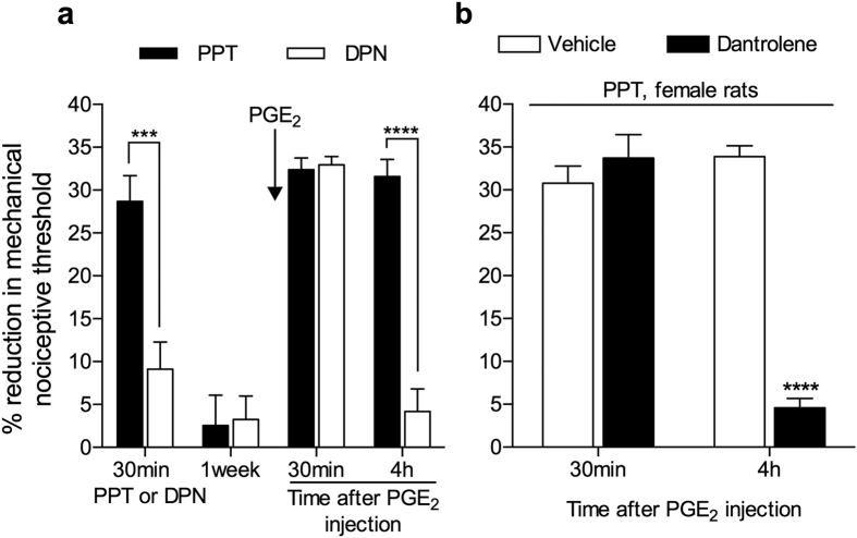
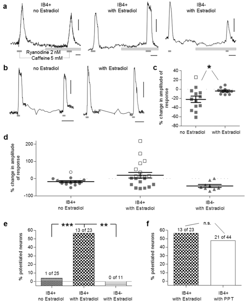
Hyperalgesic priming, an estrogen dependent model of the transition to chronic pain, produced by agonists at receptors that activate protein kinase C epsilon (PKCε), occurs in male but not in female rats. However, activation of second messengers downstream of PKCε, such as the ryanodine receptor, induces priming in both sexes. Since estrogen regulates intracellular calcium, we investigated the interaction between estrogen and ryanodine in the susceptibility to develop priming in females. The lowest dose of ryanodine able to induce priming in females (1 pg) is 1/100,000(th) that needed in males (100 ng), an effect dependent on the activation of ryanodine receptors. Treatment of female rats with antisense to estrogen receptor alpha (ERα), but not beta (ERβ), mRNA, prevented the induction of priming by low dose ryanodine, and the ERα agonist, PPT, induced ryanodine receptor-dependent priming. In vitro application of ryanodine in low concentration (2 nM) to small DRG neurons cultured from females, significantly potentiated calcium release via ryanodine receptors induced by caffeine. This effect was only observed in IB4+ neurons, cultured in the presence of β-estradiol or PPT. Our results demonstrate a profound regulatory role of ERα in ryanodine receptor-dependent transition to chronic pain.


Figures

Figure 1
Figure 1. Dose response relationship for ryanodine-induced hyperalgesic priming in male and female rats.
Different doses of ryanodine were injected on the dorsum of the hind paw in different groups of female (open circles; 0.1 pg; 1 pg; 10 pg; 30 pg; 100 pg; 10 ng) and male (black circles; 100 pg; 10 ng; 30 ng; 100 ng) rats. No change in the mechanical nociceptive threshold was observed after injections of ryanodine (see Supplementary Fig. S1). PGE2 (100 ng) was injected at the same site, 5 days later, and the mechanical hyperalgesia was evaluated by the Randall-Sellitto paw-withdrawal test. Importantly, no difference in the mechanical nociceptive threshold, compared to the baseline mechanical threshold (before injection of ryanodine) was observed (see Supplementary Table S1). The figure shows the mechanical hyperalgesia at the 4th h after the injection of PGE2; the presence of hyperalgesia at this time point was used to confirm the induction of priming by the previous treatment with ryanodine. In the groups of female rats that had received doses of ryanodine 1 pg and higher, but not 0.1 pg, and in the group of male rats previously treated with 100 ng of ryanodine, but not with 100 pg, 10 ng or 30 ng, the hyperalgesia induced by PGE2 was still present at the 4th h (female rats: 0.1 pg, t5 = 1.170, p = 0.1479 (NS); 1 pg, t5 = 5.509, p = 0.0027 (**); 10 pg, t5 = 11.59, p < 0.0001 (****); 30 pg, t5 = 20.07, p < 0.0001 (****); 100 pg, t5 = 12.25, p < 0.0001 (****); 10 ng, t5 = 15.41, p < 0.0001 (****); male rats: 100 pg, t5 = 1.151, p = 0.3019 (NS); 10 ng, t5 = 2.534, p = 0.0522 (NS); 30 ng, t5 = 0.0, p > 0.9999 (NS); 100 ng, t5 = 28.76, p < 0.0001 (****), when the mechanical nociceptive thresholds before and 4 h after the injection of PGE2, for each group, are compared, paired Student’s t-test). These results indicate that nociceptors in the female are significantly more sensitive to induction of priming by ryanodine, since a dose much lower was required to induce priming in the female rat. (N = 6 paws per group).
Figure 2
Figure 2. Induction of hyperalgesic priming by ryanodine is dependent on the ryanodine receptor.
The ryanodine receptor antagonist dantrolene (1 μg, upper panels, black bars), or the endoplasmic reticulum calcium pump inhibitor thapsigargin (1 μg, lower pannels, black bars), or their respective vehicles (white bars) were injected on the dorsum of the hind paw of male (left panels) and female (right panels) rats. 10 min later, the smallest doses of ryanodine that induced priming (100 ng in male and 1 pg in female) were injected at the same site as the inhibitor or its vehicle. No significant change in the mechanical nociceptive threshold was observed after injection of ryanodine (see Supplementary Fig. S1). Five days later, testing for the presence of priming was performed by intradermal injection of PGE2 (100 ng) at the same site as ryanodine and evaluation of the mechanical hyperalgesia, by the Randall-Selitto paw withdrawal test, 30 min and 4 h later. No significant difference (NS) in the mechanical thresholds before the injection of ryanodine and before injection of PGE2 was observed (see Supplementary Table S1). Two-way repeated measures ANOVA followed by Bonferroni post-hoc test showed that although there was no difference in the hyperalgesia induced by PGE2 30 min after the injection in the vehicle- and inhibitors-treated groups (NS for all groups), at the 4th h its magnitude was significantly smaller in the groups that received the inhibitors before ryanodine injection, 5 days previously (upper panels, males: F1,10 = 33.10; ***p = 0.0002; females: F1,10 = 97.21; ****p < 0.0001; upper panels, males: F1,10 = 27.97; ###p = 0.0004; females: F1,10 = 41.83; ****p < 0.0001, when the hyperalgesia in the vehicle- and inhibitors-treated groups is compared at the 4th h), indicating that the induction of priming by ryanodine is dependent on the activation of ryanodine receptor. (N = 6 paws per group).
Figure 3
Figure 3. Estrogen receptor alpha (ERα) regulates the induction of hyperalgesic priming by ryanodine in female rats.
(a) Female rats were treated with ODN antisense (black bars) or mismatch (white bars) for estrogen receptor alpha (ERα, left panel) or beta (ERβ, right panel), for 6 consecutive days. Ryanodine (1 pg) was injected on the dorsum of the hind paw on the 4th day of ODN treatment. On the 7th day, PGE2 (100 ng) was injected at the same site as ryanodine, and the mechanical nociceptive threshold was evaluated, 30 min and 4 h later. No difference (NS) was observed in the mechanical nociceptive thresholds before the injection of ryanodine and immediately before injection of PGE2 (see Supplementary Table S1). PGE2-induced hyperalgesia was still present 4 h after injection in all groups, except in the group treated with ODN antisense for ERα (F1,10 = 16.22; **p = 0.0024, when the groups treated with ERα ODN are compared; F1,10 = 0.2610; p =s0.6205, NS, when the groups treated with ERβ ODN are compared; two-way repeated measures ANOVA followed by Bonferroni post-hoc test); (b) The dose of ryanodine that induced priming in male rats (100 ng) was injected on the dorsum of the hind paw of female rats that have been treated intrathecally with ODN antisense or mismatch for ERα. The ODN treatment was performed for 6 consecutive days, and ryanodine was injected on the 4th day. PGE2 (100 ng) was injected at the same site as ryanodine on the 7th day, and the mechanical hyperalgesia was evaluated 30 min and 4 h later. No significant difference in the mechanical thresholds before the injection of ryanodine and before injection of PGE2 was observed (see Supplementary Table S1). We found that the hyperalgesia induced by PGE2 was still present 4 h after injection, with no attenuation, in both groups (F1,10 = 0.3839; p = 0.5494, NS, when both groups are compared). Together, these results support the suggestion that ERα regulates the capacity of ryanodine to induce priming in the female rat. (N = 6 paws, all groups).
Figure 4
Figure 4. Activation of estrogen receptor alpha (ERα) induces hyperalgesic priming in female rats.
(a) Female rats received an intradermal injection of PPT, a specific ERα agonist (1 μg, black bars), or DPN, a specific ERβ agonist (1 μg, white bars), on the dorsum of the hind paw. Evaluation of the mechanical thresholds 30 min later showed that PPT, but not DPN, induced significant hyperalgesia (***p < 0.0001). After 1 week, a time point when the mechanical thresholds were not different from before the injection of the estrogen receptors agonists (see Supplementary Table S1), testing for the presence of hyperalgesic priming was performed by injecting PGE2 (100 ng) at the same site. We observed significant mechanical hyperalgesia, 30 min after PGE2 injection, in both groups. However, when the mechanical nociceptive thresholds were evaluated at the 4th h, only the group previously treated with PPT showed hyperalgesia, indicating that PPT, but not DPN, had induced priming (F1,10 = 36.33; ****p = 0.0001, when both groups are compared; two-way repeated measures ANOVA followed by Bonferroni post-hoc test); (b) The ryanodine receptor antagonist dantrolene (1 μg, black bars), or its vehicle (white bars), was injected on the dorsum of the hind paw of female rats. 10 min later, PPT (1 μg) was injected at the same site. 1 week later, testing for the presence of priming was performed by injecting PGE2 (100 ng) at the same site as PPT, and evaluating the mechanical nociceptive threshold 30 min and 4 h later (see Supplementary Table S1, for data about the mechanical nociceptive thresholds before injection of PPT and before injection of PGE2). In the group treated with vehicle significant mechanical hyperalgesia was observed at both 30 min and 4 h. However, in the group that received dantrolene the hyperalgesia was no longer present at the 4th h, indicating that the priming induced by PPT was dependent on the ryanodine receptor (F1,10 = 35.94; ****p = 0.0001, when both groups are compared; two-way repeated measures ANOVA followed by Bonferroni post-hoc test). (N = 6 paws, all groups).
Figure 5
Figure 5. ERα activation is required for potentiation of calcium response in IB4+ small female DRG neurons by low concentration of ryanodine.
(a) Recordings of [Ca2+]i transients in IB4+ and IB4− small DRG neurons, incubated without (left panel) or with (middle and right panels) β-estradiol (100 nM). Ryanodine (2 nM) was applied for 10 min when [Ca2+]i returned to baseline after the first application of caffeine (5 mM). After that, caffeine administration was repeated. Whereas no response was produced by ryanodine itself, a significant potentiation of the response to caffeine after ryanodine application occurred, exclusively in IB4+ neurons and predominantly in cultures incubated with β-estradiol; (b) Recordings of calcium transients induced by two subsequent applications of caffeine (5 mM) in the absence of ryanodine in IB4+ small DRG neurons incubated without (left panel) or with (right panel) β-estradiol; (c) Pooled relative changes in amplitude of the second response to caffeine without ryanodine application, compared as percentage of the first response, in cultures incubated with β-estradiol or vehicle (*p < 0.05, two-tailed unpaired Student’s t-test with Welch’s correction: t14 = 2.56, p = 0.02); (d) Pooled relative changes in amplitude of the response to caffeine after ryanodine application, compared as percentage of the response before ryanodine application, as described in (a). Potentiated (above the 8% cut-off) and non-potentiated neuron activation are plotted as white and grey symbols, respectively; (e) Percentage of potentiated neurons in different groups of cultured neurons (IB4+ without estradiol; IB4+ with estradiol; IB4− with estradiol), compared by Exact Fisher’s test (**p < 0.01, ***p < 0.001, when the IB4+ with estradiol is compared to the other groups: p < 0.0001 vs IB4+ without β-estradiol; p = 0.0018 vs IB4−). Of note, there is a remarkably higher number of potentiated cells among the IB4+ neurons incubated with β-estradiol; (f) Percentage of potentiated IB4+ neurons in culture incubated with β-Estradiol (100 nM) or PPT (100 nM). No significant difference was observed between two groups (p = 0.61 > 0.05, exact Fisher’s test). For panels (a) and (b), the horizontal scale bars correspond to 100 s and, the vertical ones correspond to 0.1 a.u. (arbitrary units of the fluorescence ratio F340/F380).

Similar articles

Cited by

References

    1. Buse D. C. et al.. Sex differences in the prevalence, symptoms, and associated features of migraine, probable migraine and other severe headache: results of the American Migraine Prevalence and Prevention (AMPP) Study. Headache 53, 1278–1299 (2013). - PubMed
    1. de Mos M., Huygen F. J., Stricker B. H., Dieleman J. P. & Sturkenboom M. C. Estrogens and the risk of complex regional pain syndrome (CRPS). Pharmacoepidemiol Drug Saf 18, 44–52 (2009). - PubMed
    1. LaCroix-Fralish M. L., Tawfik V. L. & DeLeo J. A. The organizational and activational effects of sex hormones on tactile and thermal hypersensitivity following lumbar nerve root injury in male and female rats. Pain 114, 71–80 (2005). - PMC - PubMed
    1. Ouyang A. & Wrzos H. F. Contribution of gender to pathophysiology and clinical presentation of IBS: should management be different in women? Am J Gastroenterol 101, S602–9 (2006). - PubMed
    1. Staud R. Biology and therapy of fibromyalgia: pain in fibromyalgia syndrome. Arthritis Res Ther 8, 208 (2006). - PMC - PubMed

Publication types