. 2016 Aug 9;113(32):E4688-97.
doi: 10.1073/pnas.1523597113. Epub 2016 Jul 21.

Deubiquitinase Usp8 regulates α-synuclein clearance and modifies its toxicity in Lewy body disease

Affiliations

Deubiquitinase Usp8 regulates α-synuclein clearance and modifies its toxicity in Lewy body disease

Zoi Alexopoulou et al. Proc Natl Acad Sci U S A. .

Abstract

In Parkinson's disease, misfolded α-synuclein accumulates, often in a ubiquitinated form, in neuronal inclusions termed Lewy bodies. An important outstanding question is whether ubiquitination in Lewy bodies is directly relevant to α-synuclein trafficking or turnover and Parkinson's pathogenesis. By comparative analysis in human postmortem brains, we found that ubiquitin immunoreactivity in Lewy bodies is largely due to K63-linked ubiquitin chains and markedly reduced in the substantia nigra compared with the neocortex. The ubiquitin staining in cells with Lewy bodies inversely correlated with the content and pathological localization of the deubiquitinase Usp8. Usp8 interacted and partly colocalized with α-synuclein in endosomal membranes and, both in cells and after purification, it deubiquitinated K63-linked chains on α-synuclein. Knockdown of Usp8 in the Drosophila eye reduced α-synuclein levels and α-synuclein-induced eye toxicity. Accordingly, in human cells, Usp8 knockdown increased the lysosomal degradation of α-synuclein. In the dopaminergic neurons of the Drosophila model, unlike knockdown of other deubiquitinases, Usp8 protected from α-synuclein-induced locomotor deficits and cell loss. These findings strongly suggest that removal of K63-linked ubiquitin chains on α-synuclein by Usp8 is a critical mechanism that reduces its lysosomal degradation in dopaminergic neurons and may contribute to α-synuclein accumulation in Lewy body disease.

Keywords: Parkinson’s disease; endosome; neurodegeneration; ubiquitin; ubiquitin ligase.


Conflict of interest statement

The authors declare no conflict of interest.

Figures
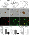
Fig. 1.
Fig. 1.
K63-linked ubiquitin conjugates are detected in α-synuclein–positive inclusions and are reduced in the substantia nigra. (A) Schematic view of the studied brain regions and corresponding light microscopy images showing K63-linked ubiquitinated inclusions. [Scale bars, 5 mm (schemes) and 20 mm (images).] (B) Confocal immunofluorescence showing colocalization of K63-linked ubiquitin chains and α-synuclein in Lewy bodies and Lewy neurites in nigral neurons. (C) Quantification of ubiquitin-positive inclusions as a percentage of α-synuclein–positive inclusions (Ub/α-Syn) in serial sections of the AC, EC, and SN; ***P < 0.0001, n = 14. (D) Quantification of K63-linked ubiquitinated inclusions as a percentage of ubiquitin–positive inclusions (K63-Ub/Ub) in serial sections of the AC, EC, and SN; ***P < 0.0001, n = 14. (E) The percentage of ubiquitinated inclusions in nigral neurons was low irrespective of the stage of disease and lower in incidental Lewy body compared with Lewy body disease cases. Ub/Syn, ***P < 0.0001, n = 14; K63/Syn, *P < 0.05, n = 14. AQ, central aqueduct; CA1–4, cornu ammonis 1–4; CC, corpus callosum; CP, corticopontine and pyramidal tracts; DG, dentate gyrus; IC, inferior colliculus; LAT.V., lateral ventricle; Ncl IV, trochlear nucleus; SCP, superior cerebellar peduncle. Error bars correspond to standard error of the mean.
Fig. S1.
Fig. S1.
(A) Correlation of K63-specific ubiquitin antibody-positive inclusions as a percentage of pan-ubiquitin antibody-positive inclusions in the AC, EC, and SN. Minimal number of analyzed sections of one brain region, n = 14. ρ = 0.4990, *P < 0.0004, Spearman’s rho. (B) Pan-ubiquitin antibody-positive Lewy bodies and Lewy neurites in the substantia nigra. (C) Intranuclear inclusions in a nigral neuron (Marinesco bodies are indicated by an arrowhead) and dystrophic neurites stained with a K63-specific ubiquitin antibody. (Scale bar, 20 µm.)
Fig. S2.
Fig. S2.
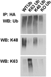
Wild-type or single-lysine HA-tagged ubiquitin was expressed in HEK-293T cells. Ubiquitinated proteins were immunoprecipitated with anti-HA antibodies and blotted with anti–K63-specific or anti–K48-specific antibodies as well as antibodies against pan-ubiquitin. Note that both antibodies and single-lysine ubiquitin constructs have the expected specificity without any cross-reactivity detected.
Fig. 2.
Fig. 2.
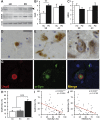
Usp8 expression and localization inversely correlate with ubiquitinated inclusions in α-synucleinopathies. (A) Representative immunoblot showing increased levels of Usp8 but not AMSH relative to the actin loading control in the SN from patients with LB disease; a specific band is indicated by an arrowhead. (B) Quantification of Usp8 protein level in the human brain showed a significant increase in the SN but not AC of patients with Lewy body disease compared with controls HC, healthy control (**P = 0.0028, n = 8). Quantification of AMSH protein levels did not show a significant difference (C). Usp8-positive LBs and Lewy neurites in the AC (D) and SN (E). (Scale bar, 20 µm.) (F) No AMSH staining was seen in nigral LBs, as indicated by the arrowhead. (G) Double immunofluorescence and confocal imaging confirmed the colocalization of Usp8 (red) and α-synuclein (green) in nigral LBs. DAPI indicates nuclear staining in blue. (H) Quantification of Usp8-positive as a percentage of α-synuclein–positive inclusions (Usp8/α-Syn) in serial sections showed a significant increase in the SN compared with cortical areas (AC, EC) (***P = 0.0001, n = 14). (I) Negative correlation between Usp8-positive and ubiquitin-positive inclusions in the SN (***P = 0.0002, ρ = −0.5044). (J) Negative correlation of Usp8-positive and K63-linked ubiquitinated inclusions shown as the ratio K63/Ub (**P = 0.0038, ρ = −0.4186). Error bars correspond to standard error of the mean.
Fig. S3.
Fig. S3.
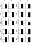
Targeted screen for proteins that are known to interact with or regulate the assembly of K63-linked ubiquitin chains in the endosomal and autophagic pathways. Total lysates from the SN and AC were compared across regions of the same brain and between healthy and LB-containing regions (n = 8 cases per region). The detection of a significantly increased expression of Usp8 in the SN with LBs despite the use of crude lysates strongly implicates this enzyme in disease pathogenesis. HC, healthy control, **P = 0.0028. Error bars correspond to standard error of the mean.
Fig. 3.
Fig. 3.
Usp8 interaction and colocalization with α-synuclein. (A) Wild-type (Usp8WT) or catalytically inactive (Cys786 to Ala) Usp8 was immunoprecipitated with untagged α-synuclein when expressed in HEK-293T cells. (B) Usp8 activity as measured by binding to HA-Ub-Br2 is not affected by increasing α-synuclein levels or expression of α-synuclein mutants. EV, empty vector; IP, immunoprecipitation; WCL, whole-cell extract. (C) Bimolecular fluorescence complementation signifying Usp8–α-synuclein interaction and expression of endosomal Rabs (Rab5, Rab7, and Rab11) tagged to a red fluorescent protein. Schematic depicting the bimolecular fluorescence complementation assay used in this study. Human Usp8 is tagged to the N-terminal fragment of Venus fluorescent protein (VFP) and α-synuclein is tagged to the C-terminal fragment of VFP. Quantification of the percentage overlap (Manders’ colocalization coefficient) between Usp8 and α-synuclein (green) with the indicated endosomal markers (red); n = 100 cells per condition; ***P < 0.001, one-way ANOVA. (D) Localization of Usp8 in relation to endosomal markers in human iPSc-derived dopaminergic neurons: triple labeling of neurons with TH (for detection of dopaminergic neurons; first column, blue), Usp8 (second column, green), and one of the following: (i) early endosome, EEA1; (ii) late endosome, LBPA; or (iii) α-synuclein (third column, red). Quantification of the percentage overlap (MCC) of Usp8 with the indicated markers in B; n = 50 neurons per condition. (Scale bar, 10 μm.) Error bars correspond to standard error of the mean.
Fig. S4.
Fig. S4.
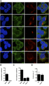
(A) Colocalization of Usp8 with various endosomal markers in HEK-293T cells. Cells were either transfected with fluorescent reporters or stained with specific antibodies: (i) early endosome, EEA1; (ii) early endosome, Rab5; (iii) late endosome, Rab7; (iv) late endosome, LBPA; and (v) recycling endosome, Rab11, and also costained for Usp8 (second column, green). (Scale bar, 10 μm.) (B) Quantification of the percentage overlap (MCC) of Usp8 with EEA1 and LBPA (endogenously expressed and stained using antibodies). (C) Quantification of the percentage overlap (MCC) of Usp8 with Rab5, Rab7, and Rab11 (fluorescent reporters transiently expressed). (D) Quantification of the size of EEA1-positive puncta corresponding to early endosomes in HEK-293T cells expressing FLAG-tagged wild-type Usp8 or an empty vector. Early endosome size was quantified by confocal microscopy [LSM 710 (Carl Zeiss)] and ImageJ, with n = 20 cells imaged per condition. The presence of untagged GFP within the vector enabled the identification of transfected cells. Error bars correspond to standard error of the mean.
Fig. S5.
Fig. S5.
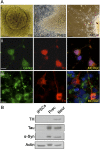
(A) Characterization of dopaminergic neurons derived from healthy control iPS cells. (A, i) Embryoid bodies (EB) were differentiated to neural precursor cells (PREC) and then manually passaged and matured to neurons (NEUR), as described in SI Materials and Methods. (A, ii and iii) Immunostaining for dopaminergic (TH and GIRK2) (ii) and neuronal (β-III tubulin) markers (iii). (Scale bars, 10 µm.) (B) The expression of neuronal markers (TH, Tau, and α-Syn) at the appropriate stage of differentiation was confirmed by immunoblotting.
Fig. 4.
Fig. 4.
Usp8 deconjugates preferentially K63-linked ubiquitin chains on α-synuclein. (A) Expression of Usp8WT caused robust deubiquitination of endogenous α-synuclein compared with expression of Usp8CA or empty vector. Quantification of ubiquitinated α-synuclein in Usp8WT- relative to Usp8CA-expressing cells shown as arbitrary units (AU; *P = 0.0410, n = 3 biological replicates). WB, Western blot. (B) Coexpression of Usp8 with either K63 or K48 single-lysine ubiquitin showed that it deconjugated preferentially K63-linked chains on α-synuclein without any obvious difference in the total amount of ubiquitinated proteins between the immunoprecipitated samples (representative of n = 3 biological replicates). (C) Expression of a deletion mutant of Usp8 lacking the MIT domain required for endosomal localization (Usp8ΔMIT) reduced its activity against K63-linked ubiquitin chains on α-synuclein compared with Usp8WT. Usp7 had reduced activity against α-synuclein compared with Usp8 in cells (n = 5 biological replicates). (D) Recombinant α-synuclein was linked to uniform K63-linked chains in vitro by purified Nedd4 and incubated with the indicated deubiquitinases (50 nM). Robust deubiquitination was observed with Usp7 and Usp8. All seven enzymes were assessed for purity with Coomassie and activity by HA-Ub-Br labeling. Immunoblotting with anti-HA revealed a shift in the molecular mass of labeled DUBs, which indicates covalent binding to HA-Ub-Br. Error bars correspond to standard error of the mean.
Fig. S6.
Fig. S6.
(A) Ubiquitinated α-synuclein was immunoprecipitated with anti-HA antibodies in M17D cells stably expressing α-synuclein and transiently transfected with HA-Ub. (B) Overexpression of wild-type but not catalytically inactive Usp8 in HEK-293T cells removes ubiquitin chains on α-synuclein, as detected by an antibody against the last 20 amino acids of the protein (C20).
Fig. S7.
Fig. S7.
(A) Time course of α-synuclein deubiquitination by recombinant Usp7 and Usp8. K63-linked ubiquitin chains were conjugated on α-synuclein using purified Nedd4 as the E3. An equal amount of ubiquitinated α-synuclein was incubated with each enzyme (50 nM). Aliquots were taken every 10 min, resolved on SDS/PAGE, and immunoblotted with anti–α-synuclein antibody (C20). (B) SH-SY5Y cells were transduced with Usp8 shRNA or Scr shRNA control. Eight days after transduction, the cells were treated for 24 h with 25 μM chloroquine or 12 h with 1 μΜ lactacystin. α-Synuclein levels were analyzed in total lysates after immunoblotting with the C20 antibody and are presented as the ratio to the actin loading control. Error bars correspond to standard error of the mean.
Fig. 5.
Fig. 5.
Usp8 regulates the degradation of α-synuclein by the lysosome. (A) Cycloheximide chase of endogenous α-synuclein showed that its rate of clearance was reduced at 7 h when wild-type Usp8 was expressed in HEK-293T cells (**P = 0.0091, n = 3 biological replicates). (B and C) Lentiviral-mediated shRNA knockdown of Usp8 in SH-SY5Y cells reduced endogenous α-synuclein levels by 35% relative to the actin loading control compared with Scr shRNA controls (**P = 0.0031, n = 5 biological replicates) (B) and increased the amount of ubiquitinated α-synuclein as evidenced by immunoblotting of immunoprecipitated α-synuclein with anti-ubiquitin and anti-K63 antibodies (C). No smear was seen when anti-HA was used as an IgG control. (D) Lysates isolated from Usp8 knockdown and Scr shRNA-treated SH-SY5Y cells were fractionated into cytosolic and membrane fractions and tested for α-synuclein levels at baseline and following 8 h of treatment with either 50 μM chloroquine (CQ) or 5 μM lactacystin (Lact). Accumulation of α-synuclein was observed in the membrane fraction, which is enriched in endosomal/autophagic–lysosomal compartments in Usp8 knockdown cells treated with chloroquine, suggesting that there is accelerated lysosomal degradation of α-synuclein in these cells (*P = 0.019, n = 3). Error bars correspond to standard error of the mean.
Fig. 6.
Fig. 6.
Knockdown of endogenous Usp8 in Drosophila protects against α-synuclein–induced toxicity. (A and B) Overexpression of α-synuclein caused a rough eye phenotype, which was more severe in flies expressing A53T mutant α-synuclein, as detected by SEM. This phenotype was rescued in double-transgenic flies with eye-specific Usp8 knockdown and either wild-type (A) or A53T mutant α-synuclein (B). *P < 0.05. Quantitative PCR did not show a reduction in α-synuclein mRNA levels between single- and double-transgenic lines to explain the phenotype. (C) Knockdown of AMSH did not rescue this phenotype, whereas knockdown of Vps28 made it worse. (D) Usp8 knockdown did not affect the expanded Ataxin 3 phenotype. (E) Representative immunoblot of fractionated lysate (C, cytosol; P, pellet) from A53T mutant α-synuclein flies and flies expressing A53T mutant α-synuclein with Usp8 knockdown (+Usp8 KD). Quantitative band densitometry showed that, relative to the actin loading control, protein levels of either wild-type or A53T mutant monomeric α-synuclein (indicated by an asterisk) were significantly reduced in flies coexpressing Usp8 RNAi (n = 4 biological replicates) in the absence of a reduction in mRNA levels, as shown in A and B. *P < 0.05; ****P < 0.0001. (F) Accelerated loss of climbing ability was seen in transgenic flies expressing human A53T mutant α-synuclein in dopaminergic neurons (ddc-GAL4 driver) with increasing age. The climbing ability of double-transgenic lines expressing A53T α-synuclein with Usp8 knockdown in dopaminergic cells was significantly improved (shown with asterisks) compared with A53T α-synuclein–expressing flies and was similar to the control genotype, ddc-GAL4/+ (*P = 0.0381 for day 10; **P = 0.01 for day 12; *P = 0.0159 for day 14; n = 50 flies per group). (G) Knockdown of JosD2 or AMSH in dopaminergic neurons slightly worsened the A53T mutant α-synuclein phenotype. (H) Expression of A53T mutant α-synuclein but not control constructs in dopaminergic neurons (ddc-GAL4 driver) led to loss of TH-immunoreactive neurons in the PPM1/2 cluster, which was prevented by concomitant Usp8 knockdown (***P < 0.001). NS, nonspecific band. Error bars correspond to standard error of the mean.
Fig. S8.
Fig. S8.
(A) The rough eye phenotype of A53T α-synuclein was not rescued in flies coexpressing in the eye RNAi to knock down AMSH, Usp14, or Usp47. (B) The rough eye phenotype of pathogenic expanded huntingtin (Htt) was not rescued in the corresponding double-transgenic line expressing RNAi against Usp8 (Htt/Usp8 KD). (C) The functional efficiency of Usp8 knockdown was confirmed by detecting the previously documented wing defect when the RNAi was expressed under the MS1096-GAL4 driver.

Similar articles

Cited by

References

    1. Braak H, et al. Staging of brain pathology related to sporadic Parkinson’s disease. Neurobiol Aging. 2003;24(2):197–211. - PubMed
    1. Spillantini MG, et al. Alpha-synuclein in Lewy bodies. Nature. 1997;388(6645):839–840. - PubMed
    1. Schneider JA, et al. Cognitive impairment, decline and fluctuations in older community-dwelling subjects with Lewy bodies. Brain. 2012;135(Pt 10):3005–3014. - PMC - PubMed
    1. Ross OA, et al. Genomic investigation of alpha-synuclein multiplication and parkinsonism. Ann Neurol. 2008;63(6):743–750. - PMC - PubMed
    1. Wirdefeldt K, et al. Expression of alpha-synuclein in the human brain: Relation to Lewy body disease. Brain Res Mol Brain Res. 2001;92(1–2):58–65. - PubMed

Publication types