. 2016 Aug 2;113(31):8783-8.
doi: 10.1073/pnas.1609057113. Epub 2016 Jul 18.

Defective HIV-1 proviruses produce novel protein-coding RNA species in HIV-infected patients on combination antiretroviral therapy

Affiliations

Defective HIV-1 proviruses produce novel protein-coding RNA species in HIV-infected patients on combination antiretroviral therapy

Hiromi Imamichi et al. Proc Natl Acad Sci U S A. .

Abstract

Despite years of plasma HIV-RNA levels <40 copies per milliliter during combination antiretroviral therapy (cART), the majority of HIV-infected patients exhibit persistent seropositivity to HIV-1 and evidence of immune activation. These patients also show persistence of proviruses of HIV-1 in circulating peripheral blood mononuclear cells. Many of these proviruses have been characterized as defective and thus thought to contribute little to HIV-1 pathogenesis. By combining 5'LTR-to-3'LTR single-genome amplification and direct amplicon sequencing, we have identified the presence of "defective" proviruses capable of transcribing novel unspliced HIV-RNA (usHIV-RNA) species in patients at all stages of HIV-1 infection. Although these novel usHIV-RNA transcripts had exon structures that were different from those of the known spliced HIV-RNA variants, they maintained translationally competent ORFs, involving elements of gag, pol, env, rev, and nef to encode a series of novel HIV-1 chimeric proteins. These novel usHIV-RNAs were detected in five of five patients, including four of four patients with prolonged viral suppression of HIV-RNA levels <40 copies per milliliter for more than 6 y. Our findings suggest that the persistent defective proviruses of HIV-1 are not "silent," but rather may contribute to HIV-1 pathogenesis by stimulating host-defense pathways that target foreign nucleic acids and proteins.

Keywords: HIV-1; activation; latency; provirus.


Conflict of interest statement

The authors declare no conflict of interest.

Figures
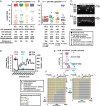
Fig. 1.
Fig. 1.
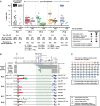
Characterization of full-length and truncated proviruses in patients with HIV-1 infection. (A) Distribution of proviral DNA lengths for four patients with pVL ≥ 40 copies per milliliter and (B) four patients with pVL < 40 copies per milliliter. Descriptions of symbols for all panels are provided in the key. Expanded proviral clones are depicted by the black rectangles. Distinct clonally expanded populations are identified by Roman numerals. (C) Agarose gel pictures depicting sizes of proviral DNA PCR fragments for one patient with pVL ≥ 40 copies per milliliter (Pt 1) and one patient with pVL < 40 copies per milliliter (Pt 5) generated using the 5′LTR-to-3′LTR PCR. Only positive PCR reactions generated at limiting dilutions are shown. The red dotted lines indicate the migration point of DNA bands corresponding to full-length HIV-1 proviruses (8.9 kb). (D) Longitudinal changes over 20 y in CD4+ T-cell counts and HIV-RNA levels for a single patient with HIV-1 infection (Pt 9, first tested positive for HIV-1 in 1985). (E) Distribution of HIV-1 proviral DNA lengths and clonal expansions for Pt 9 in 1995 and 2001. Descriptions of symbols for the panels are provided in the key in A and B. (F) The Highlighter program (v2.2.3) analyses of the sequence data from 1995 and 2001 for HIV-1 proviral DNA derived from patient 9. Of note, identical expanded provial clones (viii) were noted in 1995 and 2001.
Fig. S1.
Fig. S1.
Agarose gel pictures depicting sizes of proviral DNA fragments and the sensitivity of nested PCR used in the present study using genomic DNA from the 8E5 cell line and plasmid DNA containing full-length NL4.3 strain of HIV-1 as templates.
Fig. S2.
Fig. S2.
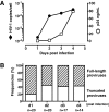
Truncated defective HIV-1 proviruses could be detected as early as 24 h following in vitro infection of primary CD4+ T cells. Purified CD4+ T cells from healthy volunteers were activated in vitro with PHA+IL-2 for 3 d and infected with HIV-1 DH12 at multiplicity of infection (MOI) = 0.01. (A) Detection of levels of HIV-1 p24 antigen in HIV-1 infected CD4+ T-cell culture supernatants by ELISA and the HIV-DNA in infected cells by nested PCR of the HIV-1 genome (using the nested PCR primers: DNA F1/R1 and DNA F2/R2). (B) Frequencies of proviral DNA lengths for HIV-1 infected CD4+ T cells. Numbers of total provirus sequences analyzed for each time point are indicated at the bottom.
Fig. S3.
Fig. S3.
Deletion junction analysis of HIV-1 proviruses. (A) Deletion junctions are plotted for each proviral DNA sequence from four patients with pVL ≥ 40 copies per milliliter (Pts 1–4), four patients with pVL < 40 copies per milliliter (Pts 5–8), and one patient (Pt 9) before and after suppressive cART. The 5′-ends of the deletion junctions are depicted by closed circles; and the 3′-ends of the deletion junctions are depicted by open circles. The sizes and locations of deletions were determined by comparing the sequences of the proviral DNA with the full-length sequence of HIV-1 HXB2. Clonally expanded populations are identified by Roman numerals and are the same numbers used in Figs. 1 B and E. Numbers in parentheses indicate the number of identical provirus sequences found for each clone. Green arrows facing in opposite directions relative to the reference HXB2 genome sequence represent a gene segment corresponding to inversion in deletion. (B) DNA sequences of truncated provirus at the deletion junctions. Two sequences from Pt 1 (pVL = 225,658 copies per milliliter) and two sequences from Pt 5 (pVL < 40 copies per milliliter) are shown. Directly repeated sequences are highlighted in red. A single nucleotide mismatch within a directly repeated sequence in patient 5 is underlined.
Fig. S4.
Fig. S4.
Schematic diagram of the HIV-1 genome, locations of nested HIV-1 primers used in the study, and locations of the splice donors (D1–D4) and acceptors (A1–A7) for HIV-1 HXB2. Amplification of HIV-DNA was performed using nested primer pairs: DNA F1/DNA R1 and DNA F2/DNA R2. An alternative second round PCR primer pair: DNA F3/DNA R3 was used to assess the possibility of PCR primer bias. Amplification of HIV-RNA was performed using nested primer pairs: RNA F1/RNA R1 and RNA F2/RNA R2 in most cases. An alternative primer pair: RNA F2/RNA R2′ was used in cases where the RNA R2 primer failed to yield an adequate band. Coordinates of PCR primers and the splice donors/acceptors are provided based on the sequence coordinates of HIV-1 HXB2.
Fig. 2.
Fig. 2.
Characterization of novel unspliced HIV-1 RNA species in patients with HIV-1 infection. (A) Distribution of usHIV-RNA lengths for one patient with pVL ≥ 40 copies per milliliter and four patients with pVL < 40 copies per milliliter. Plots for proviral DNA lengths are the same as those shown in Fig. 1 A and B and replotted here for comparison purposes. Descriptions of symbols for all panels are provided in the key. Clonally expanded populations are depicted by the black rectangles. Distinct expanded proviral clones are identified by Roman numerals. RNAs that had corresponding DNA sequences are identified by lowercase alphabets. (B) Schematic diagrams of novel usHIV-RNA transcripts for four patients with pVL < 40 copies per milliliter. Known unspliced and spliced HIV-RNA species are shown (Upper). Locations of the splice donors (D1–D4) and acceptors (A1–A7) for HIV-1 HXB2 are indicated in the HIV-1 genome structure at the top. Coordinates of the splice donors and acceptors are provided based on the sequence coordinates of HIV-1 HXB2. Descriptions of symbols used in the diagram are provided in the key. Only unique HIV-RNA sequences are shown. (C) Amino acid sequence of the ORF for the clone xi mRNA transcript that encodes a part of Gag (17 aa) and a part of Env (50 aa) derived from patient 6. The first 8 aa in the gag were deduced from the proviral DNA sequence from the same patient. The vertical black line indicates a boundary between Gag and Env coding regions.
Fig. S5.
Fig. S5.
Sequence analyses of proviral DNA and usHIV-RNA sequences from: patient 1 (pVL = 225,658 copies per milliliter), patient 5 (pVL < 40 copies per milliliter), and patient 7 (pVL < 40 copies per milliliter). All sequences were obtained from CD4+ T cells. Phylogenetic relationships among the proviral DNA and unspliced RNA sequences were estimated using the maximum-likelihood method. Bootstrap percentile values from 1,000 replications are shown at nodes defining major grouping of sequences. Statistical support of 50% or greater is shown. HIV-1 HXB2 was used as an outgroup. Sequence alignment was generated using the Highlighter program. Descriptions of symbols used in the sequence alignment are provided in the key. The key for the symbols used for the sequences in the phylogenetic trees is provided in Fig. 2A. Genome map of HIV-1 is provided on the top of the alignments. Base numbers are based on sequence coordinates for HIV-1 HXB2.
Fig. 3.
Fig. 3.
HIV-1 Western blot analysis of four patients with pVL ≥ 40 copies per milliliter (Pts 1–4), four patients with pVL < 40 copies per milliliter (Pts 5–8), and one patient (Pt 9) who exhibited clonal expansion of truncated defective provirus and persistence of the clone. HIV-1 Western blots were performed with plasma samples (diluted in 1:100). Negative and positive controls were run in parallel and are shown on the right panel. In patient 4, faint bands for p32, p51/55, and p65 proteins could be detected on the original strip but are not clearly shown in the scanned image.
Fig. S6.
Fig. S6.
Simultaneous extraction of RNA and DNA from the same populations of CD4+ T cells.
Fig. S7.
Fig. S7.
Agarose gel picture depicting precursor forms (unspliced pre-mRNA) and mature forms (fully-spliced mRNA) of GAPDH mRNA from nuclear and cytoplasmic fractions of CD4+ T cells freshly isolated from a healthy volunteer. The GAPDH pre-mRNA primer pair produced a 207-bp fragment corresponding to exon 7–intron 7 of the human GAPDH gene. The GAPDH mRNA primer pair produced two bands: a 197-bp fragment corresponding to exon 4 and exon 5 of the GAPDH gene; and a 328-bp fragment corresponding to exon 4 and exon 5 that included 131 bp of intron sequence.

Similar articles

Cited by

References

    1. DHHS Panel on Antiretoviral Guidelines for Adults and Adolescents 2015 Guidelines for the Use of Antiretroviral Agents in HIV-1-Infected Adults and Adolescents. Department of Health and Human Services. Available at https://aidsinfo.nih.gov/contentfiles/lvguidelines/adultandadolescentgl.pdf. Accessed July 1, 2016.
    1. Murray JM, et al. PINT Study Team HIV DNA subspecies persist in both activated and resting memory CD4+ T cells during antiretroviral therapy. J Virol. 2014;88(6):3516–3526. - PMC - PubMed
    1. Josefsson L, et al. The HIV-1 reservoir in eight patients on long-term suppressive antiretroviral therapy is stable with few genetic changes over time. Proc Natl Acad Sci USA. 2013;110(51):E4987–E4996. - PMC - PubMed
    1. Buzon MJ, et al. Long-term antiretroviral treatment initiated at primary HIV-1 infection affects the size, composition, and decay kinetics of the reservoir of HIV-1-infected CD4 T cells. J Virol. 2014;88(17):10056–10065. - PMC - PubMed
    1. Mendoza D, et al. Comprehensive analysis of unique cases with extraordinary control over HIV replication. Blood. 2012;119(20):4645–4655. - PMC - PubMed

Publication types

Associated data

LinkOut - more resources