. 2016 Sep 1;129(17):3231-7.
doi: 10.1242/jcs.189423. Epub 2016 Jul 18.

Sar1 localizes at the rims of COPII-coated membranes in vivo

Affiliations

Sar1 localizes at the rims of COPII-coated membranes in vivo

Kazuo Kurokawa et al. J Cell Sci. .

Abstract

The Sar1 GTPase controls coat assembly on coat protein complex II (COPII)-coated vesicles, which mediate protein transport from the endoplasmic reticulum (ER) to the Golgi. The GTP-bound form of Sar1, activated by the ER-localized guanine nucleotide exchange factor (GEF) Sec12, associates with the ER membrane. GTP hydrolysis by Sar1, stimulated by the COPII-vesicle-localized GTPase-activating protein (GAP) Sec23, in turn causes Sar1 to dissociate from the membrane. Thus, Sar1 is cycled between active and inactive states, and on and off vesicle membranes, but its precise spatiotemporal regulation remains unknown. Here, we examined Sar1 localization on COPII-coated membranes in living Saccharomyces cerevisiae cells. Two-dimensional (2D) observation demonstrated that Sar1 showed modest accumulation around the ER exit sites (ERES) in a manner that was dependent on Sec16 function. Detailed three-dimensional (3D) observation further demonstrated that Sar1 localized at the rims of the COPII-coated membranes, but was excluded from the rest of the COPII membranes. Additionally, a GTP-locked form of Sar1 induced abnormally enlarged COPII-coated structures and covered the entirety of these structures. These results suggested that the reversible membrane association of Sar1 GTPase leads to its localization being restricted to the rims of COPII-coated membranes in vivo.

Keywords: COPII-coated vesicle; ER; GTP hydrolysis; Sar1; Sec16.


Conflict of interest statement

The authors declare no competing or financial interests.

Figures
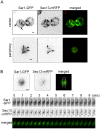
Fig. 1.
Fig. 1.
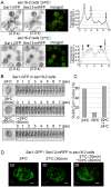
Sar1 distributes throughout the ER membrane and shows some accumulation around ERES. Wild-type cells expressing Sar1–GFP (green) and Sec13–mRFP (COPII outer coat, red) were observed with SCLIM. (A) We observed five independent cells (upper panels) and 12 independent cells (lower panels). Representative two-dimensional (2D) images near the center of a cell (upper panels) and at peripheral ER (lower panels) are shown. Sar1–GFP signal was distributed throughout the ER and appeared as punctate structures (arrows, upper left panel). These Sar1–GFP puncta were more clearly visualized in a single focal plane at the peripheral ER (arrows, lower left two panels). Most ERES colocalized with Sar1-GFP puncta (merged images). The arrowhead indicates Sar1–GFP puncta that did not colocalize with Sec13–mRFP. Scale bars: 1 μm. (B) We observed 15 independent cells. Representative 2D time-lapse images at the peripheral ER are shown. Sar1–GFP always showed a partial colocalization with Sec13–mRFP. The boxed region indicates the area shown in the lower panels. Scale bar: 1 μm.
Fig. 2.
Fig. 2.
Restricted localization of Sar1 at the rim of COPII-coated membrane. Wild-type cells expressing Sar1–GFP (green) and Sec13–mRFP (COPII outer coat, red) were observed with SCLIM. Optical slices were taken 100 nm apart. (A) We observed 12 independent cells. Representative xy, xz and yz images of a single cell are shown. (B) 3D reconstructed images of a cell hemisphere (boxed area of A) viewed from both outside and inside of the cell. (C) Magnified images of Sar1–GFP and Sec13–mRFP from the boxed area in B are shown (upper panels). Regions of colocalization between GFP and mRFP fluorescent signals are also shown (lower panels). These regions are restricted to the rims of COPII-coated membranes. Dotted ellipses show the area of mRFP signals. Dotted squares in these magnified images are further enlarged. The merged image shows the colocalized region; the Sec13–mRFP image and the Sar1–GFP image are also shown individually. These images are viewed from the side of the ER membrane and from the ER sheets. Dotted ellipses on the colocalization region images show the area of mRFP signals. Dotted ellipses on the mRFP and GFP images show the colocalization regions. Sar1–GFP and Sec13–mRFP colocalization is restricted to the rim regions of COPII-coated membranes. (D) A total of 10 wild-type cells expressing Sar1–GFP (green) and Sec31–mRFP (COPII outer coat, red) were observed with SCLIM. Four optical slices were taken at 0.2 μm apart around the center of cell. Representative dual-color 3D reconstructed time-lapse images (boxed areas) are shown in the right panels. COPII-coated vesicles labeled with Sec31–mRFP grew repeatedly on the ER membrane where Sar1–GFP signal accumulated. Regions of colocalization between Sar1–GFP and Sec31–mRFP were restricted to the rims of COPII membrane. Scale bars: 1 µm (A,B,D), 200 nm (C).
Fig. 3.
Fig. 3.
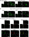
Assembly of Sar1 around ERES depends on Sec16 function. (A) sec16-2 cells expressing Sar1–GFP and Sec13–mRFP were observed at the permissive (24°C) and the restrictive (37°C) temperatures. Representative 2D images (exposure time, 2 s) from the center of a cell are shown (upper panels). The intensity of GFP signals across a cell is also shown as a linescan (right panels). Arrows indicate the positions of the ER membrane (left and right arrows in upper panel, both arrows in lower panel) and the nuclear envelope (center arrow in upper panel); arrowheads indicate the position of the cytoplasm. Upon a shift to the restrictive temperature, the fluorescent signals from Sar1–GFP decreased in the ER membrane and increased in the cytoplasm. Sec13–mRFP dispersed in the cytoplasm. Scale bar: 5 μm. We conducted at least five independent experiments and representative images are shown. (B,C) At least 10 independent sec16-2 cells expressing Sar1–GFP were observed. Representative 2D time-lapse images (exposure time, 0.5 s) at the peripheral ER are shown in B. Scale bar: 1 μm. Percentages of cells which have Sar1–GFP clusters stably localizing at their peripheral ER over 4 s are shown in C. Sar1–GFP clusters were evident in the cells cultured at the permissive temperature (24°C). Upon temperature-shift to the restrictive temperature (37°C) for 45 min, Sar1–GFP clusters on the peripheral ER disappeared. Cells were then shifted from 37°C back to 24°C and cultured for 90 min; after these 90 min, Sar1–GFP clusters were evident again. (D) We observed at least eight independent sec16-2 cells expressing Sar1–GFP (green) and Sec13–mRFP (COPII outer coat, red). Representative 3D images were reconstructed from optical slices taken 0.1 µm apart. Upon temperature shift to the restrictive temperature (37°C), weak fluorescent signals from Sar1–GFP evidently localized to the ER membrane; however, COPII-coated vesicles labeled with Sec13–mRFP vanished from the ER membrane. Cells were then shifted from 37°C back to 24°C and cultured for 90 min; after these 90 min, COPII-coated vesicle formation and restricted colocalizaton between Sar1–GFP and Sec13–mRFP to the rim of COPII membrane were evident again. Scale bar: 2 μm.
Fig. 4.
Fig. 4.
Restriction of Sar1 localization to the rims of COPII-coated membranes depends on Sar1 hydrolysis of GTP. (A) Wild-type cells expressing Sec13–GFP (COPII outer coat) with or without pGAL-Sar1H77L (GTPase-deficient mutant form) were cultured in galactose medium for 2 h at 24°C. Optical slices were taken 0.1 μm apart. We observed at least 12 independent cells and representative xy, xz and yz images, and 3D reconstructed images are shown. Expression of Sar1H77L induced formation of enlarged structures labeled with Sec13–GFP. (B) sec23-1 cells expressing Sec31–GFP (COPII outer coat) were cultured at the permissive (24°C) and the restrictive (39°C) temperatures. Optical slices were taken 0.1 μm apart; xy, xz and yz images and 3D reconstructed images are shown. Abnormal elongated vesicular structures labeled with Sec31–GFP were observed in the cells that had been cultured for 60 min at the restrictive temperature. We observed four independent cells at 24°C and nine independent cells at 39°C. (C) A total of 14 independent wild-type cells expressing Sec13–mRFP (COPII coat, red) and Sar1H77L–GFP were observed. Sar1H77L–GFP is expressed under the control of the heat-shock promoter when cells were cultured at 37°C for 60 min. Optical slices were taken 0.1 µm apart, and representative xy, xz and yz images and 3D reconstructed images are shown. The GTP-locked form of Sar1–GFP accumulated on the entirety of these enlarged structures coated with COPII coat protein. Dotted circles in the images highlight the edges of cells. Scale bars: 1 μm.

Similar articles

Cited by

References

    1. Antonny B., Madden D., Hamamoto S., Orci L. and Schekman R. (2001). Dynamics of the COPII coat with GTP and stable analogues. Nat. Cell Biol. 3, 531-537. 10.1038/35078500 - DOI - PubMed
    1. Aridor M., Bannykh S. I., Rowe T. and Balch W. E. (1995). Sequential coupling between COPII and COPI vesicle coats in endoplasmic reticulum to Golgi transport. J. Cell Biol. 131, 875-893. 10.1083/jcb.131.4.875 - DOI - PMC - PubMed
    1. Aridor M., Fish K. N., Bannykh S., Weissman J., Roberts T. H., Lippincott-Schwartz J. and Balch W. E. (2001). The Sar1 GTPase coordinates biosynthetic cargo selection with endoplasmic reticulum export site assembly. J. Cell Biol. 152, 213-229. 10.1083/jcb.152.1.213 - DOI - PMC - PubMed
    1. Bacia K., Futai E., Prinz S., Meister A., Daum S., Glatte D., Briggs J. A. G. and Schekman R. (2011). Multibudded tubules formed by COPII on artificial liposomes. Sci. Rep. 1, 17 10.1038/srep00017 - DOI - PMC - PubMed
    1. Bannykh S. I., Rowe T. and Balch W. E. (1996). The organization of endoplasmic reticulum export complexes. J. Cell Biol. 135, 19-35. 10.1083/jcb.135.1.19 - DOI - PMC - PubMed

MeSH terms

Substances