

Identifying Small Coronary Calcification in Non-Contrast 0.5-mm Slice Reconstruction to Diagnose Coronary Artery Disease in Patients with a Conventional Zero Coronary Artery Calcium Score
- PMID: 27397477
- PMCID: PMC5221495
- DOI: 10.5551/jat.35808
Identifying Small Coronary Calcification in Non-Contrast 0.5-mm Slice Reconstruction to Diagnose Coronary Artery Disease in Patients with a Conventional Zero Coronary Artery Calcium Score
Abstract
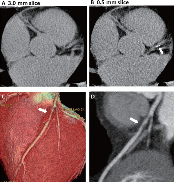
Aims: In a new-generation computed tomography (CT) scanner, coronary artery calcium (CAC) scores were measured using 3.0-mm slice reconstruction images originally acquired with 0.5 mm thickness scans in a single beat. This study investigated the usefulness of thin-slice (0.5 mm) reconstruction for identifying small calcifications in coronary arteries and evaluated the association with coronary plaques and stenosis compared to conventional 3.0-mm reconstruction images.
Methods: We evaluated 132 patients with zero CAC scores in conventional 3.0-mm Agatston method using a 320-slice CT. Then, 0.5-mm slice reconstruction was performed to identify small calcifications. The presence of stenosis and coronary plaques was assessed using coronary CT angiography.
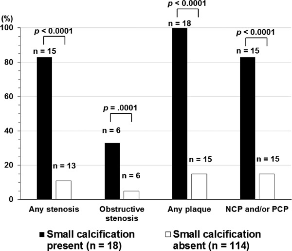
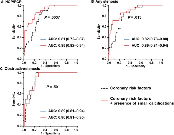
Results: In total, 22 small calcifications were identified in 18 patients. There were 28 (21%) patients with any (≥ 25%) stenosis (34 lesions). Forty-seven coronary plaques were found in 33 patients (25%), including 7 calcified plaques in 7 patients (5%), 34 noncalcified plaques in 27 patients (20%), and 6 partially calcified plaques in 5 patients (4%). Patients with small calcifications had a significantly higher prevalence of noncalcified or partially calcified plaques (83% vs 14%; p<0.001) and obstructive stenosis (33% vs 5.2%; p<0.001) compared to those without small calcifications. The addition of small calcifications to the coronary risk factors when diagnosing stenosis significantly improved the diagnostic value.
Conclusion: Small calcifications detected by thin-slice 0.5-mm reconstruction are useful for distinguishing coronary atherosclerotic lesions in patients with zero CAC scores from conventional CT reconstruction.
Figures
Similar articles
-
Assessment of isotropic calcium using 0.5-mm reconstructions from 320-row CT data sets identifies more patients with non-zero Agatston score and more subclinical atherosclerosis than standard 3.0-mm coronary artery calcium scan and CT angiography.J Cardiovasc Comput Tomogr. 2014 Jan-Feb;8(1):58-66. doi: 10.1016/j.jcct.2013.12.007. Epub 2014 Jan 11. J Cardiovasc Comput Tomogr. 2014. PMID: 24582044
-
Prevalence of noncalcified coronary plaques by 64-slice computed tomography in patients with an intermediate risk for significant coronary artery disease.J Am Coll Cardiol. 2006 Jul 18;48(2):312-8. doi: 10.1016/j.jacc.2006.02.064. Epub 2006 Jun 22. J Am Coll Cardiol. 2006. PMID: 16843181
-
Coronary artery calcium scoring: influence of reconstruction interval and reconstruction increment using 64-MDCT.AJR Am J Roentgenol. 2007 Apr;188(4):1063-8. doi: 10.2214/AJR.05.1369. AJR Am J Roentgenol. 2007. PMID: 17377048 Clinical Trial.
-
Scoring of coronary artery calcium scans: history, assumptions, current limitations, and future directions.Atherosclerosis. 2015 Mar;239(1):109-17. doi: 10.1016/j.atherosclerosis.2014.12.040. Epub 2015 Jan 2. Atherosclerosis. 2015. PMID: 25585030 Review.
-
[Noninvasive computed tomographic coronary angiography as a complement to coronary calcium quantification in symptomatic patients].Herz. 2003 Mar;28(2):106-18. doi: 10.1007/s00059-003-2452-5. Herz. 2003. PMID: 12669224 Review. German.
Cited by
-
Measurement of coronary artery calcium volume using ultra-high-resolution computed tomography: A preliminary phantom and cadaver study.Eur J Radiol Open. 2020 Sep 8;7:100253. doi: 10.1016/j.ejro.2020.100253. eCollection 2020. Eur J Radiol Open. 2020. PMID: 32964073 Free PMC article.
-
A CNN-based denoising method trained with images acquired with electron density phantoms for thin-sliced coronary artery calcium scans.J Appl Clin Med Phys. 2024 Mar;25(3):e14287. doi: 10.1002/acm2.14287. Epub 2024 Feb 12. J Appl Clin Med Phys. 2024. PMID: 38346094 Free PMC article.
-
Influence of computed tomography slice thickness on deep learning-based, automatic coronary artery calcium scoring software performance.Quant Imaging Med Surg. 2023 Jul 1;13(7):4257-4267. doi: 10.21037/qims-22-835. Epub 2023 Jan 5. Quant Imaging Med Surg. 2023. PMID: 37456306 Free PMC article.
-
A New Factor for Vascular Calcification in Chronic Kidney Disease: Computed Tomography-Based Renal Parenchymal Volume.J Atheroscler Thromb. 2017 Nov 1;24(11):1085-1087. doi: 10.5551/jat.ED081. Epub 2017 Jul 12. J Atheroscler Thromb. 2017. PMID: 28701626 Free PMC article. No abstract available.
-
Tumor Necrosis Factor-α Gene Expression in Epicardial Adipose Tissue is Related to Coronary Atherosclerosis Assessed by Computed Tomography.J Atheroscler Thromb. 2018 Mar 1;25(3):269-280. doi: 10.5551/jat.41178. Epub 2017 Sep 20. J Atheroscler Thromb. 2018. PMID: 28931782 Free PMC article.
References
-
- Agatston AS, Janowitz WR, Hildner FJ, Zusmer NR, Viamonte M, Jr, Detrano R: Quantification of coronary artery calcium using ultrafast computed tomography. J Am Coll Cardiol, 1990; 15: 827-832 - PubMed
-
- Budoff MJ, Georgiou D, Brody A, Agatston AS, Kennedy J, Wolfkiel C, Stanford W, Shields P, Lewis RJ, Janowitz WR, Rich S, Brundage BH: Ultrafast computed tomography as a diagnostic modality in the detection of coronary artery disease: a multicenter study. Circulation, 1996; 93: 898-904 - PubMed
-
- Yamamoto H, Kitagawa T, Kihara Y: Clinical implications of the coronary artery calcium score in Japanese patients. J Atheroscler Thromb, 2014; 21: 1101-1108 - PubMed
-
- Detrano R, Guerci AD, Carr JJ, Bild DE, Burke G, Folsom AR, Liu K, Shea S, Szklo M, Bluemke DA, O'Leary DH, Tracy R, Watson K, Wong ND, Kronmal RA: Coronary calcium as a predictor of coronary events in four racial or ethnic groups. New Engl J Med, 2008; 358: 1336-1345 - PubMed
-
- Budoff MJ, Shaw LJ, Liu ST, Weinstein SR, Mosler TP, Tseng PH, Flores FR, Callister TQ, Raggi P, Berman DS: Long-term prognosis associated with coronary calcification: observations from a registry of 25,253 patients. J Am Coll Cardiol, 2007; 49: 1860-1870 - PubMed
MeSH terms
Substances
LinkOut - more resources
Full Text Sources
Other Literature Sources
Medical