. 2016 Sep;15(9):2119-29.
doi: 10.1158/1535-7163.MCT-16-0197. Epub 2016 Jul 7.

Dual and Specific Inhibition of NAMPT and PAK4 By KPT-9274 Decreases Kidney Cancer Growth

Affiliations

Dual and Specific Inhibition of NAMPT and PAK4 By KPT-9274 Decreases Kidney Cancer Growth

Omran Abu Aboud et al. Mol Cancer Ther. 2016 Sep.

Abstract

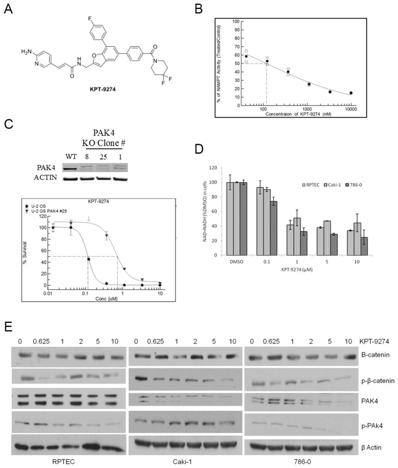
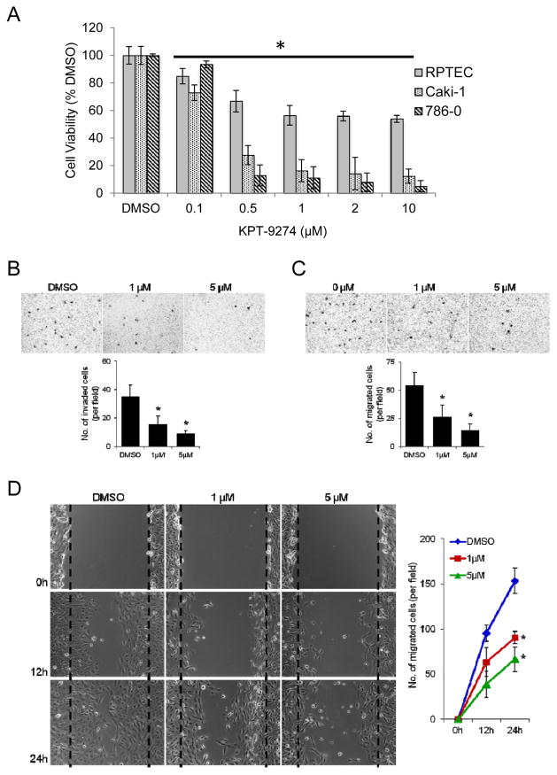
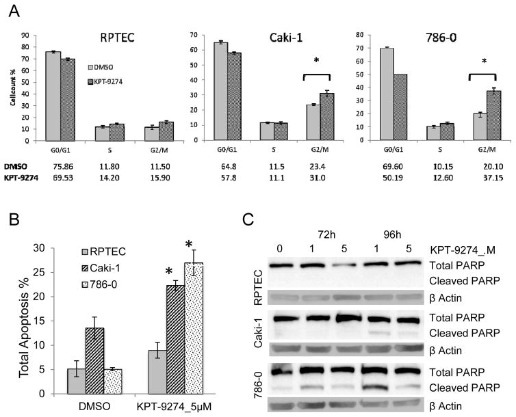
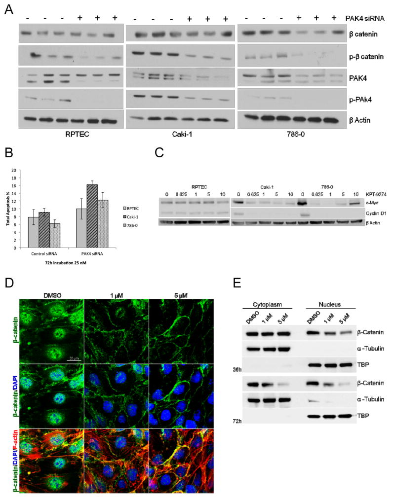
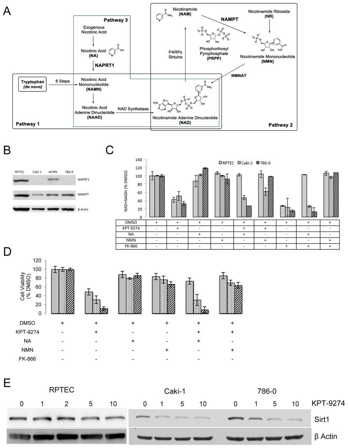
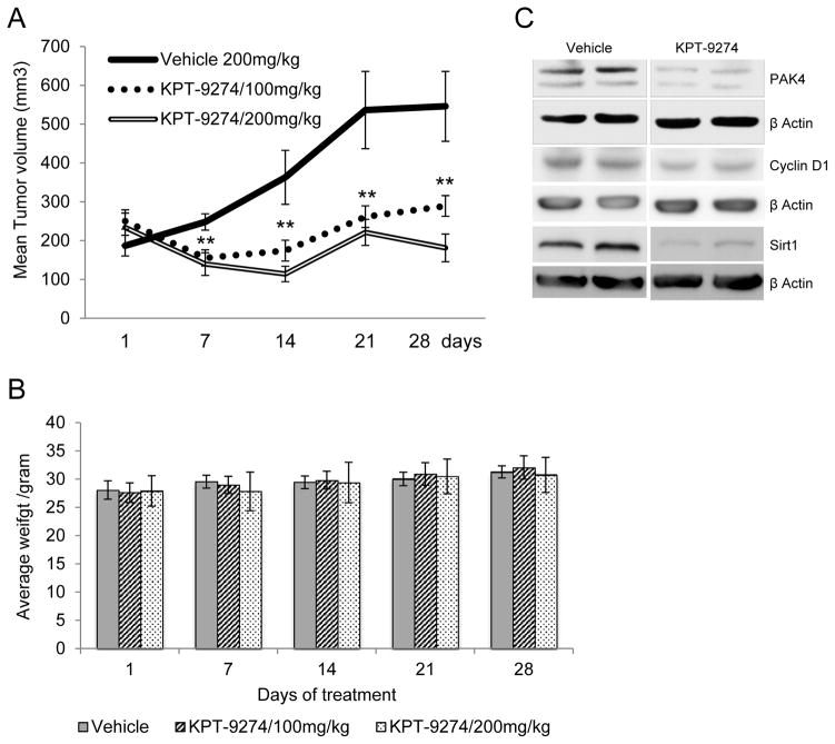
Kidney cancer (or renal cell carcinoma, RCC) is the sixth most common malignancy in the United States and one of the relatively few whose incidence is increasing. Because of the near universal resistance which occurs with the use of current treatment regimens, reprogrammed metabolic pathways are being investigated as potential targets for novel therapies of this disease. Borrowing from studies on other malignancies, we have identified the PAK4 and NAD biosynthetic pathways as being essential for RCC growth. We now show, using the dual PAK4/NAMPT inhibitor KPT-9274, that interference with these signaling pathways results in reduction of G2-M transit as well as induction of apoptosis and decrease in cell invasion and migration in several human RCC cell lines. Mechanistic studies demonstrate that inhibition of the PAK4 pathway by KPT-9274 attenuates nuclear β-catenin as well as the Wnt/β-catenin targets cyclin D1 and c-Myc. Furthermore, NAPRT1 downregulation, which we show occurs in all RCC cell lines tested, makes this tumor highly dependent on NAMPT for its NAD requirements, such that inhibition of NAMPT by KPT-9274 leads to decreased survival of these rapidly proliferating cells. When KPT-9274 was administered in vivo to a 786-O (VHL-mut) human RCC xenograft model, there was dose-dependent inhibition of tumor growth with no apparent toxicity; KPT-9274 demonstrated the expected on-target effects in this mouse model. KPT-9274 is being evaluated in a phase I human clinical trial in solid tumors and lymphomas, which will allow this data to be rapidly translated into the clinic for the treatment of RCC. Mol Cancer Ther; 15(9); 2119-29. ©2016 AACR.


Conflict of interest statement

W.S., E.B., and C.A. are employees of Karyopharm and made suggestions on the manuscript but did not influence the direction of the research reported here. Some of the reagents in this study were ordered by Karyopharm and gifted to the Weiss laboratory.

Figures

Figure 1
Figure 1
KPT-9274 inhibits NAMPT and PAK4 and associated signaling pathways in RCC cells. A. Molecular structure of KPT-9274 B. Cell-free assay of NAMPT activity as a function of KPT-9274 concentration C. CRISPR-Cas9 splicing out of PAK4 in U-2 osteosarcoma cells showing the expected immunoblot and a shift to the right in the survival curve assessed by MTT assay. D. NAD+NADH assay: 300 cells/well were plated in 96 well plates (n=4) and incubated for 48h with DMSO or KPT-9274. Assays of total NAD+NADH were performed in RCC cell lines as well as a normal primary renal proximal tubular epithelial cell (RPTEC) line as described in Materials and Methods. Error bars are SD. *p<0.05 compared to DMSO treated controls. The black sold line indicates treatments significantly different compared to DMSO alone. E. Immunoblotting of the cell lysates with the indicated antibodies was performed. Cells were plated in 6-well (1000 cells/well) and incubated with different concentrations of KPT-9274 for 72h before they lysed for protein extraction. The experiments shown are representative of at least three independent repeats.
Figure 2
Figure 2
KPT-9274 preferentially attenuates RCC cell viability and decreases RCC invasion and migration. A. Both RCC cell lines in addition to a normal primary renal primary proximal tubular epithelial cell line (RPTEC) were plated in 96 well plates (3000 cells/well, n=8) and incubated 72h with DMSO or KPT-9274 before MTT was performed. Error bars are SD. *p<0.05 compared to DMSO treated controls. The black sold line indicates treatments significantly different compared to DMSO alone. The experiment shown is representative of at least three independent repeats. B–D: 786-O cells were treated with DMSO, 1μM or 5μM KPT-9274 for 24 hours and then subjected to Matrigel invasion (B), transwell migration (C) and scratch/wound healing (D) assays. (B) DMSO- or KPT-9274-treated cells were seeded on Matrigel-coated transwells with DMSO or KPT-9274; 20 hours later, migrated cells were fixed, stained, and counted using light microscopy. A representative picture of each group is shown in the top. Bottom, quantification of migrated cells to the lower chamber. Data were presented as the mean value from six different fields ± SD (n = 3), *p < 0.05 as compared to DMSO-treated cells. (C) Migration assay in transwell chambers. Cells that migrated from the upper well of a transwell chamber into the lower well were stained, photographed (top) and counted (bottom). n = 3, *p < 0.05 versus DMSO group. (D) Confluent cultures of these cells were scratched and wound healing repair was monitored microscopically examined at 12 and 24 hours after the scratch and the addition of DMSO or KPT-9274. Left, representative phase contrast pictures. Right, numbers of cells migrated to the wound area were quantified at 0, 12 and 24 hours post-scratching (n = 3, *p < 0.05 versus DMSO-treated cells).
Figure 3
Figure 3
KPT-9274 decreases G2/M transit and causes apoptosis preferentially in RCC cells. Both RCC cell lines in addition to a normal primary renal proximal tubular epithelial cell line (RPTEC) were grown to 50% confluence and subjected to cell cycle analysis or apoptosis assays: a. Cells were incubated with DMSO or KPT-9274 (5 μM) for 72h and analyzed with the Muse Analyzer for cell cycle analysis as described in Materials and Methods. The percentage of cells in each cell cycle phase was plotted. *p<0.05 KPT-9274 as compared to DMSO treatment b. After incubation with KPT-9274 for 72h cells, annexin-V staining was used to measure total apoptosis by the Muse Analyzer as described in Materials and Methods. *p<0.05 as compared to DMSO treatment. c. Immunoblotting of total and cleaved PARP in RPTEC and RCC cells KPT-9274 treated after 72h and 96h. All results represent at least three independent experiments.
Figure 4
Figure 4
KPT-9274 shows specificity for attenuation of PAK4 targets preferentially in RCC cells. Both RCC cell lines in addition to a normal primary renal proximal tubular epithelial cell line (RPTEC) were grown to 50% and transfected with an siRNA specific to PAK4 or a scrambled sequence control siRNA, then subjected to: A. immunoblotting and B. apoptosis assay by flow cytometry. C. Whole cell lysates were immunoblotted with the indicated antibodies after incubation of the cells with the indicated concentrations of KPT-9274. D. The subcellular localization of β-catenin in 786-O cells was determined by immunofluorescent staining after 24 hours of treatment with DMSO, 1μM or 5μM KPT-9274. The fluorescence of FITC-conjugated β-catenin (green), TRITC-conjugated phalloidin (F-actin stained: red) and DAPI (nucleus counter-stained: blue) was visualized under a confocal laser-scanning microscope. Scale bar: 20 μm E. Western blot analysis of β-catenin in the cytosolic and nuclear portions of 786-O cells exposed to DMSO or KPT-9274 for 36 and 72 hours. TBP=TATA-Binding Protein, a nuclear constituent. Please see Materials and Methods for details.
Figure 5
Figure 5
KPT-9274 shows specificity for attenuation of NAD biosynthesis targets preferentially in RCC cells. A. Schema of NAD biosynthesis from de novo (Pathway 1) and salvage pathways (Pathways 2 and 3). B. Whole cell lysates from non-treated RCC and RPTEC cells were immunoblotted with the indicated antibodies. C. Cells were grown in 96 well plates (300 cells/well), incubated as indicated, and subjected to assays for NAD+NADH as described in the Materials and Methods section. D. Cells were grown in 96 well plates (300 cells/well, n=4), incubated as indicated, and subjected to MTT assays as described in the Materials and Methods section.. E. Cells were grown in 6 well plates (10,000 cells/well) exposed to KPT-9274 at the indicated concentrations (μM) for 72h and whole cell lysates were immunoblotted with sirt1 antibody.
Figure 6
Figure 6
KPT-9274 attenuates xenograft growth in vivo with minimal toxicity and shows the expected on-target effects. A. Nude mice (n=8 per condition) were xenografted subcutaneously with 786-O cells and gavaged with KPT-9274 (100mg/kg or 200 mg/kg twice a day). Tumors were measured with calipers weekly at the times indicated and the mean tumor volumes ± SEM was calculated as described in Materials and Methods. **p<0.05 indicates significant decrease in tumor volume compared to vehicle only group. B. Mouse weights were determined at the time points indicated. C. Tumors were harvested at sacrifice, pooled, and immunoblotted with the target antibodies indicated as well as β-actin as loading control.

Similar articles

Cited by

References

    1. Wettersten HI, Weiss RH. Potential biofluid markers and treatment targets for renal cell carcinoma. Nat Rev Urol. 2013;10:336–44. - PubMed
    1. Wettersten HI, Hakimi AA, Morin D, Bianchi C, Johnstone ME, Donohoe DR, et al. Grade-dependent metabolic reprogramming in kidney cancer revealed by combined proteomics and metabolomics analysis. Cancer Res. 2015;75:2541–52. - PMC - PubMed
    1. Hakimi AA, Reznik E, Lee CH, Creighton CJ, Brannon AR, Luna A, et al. An Integrated Metabolic Atlas of Clear Cell Renal Cell Carcinoma. Cancer Cell. 2016;29:104–16. - PMC - PubMed
    1. Radu M, Semenova G, Kosoff R, Chernoff J. PAK signalling during the development and progression of cancer. Nat Rev Cancer. 2014;14:13–25. - PMC - PubMed
    1. Dart AE, Wells CM. P21-activated kinase 4--not just one of the PAK. Eur J Cell Biol. 2013;92:129–38. - PubMed

Publication types

MeSH terms