. 2016 Jul 12;16(2):392-404.
doi: 10.1016/j.celrep.2016.05.088. Epub 2016 Jun 23.

RBPJ Controls Development of Pathogenic Th17 Cells by Regulating IL-23 Receptor Expression

Affiliations

RBPJ Controls Development of Pathogenic Th17 Cells by Regulating IL-23 Receptor Expression

Gerd Meyer Zu Horste et al. Cell Rep. .

Abstract

Interleukin-17 (IL-17)-producing helper T cells (Th17 cells) play an important role in autoimmune diseases. However, not all Th17 cells induce tissue inflammation or autoimmunity. Th17 cells require IL-23 receptor (IL-23R) signaling to become pathogenic. The transcriptional mechanisms controlling the pathogenicity of Th17 cells and IL-23R expression are unknown. Here, we demonstrate that the canonical Notch signaling mediator RBPJ is a key driver of IL-23R expression. In the absence of RBPJ, Th17 cells fail to upregulate IL-23R, lack stability, and do not induce autoimmune tissue inflammation in vivo, whereas overexpression of IL-23R rescues this defect and promotes pathogenicity of RBPJ-deficient Th17 cells. RBPJ binds and trans-activates the Il23r promoter and induces IL-23R expression and represses anti-inflammatory IL-10 production in Th17 cells. We thus find that Notch signaling influences the development of pathogenic and non-pathogenic Th17 cells by reciprocally regulating IL-23R and IL-10 expression.

Keywords: IL-23R; Notch; RBPJ; Th17 cells; pathogenicity.


Figures

Figure 1
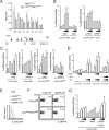
Figure 1. RBPJ in T cells maintains pathogenicity of Th17 cells
(A) Naïve CD4+CD62LhighCD44lowCD25 T cells were sorted from RBPJflox/flox and CD4CreRBPJflox/flox mice, differentiated with TGF-β1 and IL-6, and analyzed by intracellular cytokine staining after 4 days. (B) Cytokine ELISA of cultures described in A. (C) Naïve T cells were differentiated with IL-1β, IL-6, and IL-23. (D) Cytokine ELISA of cultures described in C. (E) CD4+CD62LlowCD44highCD25 memory T cells were sorted from RBPJflox/flox and CD4CreRBPJflox/flox mice and cultured with IL-23 for 5 days and stained for intracellular cytokines. One representative out of five independent experiments is shown in A–E; nd not detected. (F) Active EAE was induced in RBPJflox/flox (n = 26) and CD4CreRBPJflox/flox (n = 26) mice by subcutaneous immunization with 100μg of MOG35–55 peptide in complete Freund’s adjuvant together with intraperitoneal injection of pertussis toxin (200 ng) on day 0 and 2. (G) Regression analysis of the clinical scores observed between day 12 and day 28. (H) CNS infiltrating mononuclear cells were extracted and stained for intracellular cytokines at day 15 after immunization (d15). The percentage of CNS infiltrating cytokine producing CD4+ cells was calculated. (I) The cytokine production by CNS infiltrating CD4+ cells was assessed at d28 and the proportion of IL-10 producing CD4+ T cells was calculated. (J) CNS infiltrating CD4+ T cells were sorted from RBPJflox/flox (n = 3) and CD4CreRBPJflox/flox (n = 3) mice and relative Il10 expression was measured by qPCR using GAPDH as housekeeping gene. Results are summed from three independent experiments in F–I with three indidivual mice analyzed per group in H and I. See also Figure S1.
Figure 2
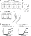
Figure 2. RBPJ is required in Th17 cells to maintain their pathogenicity
(A) Naïve CD4+CD62LhighCD44lowCD25 T cells were sorted from RBPJflox/flox and IL17ACreRBPJflox/flox mice, differentiated with TGF-β1 and IL-6, and stained for intracellular cytokines after 4 days. (B) Cytokine ELISA of cultures described in A. (C) Naïve T cells were differentiated in vitro with IL-1β, IL-6, and IL-23 and analyzed as in A. (D) Cytokine ELISA of cultures described in C. One representative of five independent experiments is shown in A–D. (E) RBPJflox/flox (n = 43) and IL17ACreRBPJflox/flox (n = 28) mice were immunized subcutaneously with MOG35–55 peptide emulsified in complete Freund’s adjuvant with intraperitoneal injections of pertussis toxin. (F) Regression analysis of the clinical scores observed between day 12 and day 28. Data in E and F are summed from five independent experiments. (G) CNS infiltrating cells were extracted and analyzed by intracellular cytokine staining at day 15 after immunization (d15). (H) CNS infiltrating lymphocytes were stained for intracellular cytokines at d28 and the percentage of IL-10+ CD4+ cells was calculated. (I) Draining lymph node cells of mice at d15 were restimulated in vitro with 10μg/ml MOG35–55 peptide and IL-23 for five days. Cytokine concentrations were measured by ELISA, nd not detected. Data in G–I are summed from three independent experiments with three to five mice per group. See also Figure S2. (J) Naïve CD4+CD62LhighCD44lowCD25 T cells were sorted from wildtype 2D2 and CD4CreRBPJfl/fl2D2 mice and differentiated in vitro with TGF-β1, IL-6 and IL-23 and after 7 days of culture 5*106 cytokine producing T cells were intravenously injected into C57BL/6 wildtype recipients.
Figure 3
Figure 3. RBPJ-deficient Th17 cells show non-pathogenic gene expression and are unstable in vitro due to a lack of IL-23R expression
(A) Naïve CD4+CD62LhighCD44lowCD25 T cells were sorted from IL17ACreRBPJflox/wt and IL17ACreRBPJflox/flox mice and differentiated with TGF-β1 and IL-6. Expression of a predefined set of Th17-relevant genes was quantified using Nanostring nCounter after 4 days of culture. (B) Identical experiment as in A using naïve CD4+ cells differentiated with IL-1β, IL-6, and IL-23. Average values from three independent experiments with technical duplicates per sample are depicted in A and B as fold change of IL17ACreRBPJflox/flox versus IL17ACreRBPJflox/wt cells. Please note that Il10 is upregulated 1.47-fold (data not shown), but does not reach the defined cut-off for minimum read counts i.e. transcript abundance. Cut-off for differentially regulated genes was ≥1.25-fold regulation. (C) Naïve T cells from IL17ACreRBPJflox/wtR26RFP and IL17ACreRBPJflox/floxR26RFP mice were differentiated in vitro with TGF-β1 and IL-6 or IL-1β, IL-6, and IL-23 for 4 days and the proportion of CD4+RFP+ cells was assessed by flow cytometry. (D) CD4+RFP+ cells were sorted from cultures differentiated with TGF-β1 and IL-6 described in (C) and analyzed for gene expression using Nanostring nCounter. Expression is depicted as fold-change of IL17ACreRBPJflox/floxR26RFP versus IL17ACreRBPJflox/wtR26RFP cells. Cut-off for differentially regulated genes was ≥1.25-fold and changes are averaged from three independent experiments. (E) Naïve T cells were differentiated with TGF-β1 and IL-6 from IL17ACreRBPJflox/wtR26RFP and IL17ACreRBPJflox/floxR26RFP mice as in D. Live CD4+RFP+ cells were sorted from the cultures and restimulated in vitro for 3 days with IL-23 only. Cytokine production was assessed before (d4) and after (d7) restimulation after gating on live RFP+ cells. (F) IL17ACreRBPJflox/wtR26RFP (Rfl/wt) and IL17ACreRBPJflox/floxR26RFP (Rfl/fl) mice were immunized with MOG35–55 and at peak of EAE, draining lymph nodes cells were cultured in the presence of 1 μg/ml MOG and IL-23 for 5 days. Intracellular cytokine production was assessed after gating on CD4+ (top) and on CD4+RFP+ (bottom) cells. Data in F are summarized from 4 independent mice in three independent experiments. (G) Il23r promoter driven GFP fluorescence was quantified in naïve T cells differentiated for 4 days without cytokines from IL17ACreRBPJflox/wtIL23RGFP/wt mice (red histogram), or with IL-1β, IL-6, and IL-23 from IL17ACreRBPJflox/wtIL23RGFP/wt mice (blue histogram) and IL17ACreRBPJflox/floxIL23RGFP/wt mice (orange histogram). (H) Naïve CD4+ T cells were sorted from RBPJflox/flox and CD4CreRBPJflox/flox mice, differentiated with IL-1β, IL-6, and IL-23 for 4 days, and IL-23R was detected by cell-surface staining for IL-23R. Data in C, E, G, H are representative of three independent experiments. See also Figure S3.
Figure 4
Figure 4. RBPJ binds and transactivates the Il23r promoter and induces IL-23R expression in cooperation with RORγt
Naïve CD4+ T cells were sorted from RBPJflox/flox and CD4CreRBPJflox/flox mice and differentiated with TGF-β1 and IL-6 for 4 days followed by IL-23 for 3 days. Cross-linked protein-DNA complexes were immunoprecipitated using an anti-RBPJ antibody. DNA was purified and used for qPCR with primers spanning parts of the Il23r promoter predicted to contain RBPJ binding sites indicated as horizontal bars in the scheme of the Il23r locus; in intron, p promoter. DNA binding was calculated as percentage of input DNA. One representative out of three independent experiments with three replicate samples per IP is shown. (B) Il23r promoter activity was measured in HEK293T cells transfected with a luciferase vector driven by 2.7kb of the IL23R promoter (pGL4-IL23R) and increasing amounts of constructs encoding c-Maf and consitutively active (ca)RBPJ (RBPJ-VP16). (C) IL23r promoter activity was measured after transfection with a luciferase vector driven by 1.5kb of the IL23R promoter (pTA-IL23R) and constructs encoding c-Maf and caRBPJ (RBPJ-VP16). Luciferase activity induced by c-Maf and RBPJ-VP16 was measured using an pTA-IL23R promoter construct with mutatetd RBPJ binding site p2 (pTA-IL23RΔp2). (D) pGL4-IL23R luciferase activity was measured after transfection with combinations of RBPJ, caRBPJ, and c-Maf constructs. Data in B–D are representative of five independent experiments. (E) Naïve T cells were sorted from IL23RGFP/wt reporter mice and transduced with either empty retrovirus (RV) (MSCV-Thy1.1) or RV encoding caRBPJ (MSCV-RBPJ-VP16-Thy1.1) and cultured without cytokines. GFP expression was detected after gating on live CD4+Thy1.1+ cells after 4 days of culture. (F) Naïve T cells were sorted from Rorγtflox/flox andCD4CreRorγtflox/flox mice and transduced with either empty RV or caRBPJ RV. Cell surface expression of IL-23R was analyzed after 4 days in culture after gating on live CD4+Thy1.1+ cells. (G) IL23r promoter activity was measured in HEK293T cells transfected with the pGL4-IL23R vector and increasing amounts of caRBPJ and Rorγt constructs or both. Luciferase activities were calculated as fold empty vector in B–D, G. One representative out of three independent experiments is shown in E–G.
Figure 5
Figure 5. RBPJ is required to promote IL-23R expression rather than IL-17 in Th17 cells
(A) Naïve T cells from IL17ACreRBPJflox/wt and IL17ACreRBPJflox/flox mice were transduced with either empty retrovirus (RV) or with RV encoding mouse IL-23R and stained for intracellular cytokines after differentiation with IL-1β, IL-6, and IL-23 for 4 days. Gating was on live CD4+GFP+ cells. (B) The percentage of IL-17+ cells was calculated, defining empty RV in IL17ACreRBPJflox/wt cells as 100% in each experiment. Significance is calculated using Student’s t-test for paired samples. Data in A are representative and in B are summarized from 4 independent experiments. (C) Naïve T cells were sorted from IL17ACreRBPJflox/wtIL23RGFP/GFP and IL17ACreRBPJflox/floxIL23RGFP/GFP mice and differentiated with IL-1β, IL-6, and IL-23. Intracellular cytokine staining was performed after 4 days. (D) Cytokine ELISA of cell culture supernatants described in C. One representative of three independent experiments is shown in C and D. (E) Naïve T cells were sorted from wildtype 2D2 and CD4CreRBPJfl/fl2D2 mice and during Th17 differentiation were transduced with either empty or IL-23R encoding RV. After 7 days of culture, 6*106 cytokine producing T cells were intravenously injected into C57BL/6 wildtype recipients. The clinical score was assessed daily. (F) Naïve T cells were sorted from wildtype 2D2 and CD4CreRBPJfl/fl2D2 mice and during Th17 differentiation in vitro were transduced with a lentivirus (LV) encoding both Cas9 and an IL-10 targeting CRISPR guide RNA or Cas9 only (empty). After 7 days of culture, 8*106 cytokine producing T cells were intravenously injected into C57BL/6 wildtype recipients.
Figure 6
Figure 6. RBPJ represses c-Maf induced IL-10 production in Th17 cells
(A) Sorted naïve RBPJflox/flox and CD4CreRBPJflox/flox CD4+ T cells were transduced with either empty MSCV-GFP or MSCV-cMaf-GFP retrovirus (RV) and cultured with TGF-β1 and IL-6 for 4 days and analyzed by intracellular cytokine staining. (B) Cell culture supernatants from cultures described in A were analyzed by ELISA. (C) Naïve CD4+ T cells were sorted from RBPJflox/flox and CD4CreRBPJflox/flox mice and differentiated in vitro with TGF-β1 and IL-6 for 4 days followed by IL-23 for 3 days. Cross-linked protein-DNA complexes were immunoprecipitated with an anti-RBPJ antibody. DNA was purified and used for qPCR spanning predicted RBPJ binding sites in the IL-10 promoter. DNA binding was calculated as percentage of input DNA. Predicted RBPJ-binding sites are indicated as p1 to p8 and previously identified MARE-sites (Apetoh et al., 2011) are indicated as M1 to M4. One representative out of four independent experiments is shown in C with three replicate IPs per sample. (D) Il10 promoter activity was measured in HEK293T cells transfected with the pGL4-IL10-1.5kb luciferase vector and constructs encoding c-Maf and RBPJ. Luciferase activities were calculated as fold change compared to empty vector. One representative out of three independent experiments is shown in A, B, and D.

Similar articles

Cited by

References

    1. Abromson-Leeman S, Bronson RT, Dorf ME. Encephalitogenic T cells that stably express both T-bet and ROR gamma t consistently produce IFNgamma but have a spectrum of IL-17 profiles. Journal of neuroimmunology. 2009;215:10–24. - PMC - PubMed
    1. Amsen D, Antov A, Flavell RA. The different faces of Notch in T-helper-cell differentiation. Nature reviews Immunology. 2009;9:116–124. - PubMed
    1. Apetoh L, Quintana FJ, Pot C, Joller N, Xiao S, Kumar D, Burns EJ, Sherr DH, Weiner HL, Kuchroo VK. The aryl hydrocarbon receptor interacts with c-Maf to promote the differentiation of type 1 regulatory T cells induced by IL-27. Nature immunology. 2011;11:854–861. - PMC - PubMed
    1. Awasthi A, Carrier Y, Peron JP, Bettelli E, Kamanaka M, Flavell RA, Kuchroo VK, Oukka M, Weiner HL. A dominant function for interleukin 27 in generating interleukin 10-producing anti-inflammatory T cells. Nature immunology. 2007;8:1380–1389. - PubMed
    1. Awasthi A, Riol-Blanco L, Jager A, Korn T, Pot C, Galileos G, Bettelli E, Kuchroo VK, Oukka M. Cutting edge: IL-23 receptor gfp reporter mice reveal distinct populations of IL-17-producing cells. J Immunol. 2009;182:5904–5908. - PMC - PubMed

MeSH terms

Substances