

Merestinib blocks Mnk kinase activity in acute myeloid leukemia progenitors and exhibits antileukemic effects in vitro and in vivo
- PMID: 27307295
- PMCID: PMC4957163
- DOI: 10.1182/blood-2016-02-698704
Merestinib blocks Mnk kinase activity in acute myeloid leukemia progenitors and exhibits antileukemic effects in vitro and in vivo
Abstract
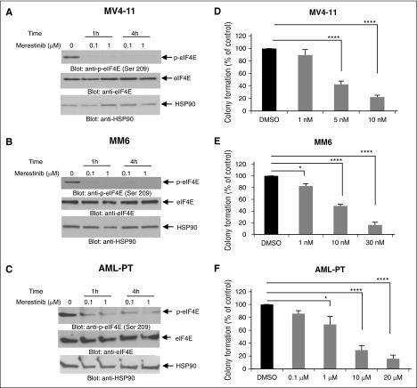
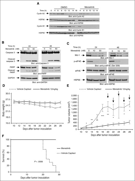
Mitogen-activated protein kinase interacting protein kinases (Mnks) play important roles in the development and progression of acute myeloid leukemia (AML) by regulating eukaryotic translation initiation factor 4E (eIF4E) activation. Inhibiting Mnk1/2-induced phosphorylation of eIF4E may represent a unique approach for the treatment of AML. We provide evidence for antileukemic effects of merestinib, an orally bioavailable multikinase inhibitor with suppressive effects on Mnk activity. Our studies show that merestinib effectively blocks eIF4E phosphorylation in AML cells and suppresses primitive leukemic progenitors from AML patients in vitro and in an AML xenograft model in vivo. Our findings provide evidence for potent preclinical antileukemic properties of merestinib and support its clinical development for the treatment of patients with AML.
Figures
Similar articles
-
Inhibition of Mnk kinase activity by cercosporamide and suppressive effects on acute myeloid leukemia precursors.Blood. 2013 May 2;121(18):3675-81. doi: 10.1182/blood-2013-01-477216. Epub 2013 Mar 18. Blood. 2013. PMID: 23509154 Free PMC article.
-
The MNK-eIF4E Signaling Axis Contributes to Injury-Induced Nociceptive Plasticity and the Development of Chronic Pain.J Neurosci. 2017 Aug 2;37(31):7481-7499. doi: 10.1523/JNEUROSCI.0220-17.2017. Epub 2017 Jul 3. J Neurosci. 2017. PMID: 28674170 Free PMC article.
-
Phosphorylation of eIF4E by MNKs supports protein synthesis, cell cycle progression and proliferation in prostate cancer cells.Carcinogenesis. 2008 Dec;29(12):2279-88. doi: 10.1093/carcin/bgn221. Epub 2008 Sep 22. Carcinogenesis. 2008. PMID: 18809972
-
Dual targeting of eIF4E by blocking MNK and mTOR pathways in leukemia.Cytokine. 2017 Jan;89:116-121. doi: 10.1016/j.cyto.2016.01.024. Epub 2016 Apr 16. Cytokine. 2017. PMID: 27094611 Free PMC article. Review.
-
Progress in developing MNK inhibitors.Eur J Med Chem. 2021 Jul 5;219:113420. doi: 10.1016/j.ejmech.2021.113420. Epub 2021 Apr 2. Eur J Med Chem. 2021. PMID: 33892273 Review.
Cited by
-
Inhibiting TRK Proteins in Clinical Cancer Therapy.Cancers (Basel). 2018 Apr 4;10(4):105. doi: 10.3390/cancers10040105. Cancers (Basel). 2018. PMID: 29617282 Free PMC article. Review.
-
MNK1/2 inhibition limits oncogenicity and metastasis of KIT-mutant melanoma.J Clin Invest. 2017 Nov 1;127(11):4179-4192. doi: 10.1172/JCI91258. Epub 2017 Oct 16. J Clin Invest. 2017. PMID: 29035277 Free PMC article.
-
AXL Receptor Tyrosine Kinase as a Therapeutic Target in Hematological Malignancies: Focus on Multiple Myeloma.Cancers (Basel). 2019 Nov 5;11(11):1727. doi: 10.3390/cancers11111727. Cancers (Basel). 2019. PMID: 31694201 Free PMC article. Review.
-
MNK Proteins as Therapeutic Targets in Leukemia.Onco Targets Ther. 2023 Apr 21;16:283-295. doi: 10.2147/OTT.S370874. eCollection 2023. Onco Targets Ther. 2023. PMID: 37113687 Free PMC article. Review.
-
Translational control in the tumor microenvironment promotes lung metastasis: Phosphorylation of eIF4E in neutrophils.Proc Natl Acad Sci U S A. 2018 Mar 6;115(10):E2202-E2209. doi: 10.1073/pnas.1717439115. Epub 2018 Feb 20. Proc Natl Acad Sci U S A. 2018. PMID: 29463754 Free PMC article.
References
-
- Gilliland DG, Tallman MS. Focus on acute leukemias. Cancer Cell. 2002;1(5):417–420. - PubMed
-
- Dash A, Gilliland DG. Molecular genetics of acute myeloid leukaemia. Best Pract Res Clin Haematol. 2001;14(1):49–64. - PubMed
-
- Beauchamp EM, Platanias LC. The evolution of the TOR pathway and its role in cancer. Oncogene. 2013;32(34):3923–3932. - PubMed
MeSH terms
Substances
Grants and funding
LinkOut - more resources
Full Text Sources
Other Literature Sources
Medical
