. 2016 Jul 7;19(1):52-65.
doi: 10.1016/j.stem.2016.05.003. Epub 2016 Jun 9.

Persistent Activation of NF-κB in BRCA1-Deficient Mammary Progenitors Drives Aberrant Proliferation and Accumulation of DNA Damage

Affiliations

Persistent Activation of NF-κB in BRCA1-Deficient Mammary Progenitors Drives Aberrant Proliferation and Accumulation of DNA Damage

Andrea Sau et al. Cell Stem Cell. .

Abstract

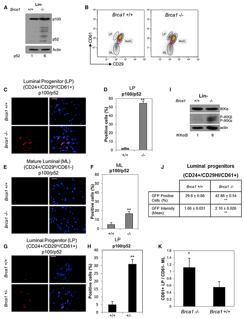
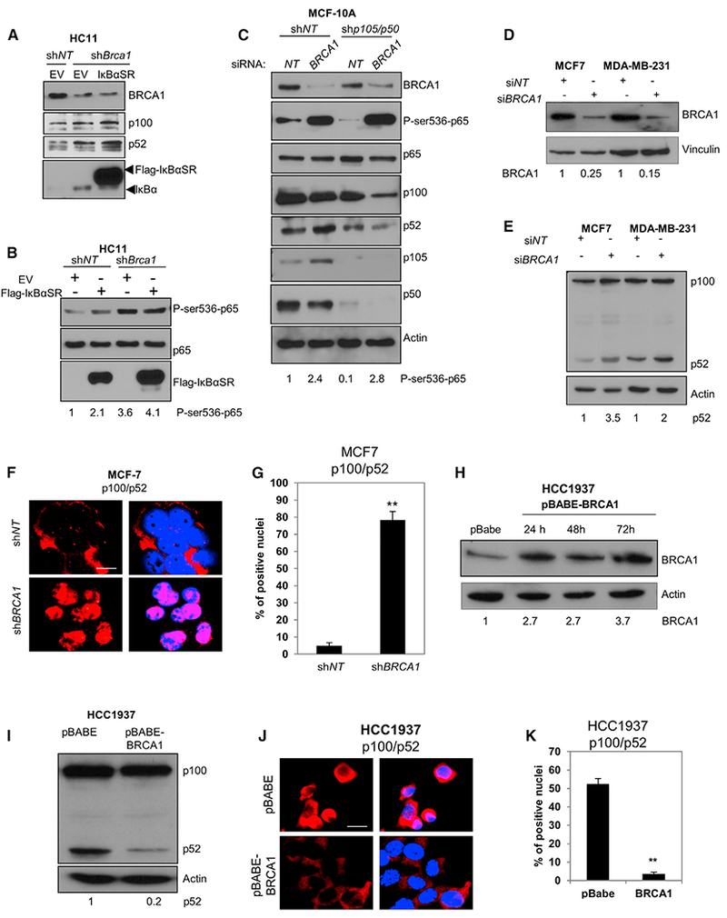
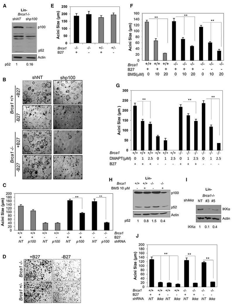
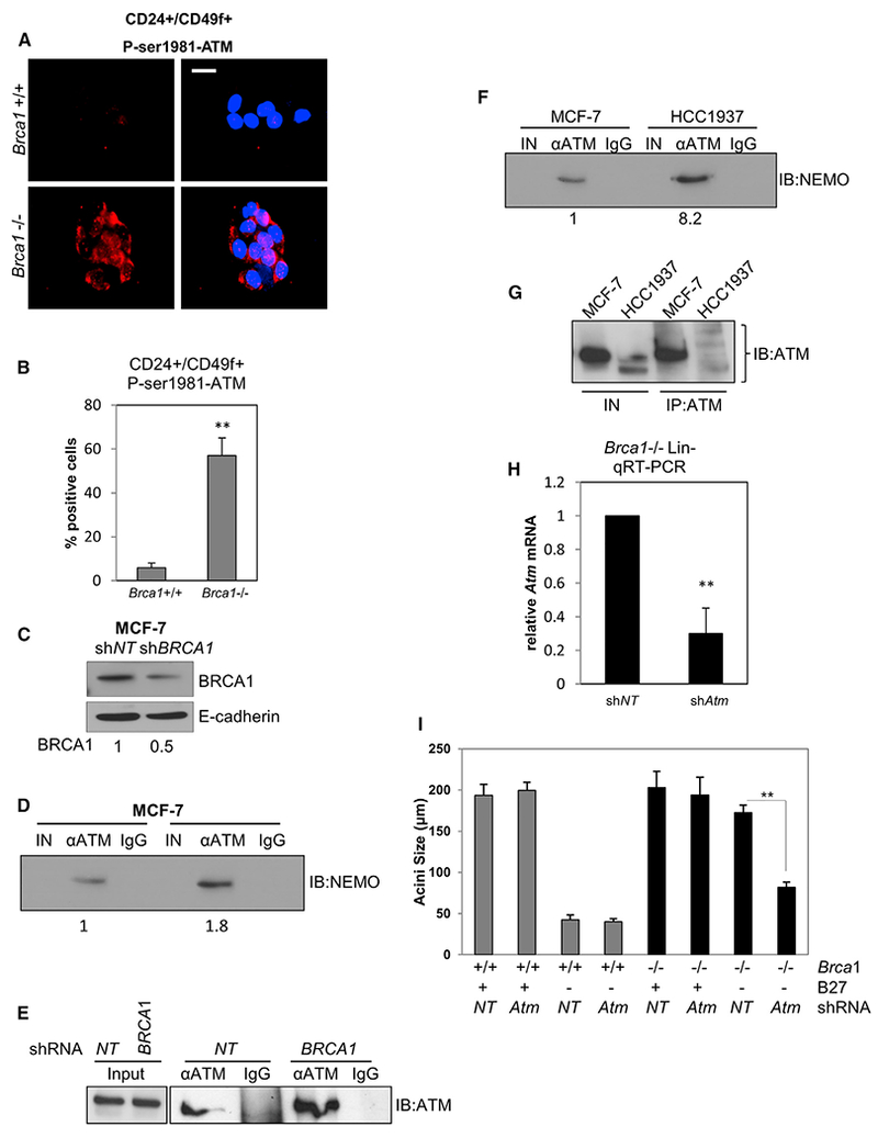
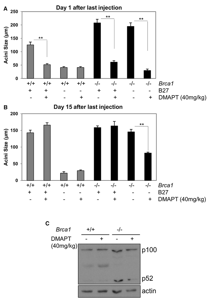
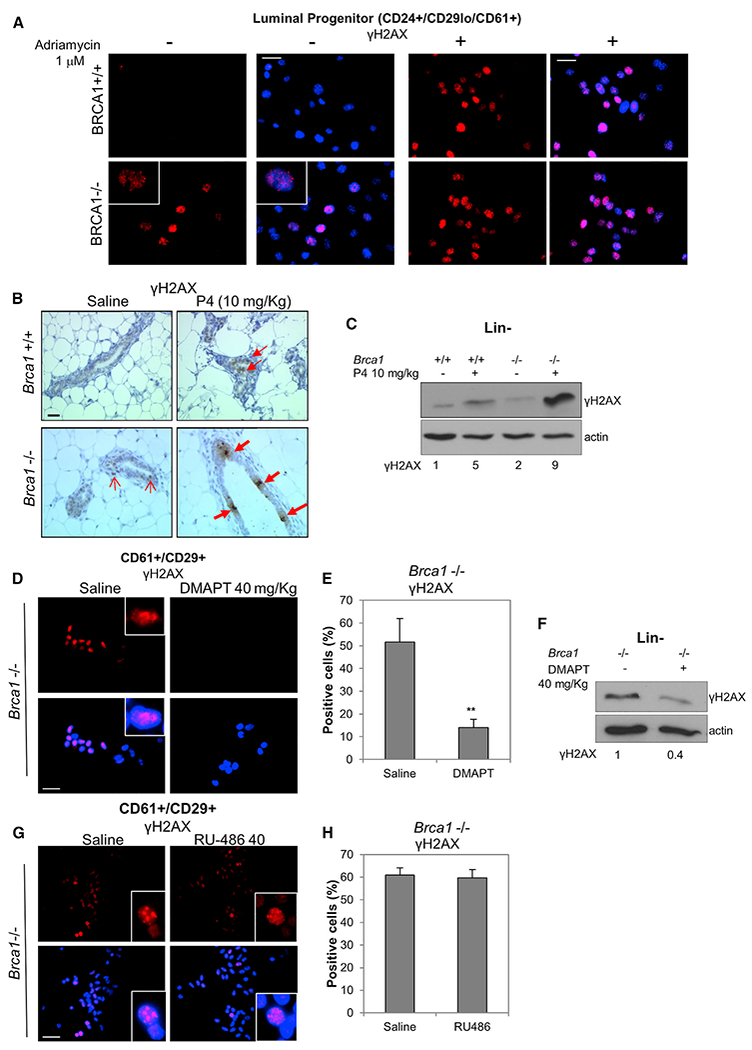
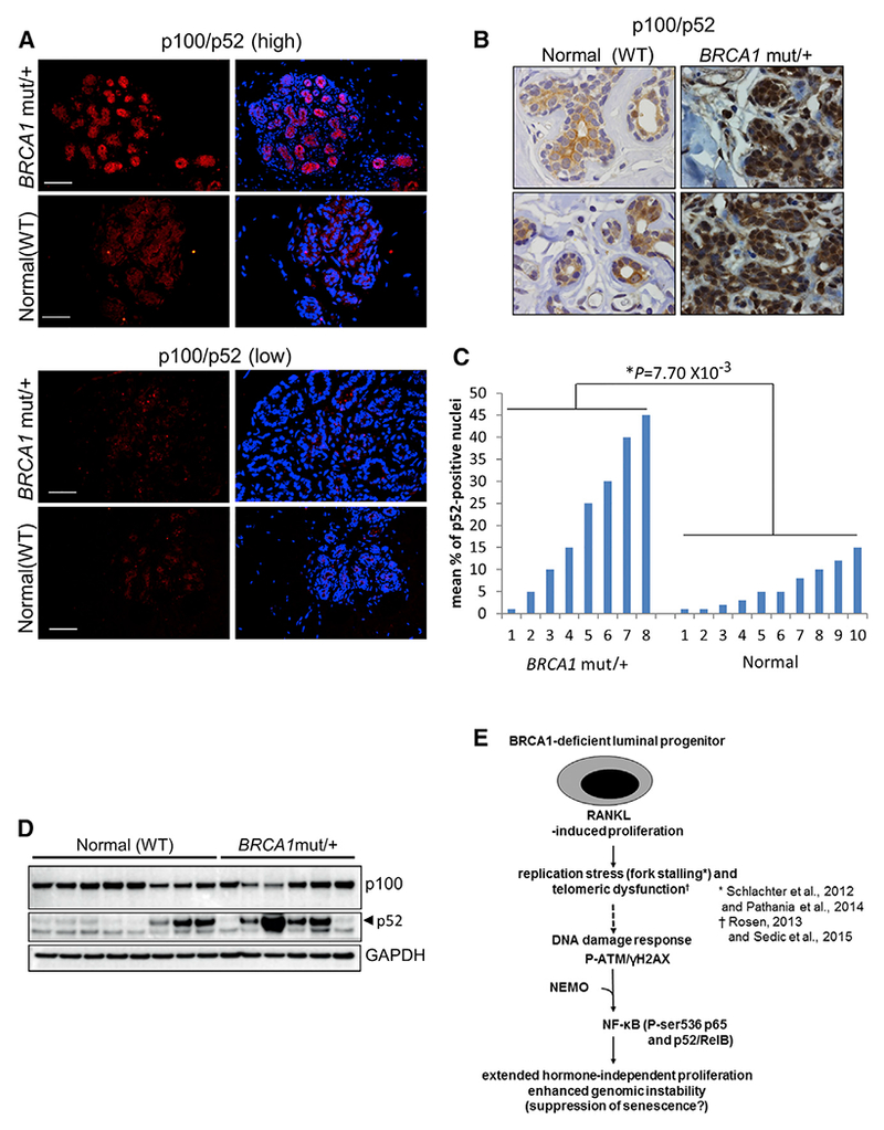
Human BRCA1 mutation carriers and BRCA1-deficient mouse mammary glands contain an abnormal population of mammary luminal progenitors that can form 3D colonies in a hormone-independent manner. The intrinsic cellular regulatory defect in these presumptive breast cancer precursors is not known. We have discovered that nuclear factor kappaB (NF-κB) (p52/RelB) is persistently activated in a subset of BRCA1-deficient mammary luminal progenitors. Hormone-independent luminal progenitor colony formation required NF-κB, ataxia telangiectasia-mutated (ATM), and the inhibitor of kappaB kinase, IKKα. Progesterone (P4)-stimulated proliferation resulted in a marked enhancement of DNA damage foci in Brca1(-/-) mouse mammary. In vivo, NF-κB inhibition prevented recovery of Brca1(-/-) hormone-independent colony-forming cells. The majority of human BRCA1(mut/+) mammary glands showed marked lobular expression of nuclear NF-κB. We conclude that the aberrant proliferative capacity of Brca1(-/-) luminal progenitor cells is linked to the replication-associated DNA damage response, where proliferation of mammary progenitors is perpetuated by damage-induced, autologous NF-κB signaling.


Conflict of interest statement

CONFLICTS OF INTEREST

The University of Kentucky holds a patent on DMAPT. A potential royalty to P.A.C. may occur consistent with University of Kentucky policy. P.A.C. is a founder of Leuchemix, Inc.

Figures

Figure 1.
Figure 1.. The Alternative NF-κB Pathway Is Constitutively Active in BRCA1-Deficient Mammary Progenitor Cells
(A) Lin Brca1+/+ and Brca1−/− cells were expanded in monolayer culture for 3 days, and protein extracts were subjected to immunoblot with anti-p100/p52. Numbers below the blots represent relative density normalized to actin. (B) Representative FACS images of primary MECs from Brca1+/+ and Brca1−/− mice enriched based on CD24/CD29/CD61 immunophenotype. Mammary stem cell (MaSC) (CD24+/CD29hi/CD61+), luminal progenitor (LP) (CD24+/CD29lo/CD61+), and mature luminal (ML) (CD24+/CD29lo/CD61) cell populations are shown. (C and E) Brca1−/− and Brca1+/+ LP and ML cell fractions were FACS sorted, cytospun, and immunostained. Representative images are shown for anti-p100/p52. Scale bar, 20 μm. (D and F) Percentage of p52-positive cells in LP- and ML-enriched populations, respectively. Bars on graphs are ±SEM (**p < 0.01, unpaired t test). (G) CD24hi/CD29lo/CD61+ primary cells from Brca1+/− mice were FACS sorted, cytospun, and immunostained with anti-p100/p52. Representative image is shown (n = 15). Scale bar, 20 μm. (H) Graph showing percentage of Brca1+/− p100/p52-positive cells ± SEM values derived from 15 fields. **p < 0.01, unpaired t test. (I) Lin cells as in (A) were immunoblotted with anti-IKKα and P-IKKα/β antibodies. Actin reactivity was used as a protein loading control. Numbers below represent relative density of P-IKKα normalized to actin. (J) FACS-sorted CD24+/CD29hi/CD61+ cells were infected for 72 hr with lenti-NF-κB reporter and GFP expression analyzed by FACS. **p < 0.01, unpaired t test. (K) Ratio of LP/ML cell populations obtained from FACS analyses of n = 5 Brca1+/+ and five Brca1−/− mice (*p < 0.05, unpaired t test). Bars on graphs are ±SEM. See also Figure S1.
Figure 2.
Figure 2.. BRCA1 Depletion Induces ser536-p65 Phosphorylation and Alternative NF-κB in Human Mammary Epithelial and Breast Cancer Cells
(A) HC11 mouse MECs stably expressing shNT or shBRCA1 were transfected with empty vector (EV) or CMV4-FLAG-IκBαSR and harvested after 72 hr. Immunoblots were reacted with anti-BRCA1, p100/p52, or IκBα. (B) HC11 cells described in (A) were immunoblotted with P-ser536-p65, p65, and IκBα antibodies. (C) MCF-10A immortalized human MECs were infected with lenti-shNT or shp105/p50 and then transfected 72 hr later with BRCA1 or NT siRNA. After an additional 72 hr, cells were harvested and immunoblotted with the indicated antibodies. Actin was used as a loading control. (D and E) MCF-7 and MDA-MB-231 cells were transfected with BRCA1 siRNA (siBRCA1) and non-targeting siRNA (siNT), and cell lysates were immunoblotted 72 hr later with anti-BRCA1 (D) or anti-p100/p52 (E). Numbers below the blots represent relative density normalized to vinculin or actin. (F) MCF-7 cells were infected with BRCA1 lenti-shRNA (shBRCA1) and non-targeting lenti-shRNA (shNT), cultured for 72 hr, and then immunostained with anti-p100/p52 and RelB antibodies, respectively. Scale bar, 10 μm. (G) Nuclear fluorescence intensities for p100/p52 and RelB, respectively, were determined using ImageJ and represented as mean intensities normalized to the control (shNT). Bars on graphs are ±SEM (**p < 0.01 unpaired t test). (H) HCC1937 BRCA1mut/− cells were infected with retrovirus pBABE-BRCA1 or empty vector (pBABE). Cell lysates were collected at indicated times and immunoblotted with anti-BRCA1 antibody. Numbers below the blots represent relative density normalized to actin. (I) Immunoblot with anti-p100/p52 antibody in extracts from pBABE-BRCA1 and pBABE-infected HCC1937 cells. Numbers below represent relative densities for the p52 band normalized to actin. (J) Representative images of HCC1937 Brca1mut/− breast cancer cells 72 hr following infection with retrovirus pBABE-BRCA1 or empty vector. Cells were immunostained with anti-p100/p52 and RelB antibodies. Scale bar, 10 μm. (K) Mean nuclear fluorescence intensity for p100/p52 and RelB in HCC1937 cells normalized to the control (pBABE) (**p < 0.01, unpaired t test). Bars on graphs are ±SEM. See also Figure S2.
Figure 3.
Figure 3.. Alternative NF-κB Is Essential for BRCA1-Deficient CFCs in the Absence of B27
(A) Immunoblot of Brca1−/− Lin cell extracts collected 72 hr after infection with p100/p52 lenti-shRNA (shp100) or non-targeting lenti-shRNA (shNT). Relative densities normalized to actin are shown below. (B) Representative images of CFC assays of Lin- cells in the presence or absence of the B27 media supplement. Cells were infected just prior to plating with lenti-shp100 or shNT. Scale bar, 200 μm. (C) Acini diameters from Lin cells infected with shp100 or shNT and plated on Matrigel. Bars on graphs are ±SEM (**p < 0.01, two-way ANOVA followed by Tukey test). (D) Representative images of CFC assays in the presence or absence of B27 supplement using Lin cells from Brca1−/− and Brca1+/− mice. (E) Diameters of acini formed by Lin- Brca1−/− and Brca1+/− MECs in the presence or absence of B27. (F) Diameters of acini formed by CFCs from Lin cells in the presence of vehicle (0) or 10 μM or 20 μM BMS. (G) Diameters of acini formed by CFCs from Lin cells in the presence of vehicle (0) or 1 or 2.5μM DMAPT. (H) Cell lysates were collected from Lin cells after 72 hr of 10 μM BMS treatment and immunoblotted with anti-p100/p52. Relative densities normalized to actin are shown below. (I) Cell lysates were collected from Brca1−/− Lin cells infected with two different IKKα lenti-shRNAs or non-targeting lenti-shRNA and immunoblotted for IKKα. Relative densities normalized to actin are shown below. (J) Acini diameters from CFCs derived from Lin cells infected with IKKα lenti-shRNAs or non-targeting lenti-shRNA and plated on Matrigel. Bars on graphs are ±SEM (**p < 0.01, two-way ANOVA followed by Tukey test). See also Figure S3.
Figure 4.
Figure 4.. ATM Is Activated, Interacts with NEMO, and Is Required for Brca1-Deficient B27-Independent Colony Formation
(A) CD24+/CD49f+ FACS-sorted cells from Brca1+/+ and Brca1−/− mice were cultured for 72 hr and then immunostained with anti-phospho ATM. Scale bar, 10 μm. (B) Nuclear fluorescence intensity for P-ser1981-ATM was determined using ImageJ and represented as mean intensities normalized to the control (Brca1+/+). **p < 0.01, unpaired t test. (C) MCF-7 cells were infected with BRCA1 lenti shRNA (shBRCA1) and non-targeting shRNA (shNT) for 72 hr. Cell lysates were collected and immunoblotted with anti-BRCA1 normalized to E-cadherin. (D) 20 μg input lysate (IN), α-ATM immunoprecipitate (IP), or non-immune IgG IP derived from MCF-7 cells infected with shBRCA1 shNT was immunoblotted (IB) to detect NEMO. Relative densities normalized to IB:ATM in (E) are shown below. (E and G) Duplicate IN and ATM IPs from cells in (D) and (F), respectively, were immunoblotted on a separate 6% polyacrylamide gel to facilitate immunoblot of high-molecular-weight ATM. (F) Twenty μg of input lysate (IN), α-ATM IP or non-immune IgG IP derived from MCF-7 cells and HCC1937 were subjected to immunoblot (IB) to detect NEMO. Results are typical of two separate experiments. (H) qRT-PCR for ATM mRNA in Lin- cells infected with lenti-shRNA to ATM (shATM) or non-targeting lenti-shRNA (shNT) normalized to the control (shNT). Bars on graphs are ±SEM (**p < 0.01, unpaired t test). (I) Acini diameters from CFC assays of Lin- cells infected with shATM or shNT. Bars on graphs are ±SEM (**p < 0.01, two-way ANOVA followed by Tukey test). See also Figure S4.
Figure 5.
Figure 5.. DMAPT Eliminates B27-Independent Progenitor Cell Proliferation In Vivo
(A and B) Brca1+/+ and Brca1−/− mice were injected i.p. with 40 mg/kg DMAPT or vehicle every 2 days for 2 weeks. Lin cells were isolated 1 day (A) or 15 days (B) after the last injection and subjected to CFC assay in the presence or absence of B27. Bars on graphs are ±SEM (**p < 0.01, two-way ANOVA followed by Tukey test). (C) Lin cells isolated as in (A) were cultured for 2 days in monolayer and lysates subjected to immunoblot to assess p100/p52.
Figure 6.
Figure 6.. DMAPT Treatment Reduces DNA Damage in BRCA1-Deficient Cells
(A) CD24+/CD29lo/CD61+ cells from Brca1+/+ and Brca1−/− mice were FACS-sorted and plated for 24 hr. γH2AX levels were assessed by IF. Adriamycin was added at 1 μM and used as positive control for γH2AX foci. Scale bar, 20 μm. (B) Brca1+/+ and Brca1−/− mice were injected daily with progesterone at 10 mg/kg or saline for 3 days. Mammary glands were collected the day after the last injection, paraffin embedded, and subjected to IHC with anti-γH2AX antibody. Open and closed arrows indicate light and intense γH2AX foci, respectively. Scale bar, 30 μm. (C) Same as in (B), but the day after the last injection, Lin cells were isolated from mouse mammary glands and plated for 72 hr. Cell lysates were then collected and immunoblotted with anti-γH2AX. Relative densities normalized to actin are shown below. (D) Brca1−/− mice were injected every 2 days over a period of 14 days with 40 mg/kg DMAPT or saline. The day after the last injection, CD29+/CD61+ cells were FACS sorted, cytospun onto slides, and subjected to IF with anti-γH2AX. Scale bar, 20 μm. (E) The graph indicates the percentage of γH2AX-positive cells from (D). Bars on graphs are ±SEM (**p < 0.01, unpaired t test). (F) Lin cells were isolated from Brca1−/− mice injected with DMAPT or saline as in (D) and plated for 72 hr. Cell lysates were immunoblotted with anti-γH2AX. Relative densities normalized to actin are shown below. (G) BRCA1−/− mice were injected for 3 days with 40 mg/kg RU-486 or saline. The day after the last injection, CD29+/CD61+ cells were FACS sorted, cytospun onto slides, and subjected to IF with anti-γH2AX. Scale bar, 20 μm. (H) The graph indicates the percentage of γH2AX-positive cells from (G). Bars on graphs are ±SEM. See also Figure S5.
Figure 7.
Figure 7.. Nuclear p52 Is Differentially Expressed in Luminal Cells of Lobules within BRCA1mut/+ Human Breast Tissue
(A) Representative anti-p100/p52 IF on lobular units in deparaffinized sections from BRCA1mut/+ breast tissue or normal breast tissue from reduction mammoplasty. Scale bar, 30 μm. (B) Examples of anti-p100/p52 IHC on sections of human mammary tissue. The WT and BRCA1mut/+ samples show a low and high percentage of nuclear p52, respectively (63× objective). (C) Mean percentages of p52-positive nuclei from BRCA1mut/+ and WT patient mammary sections. Values were determined by IHC analysis of 200–300 cells in the six lobules with the highest percentages of nuclear p52 (p = 0.0077, unpaired t test). (D) Western blot for p100/p52 showing sorted epithelial cells from frozen organoids. Lysates from ~105 cells were loaded in each lane. Samples denoted as “normal” are from reduction mammoplasties of women with verified wild-type BRCA1. BRCA1mut/+ samples are normal breast tissue from patients with no cancer who underwent a prophylactic mastectomy. (E) Hypothetical model of DDR signaling leading to activation of NF-κB in BRCA1-deficient progenitor cells. p52/RelB promotes proliferation to expand the genomically unstable LP population in the absence of P4. See text for details.

Similar articles

Cited by

References

    1. Andrechek ER, Hardy WR, Siegel PM, Rudnicki MA, Cardiff RD, and Muller WJ (2000). Amplification of the neu/erbB-2 oncogene in a mouse model of mammary tumorigenesis. Proc. Natl. Acad. Sci. U S A 97, 3444–3449. - PMC - PubMed
    1. Asselin-Labat ML, Sutherland KD, Barker H, Thomas R, Shackleton M, Forrest NC, Hartley L, Robb L, Grosveld FG, van der Wees J, et al. (2007). Gata-3 is an essential regulator of mammary-gland morphogenesis and luminal-cell differentiation. Nat. Cell Biol 9, 201–209. - PubMed
    1. Asselin-Labat ML, Vaillant F, Sheridan JM, Pal B, Wu D, Simpson ER, Yasuda H, Smyth GK, Martin TJ, Lindeman GJ, and Visvader JE (2010). Control of mammary stem cell function by steroid hormone signalling. Nature 465, 798–802. - PubMed
    1. Barham W, Chen L, Tikhomirov O, Onishko H, Gleaves L, Stricker TP, Blackwell TS, and Yull FE (2015). Aberrant activation of NF-κB signaling in mammary epithelium leads to abnormal growth and ductal carcinoma in situ. BMC Cancer 15, 647. - PMC - PubMed
    1. Baud V,and Karin M(2009). Is NF-kappaB a good target for cancer therapy? Hopes and pitfalls. Nat. Rev. Drug Discov 8, 33–40. - PMC - PubMed

Publication types

MeSH terms