The alterations in the extracellular matrix composition guide the repair of damaged liver tissue
- PMID: 27264108
- PMCID: PMC4893701
- DOI: 10.1038/srep27398
The alterations in the extracellular matrix composition guide the repair of damaged liver tissue
Abstract
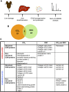
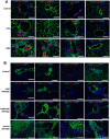
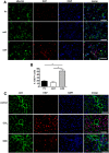
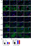
While the cellular mechanisms of liver regeneration have been thoroughly studied, the role of extracellular matrix (ECM) in liver regeneration is still poorly understood. We utilized a proteomics-based approach to identify the shifts in ECM composition after CCl4 or DDC treatment and studied their effect on the proliferation of liver cells by combining biophysical and cell culture methods. We identified notable alterations in the ECM structural components (eg collagens I, IV, V, fibronectin, elastin) as well as in non-structural proteins (eg olfactomedin-4, thrombospondin-4, armadillo repeat-containing x-linked protein 2 (Armcx2)). Comparable alterations in ECM composition were seen in damaged human livers. The increase in collagen content and decrease in elastic fibers resulted in rearrangement and increased stiffness of damaged liver ECM. Interestingly, the alterations in ECM components were nonhomogenous and differed between periportal and pericentral areas and thus our experiments demonstrated the differential ability of selected ECM components to regulate the proliferation of hepatocytes and biliary cells. We define for the first time the alterations in the ECM composition of livers recovering from damage and present functional evidence for a coordinated ECM remodelling that ensures an efficient restoration of liver tissue.
Figures


Similar articles
-
Extracellular Matrix Molecular Remodeling in Human Liver Fibrosis Evolution.PLoS One. 2016 Mar 21;11(3):e0151736. doi: 10.1371/journal.pone.0151736. eCollection 2016. PLoS One. 2016. PMID: 26998606 Free PMC article.
-
Composition and organization of the pancreatic extracellular matrix by combined methods of immunohistochemistry, proteomics and scanning electron microscopy.Curr Res Transl Med. 2017 Jan-Mar;65(1):31-39. doi: 10.1016/j.retram.2016.10.001. Epub 2016 Nov 7. Curr Res Transl Med. 2017. PMID: 28340694
-
Basic components of connective tissues and extracellular matrix: elastin, fibrillin, fibulins, fibrinogen, fibronectin, laminin, tenascins and thrombospondins.Adv Exp Med Biol. 2014;802:31-47. doi: 10.1007/978-94-007-7893-1_3. Adv Exp Med Biol. 2014. PMID: 24443019 Review.
-
Substrate stiffness and matrix composition coordinately control the differentiation of liver progenitor cells.Biomaterials. 2016 Aug;99:82-94. doi: 10.1016/j.biomaterials.2016.05.016. Epub 2016 May 12. Biomaterials. 2016. PMID: 27235994
-
The Extracellular Matrix in Ischemic and Nonischemic Heart Failure.Circ Res. 2019 Jun 21;125(1):117-146. doi: 10.1161/CIRCRESAHA.119.311148. Epub 2019 Jun 20. Circ Res. 2019. PMID: 31219741 Free PMC article. Review.
Cited by
-
Regulators, functions, and mechanotransduction pathways of matrix stiffness in hepatic disease.Front Physiol. 2023 Jan 12;14:1098129. doi: 10.3389/fphys.2023.1098129. eCollection 2023. Front Physiol. 2023. PMID: 36711017 Free PMC article. Review.
-
Label-free functional and structural imaging of liver microvascular complex in mice by Jones matrix optical coherence tomography.Sci Rep. 2021 Oct 8;11(1):20054. doi: 10.1038/s41598-021-98909-6. Sci Rep. 2021. PMID: 34625574 Free PMC article.
-
The bright side of fibroblasts: molecular signature and regenerative cues in major organs.NPJ Regen Med. 2021 Aug 10;6(1):43. doi: 10.1038/s41536-021-00153-z. NPJ Regen Med. 2021. PMID: 34376677 Free PMC article. Review.
-
Extracellular Microenvironmental Control for Organoid Assembly.Tissue Eng Part B Rev. 2022 Dec;28(6):1209-1222. doi: 10.1089/ten.TEB.2021.0186. Epub 2022 Jun 21. Tissue Eng Part B Rev. 2022. PMID: 35451330 Free PMC article. Review.
-
Advanced and Rationalized Atomic Force Microscopy Analysis Unveils Specific Properties of Controlled Cell Mechanics.Front Physiol. 2018 Aug 17;9:1121. doi: 10.3389/fphys.2018.01121. eCollection 2018. Front Physiol. 2018. PMID: 30174612 Free PMC article.
References
Publication types
MeSH terms
LinkOut - more resources
Full Text Sources
Other Literature Sources

