. 2016 May 19:7:11363.
doi: 10.1038/ncomms11363.

Suppression of KRas-mutant cancer through the combined inhibition of KRAS with PLK1 and ROCK

Affiliations

Suppression of KRas-mutant cancer through the combined inhibition of KRAS with PLK1 and ROCK

Jieqiong Wang et al. Nat Commun. .

Abstract

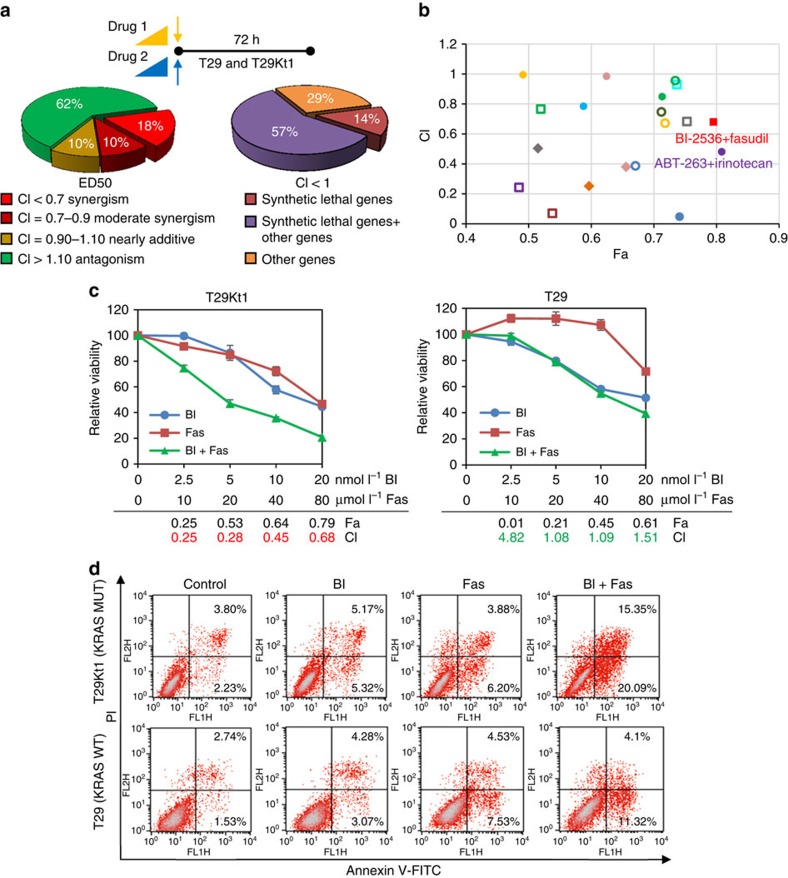
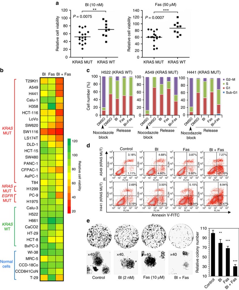
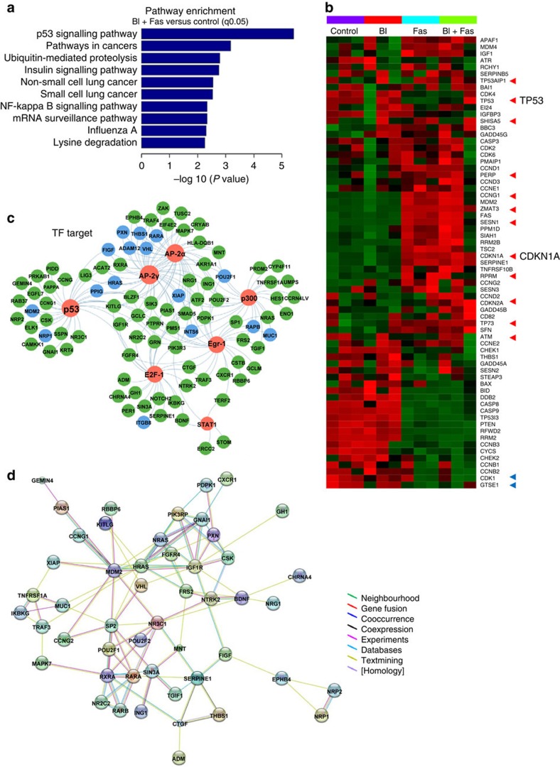
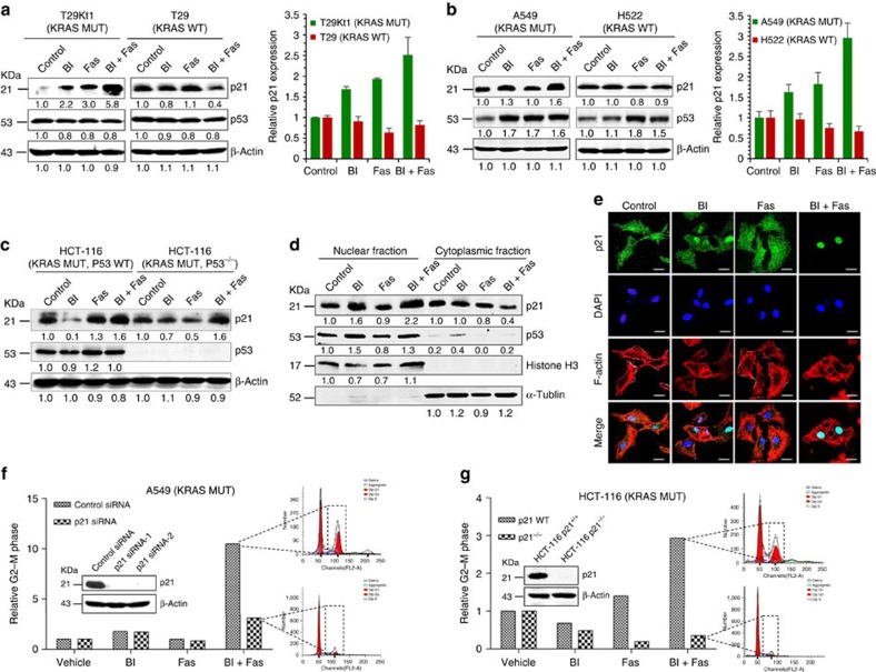
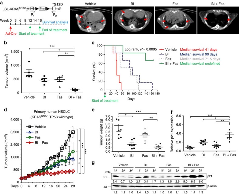
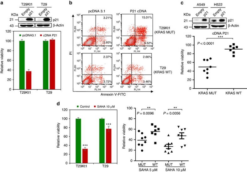
No effective targeted therapies exist for cancers with somatic KRAS mutations. Here we develop a synthetic lethal chemical screen in isogenic KRAS-mutant and wild-type cells to identify clinical drug pairs. Our results show that dual inhibition of polo-like kinase 1 and RhoA/Rho kinase (ROCK) leads to the synergistic effects in KRAS-mutant cancers. Microarray analysis reveals that this combinatory inhibition significantly increases transcription and activity of cyclin-dependent kinase inhibitor p21(WAF1/CIP1), leading to specific G2/M phase blockade in KRAS-mutant cells. Overexpression of p21(WAF1/CIP1), either by cDNA transfection or clinical drugs, preferentially impairs the growth of KRAS-mutant cells, suggesting a druggable synthetic lethal interaction between KRAS and p21(WAF1/CIP1). Co-administration of BI-2536 and fasudil either in the LSL-KRAS(G12D) mouse model or in a patient tumour explant mouse model of KRAS-mutant lung cancer suppresses tumour growth and significantly prolongs mouse survival, suggesting a strong synergy in vivo and a potential avenue for therapeutic treatment of KRAS-mutant cancers.


Figures

Figure 1
Figure 1. A synthetic lethal chemical screen reveals that KRAS-mutant cells are selectively sensitive to the combined inhibition of PLK1 and ROCK.
(a) Schematic of the combinations tested and the drug interaction signatures. Twenty clinical drugs were tested in pairwise combinations (a total of 190 pairs) in T29Kt1 cells harbouring a KRAS mutation. Drugs were added at a relevant fixed ratios (IC50 ratios, see also Supplementary Table 1) at four concentration combinations in each representative drug pair. The cell viability was determined. Left: compilation of the total number of drug pair synergies, moderate synergies, nearly additive interactions and antagonistic interactions. The combination index (CI) was calculated using CalcuSyn software (Version 2; Biosoft) as described in the Methods section. Right: the frequencies at which the drug target gene types appear in the synergy cluster (CI<1). The oncogenic KRAS synthetic lethal genes accounted for the largest proportion of synergies specific to KRAS-mutant cells. (b) The Fa–CI plot. The Fa (fraction affected by the dose) and CI value of two drugs at their combination of IC50's were listed in X and Y axes and synergistic pairs with CI<1 were shown. The combination of BI-2536 and fasudil exhibited leading therapeutic efficacy and applicable potential. (c) The cytotoxicity of BI-2536 and fasudil. T29Kt1 and T29 cells were incubated with increasing concentrations of BI-2536 (BI) and fasudil (Fas) alone or in combination for 72 h, and the cell viability was determined. The CI and Fa values for the combination of BI-2536 and fasudil were calculated. The averages and error bars represent the mean±s.d. from three independent experiments. (d) Percentage of apoptotic cells was determined by Annexin-V and propidium iodide staining after BI-2536 (10 nmol l−1) and fasudil (40 μmol l−1) treatment alone or in combination for 72 h in T29Kt1 and T29 cells.
Figure 2
Figure 2. KRAS-mutant cancer cells are significantly sensitive to the pharmacologic inhibition of PLK1 and ROCK.
(a) Seventeen KRAS-mutant (KRAS MUT) and nine wild-type (KRAS WT) cancer cell lines were treated with the indicated concentrations of BI-2536 or fasudil for 72 h; the dots represent the cell viability normalized to no drug treatment. The bars indicate the means. Student's t-tests were performed between the MUT and WT groups; **P<0.01; ***P<0.001. (b) Thirty-two cell lines carrying different KRAS genotypes were treated with BI, Fas or the combination of BI and Fas for 72 h. The percentage of viable cells was colour coded in a heatmap. (c) H522 (KRAS WT), A549 (KRAS MUT) and H441 (KRAS MUT) cells were treated with BI-2536 (2 nmol l−1), fasudil (10 μmol l−1) or the combination (BI-2536+fasudil) for 48 h after synchronization and release. The cell cycle distribution was analysed by flow cytometry using propidium iodide staining. (d) A549 and H441 (KRAS MUT) cells were treated with BI-2536 (2 nmol l−1), fasudil (10 μmol l−1) or a combination of BI and Fas for 72 h, and the percentage of apoptotic cells (Annexin positive) was determined by Annexin-V and propidium iodide staining. (e) A549 cells (1,000) were plated in 60-mm dishes and treated with dimethylsulphoxide, BI-2536 (2 nmol l−1), fasudil (10 μmol l−1) or a combination of BI and Fas for 7 days. The cell colonies were stained with crystal violet and counted. The relative number of colonies was calculated by normalization to control as 100%. The values represent the mean±s.d. of three independent assays; Student's t-tests were performed; **P<0.01; ***P<0.001. MUT, mutant; WT, wild type.
Figure 3
Figure 3. The p53 signalling pathway is involved in the sensitivity of BI-2536/fasudil in KRAS-mutant cancers.
(a) Pathway enrichment of the differentially expressed genes in A549 cells after BI-2536+fasudil treatment. For enrichment analysis, the subsets of upregulated genes were used. The bar plot shows the top 10 enrichment score (−log (P value)) value of the significant enrichment pathway. The P value cutoff was 0.05 and denotes the significance of the pathway correlated to the conditions from three independent experiments. (b) Heatmap coloured according to the Z scores of the expression value, showing the expression profile of all of the genes in the p53 pathway. The arrow points to DEGs with FC≥2 and FDR≤0.05. The red arrows represent genes that are upregulated, and the blue arrows represent genes that are downregulated. TP53 and CDKN1A, primary members of this pathway, are indicated. (c) Differentially expressed disease genes that are explained by enriched TF. Red node, TFs; blue node, non-small cell lung cancer-related genes; green node, lung cancer-related genes. (d) Protein–protein interaction networks, functional modules consisting of the genes downstream of the enriched TF.
Figure 4
Figure 4. The combined inhibition of PLK1 and ROCK leads to the overexpression of p21WAF1/CIP1 in KRAS-mutant cells but not wild-type cells.
(a) The levels of p21 protein and mRNA expression in isogenic T29Kt1/T29 cells. Cells were treated with DMSO (control), BI-2536 (4 nmol l−1), fasudil (20 μmol l−1) or BI-2536/fasudil. Equal amounts of proteins from cell lysates were subjected to western blotting analyses. The numbers underneath the blotting bands represent the normalized density quantified by densitometry using ImageJ 2 × software. The relative mRNA levels of p21WAF1/CIP1 after normalization to β-actin expression were determined by quantitative PCR. The error bars correspond to the s.d.'s from three independent experiments. (b) The levels of p21 protein and mRNA expression in treated A549 and H522 cells. The error bars correspond to the s.d.'s from three independent experiments. (c) BI-2536/fasudil-mediated p21 activation was independent of p53 regulation. p21 and p53 were probed in isogenic HCT-116 (p53+/+) and HCT-116 (p53−/−) cells. (d) Immunoblot analysis of protein levels in the nucleus and cytoplasm in response to the indicated treatments in A549 cells. Histone H3 and α-tubulin served as nuclear and cytoplasmic fraction markers, respectively. (e) A549 cells were treated as indicated for 16 h. The nuclear accumulation of p21 was determined by immunofluorescence staining. The cells were stained with the anti-p21 antibody (green), nuclei were counterstained with 4,6-diamidino-2-phenylindole (blue) and F-actin was stained with phalloidin (red). Immunofluorescence was recorded using confocal laser fluorescence microscopy. Scale bars, 20 μm. (f) A549 cells were transfected with p21 siRNA or a control siRNA for 24 h, and then exposed to single drugs or to BI-2536/fasudil 48 h. The cell cycle distribution was analysed by flow cytometry using propidium iodide staining. The efficiency of p21 knockdown was examined by immunoblotting. (g) HCT-116 (KRAS mutant and p21 wild type) cells and its p21 knockout counterparts (HCT-116 p21−/−) were exposed to single drugs or to BI-2536/fasudil respectively. The cell cycle distribution was analysed. The efficiency of p21 knockout was examined by immunoblotting.
Figure 5
Figure 5. In vivo efficacy of combined PLK1 and ROCK inhibition.
(a) Left: schematic illustration of the LSL-KRASG12D allele and drug treatment protocol. Established lung tumours in LSL-KRASG12D mice were treated with vehicle, BI-2536 (BI), fasudil (Fas) or both drugs in combination (BI+Fas). Right: after 4 weeks treatment, animals were scanned by micro-computed tomography and representative transverse images are shown. Red arrows indicate lung tumours. (b) Mean tumour volume at the end point of the indicated treatments. Each dot represents an individual mice (n=5 per group). Data represent the mean±s.e.m. *P<0.05; **P<0.01; ***P<0.001 by one-way analysis of variance (ANOVA) followed by Bonferroni multiple comparison test. (c) The survival rate was calculated by Kaplan–Meier method (n=6 per group). Statistical significance was assessed by log-rank tests, *P<0.05; **P<0.01; ***P<0.001. (d) The growth of primary NSCLC tumour xenografts (n=8 per group). Data represent the mean±s.e.m. *P<0.05; **P<0.01; ***P<0.001 by one-way ANOVA followed by Bonferroni multiple comparison test. (e) Tumour weights of the primary NSCLC tumour xenografts upon euthanasia at day 28. Each dot represents a tumour from an individual mouse. Data represent the mean±s.e.m. *P<0.05; **P<0.01; ***P<0.001 by one-way ANOVA followed by Bonferroni multiple comparison test. (f) p21 mRNA expression in the primary NSCLC tumour xenografts. Each dot represents a tumour from an individual mouse (n=6 per group). Data represent mean±s.e.m. *P<0.05; **P<0.01; ***P<0.001 by one-way ANOVA followed by Bonferroni multiple comparison test. (g) p21 protein level in the lysates from the tumours in the primary tumour xenograft mice. Each number represents a tumour from an individual mouse. See also Supplementary Fig. 3.
Figure 6
Figure 6. P21WAF1/CIP1 overexpression impairs the survival of KRAS-mutant cancer cells.
(a) T29Kt1 and T29 cells were transfected with p21 cDNA plasmid or pcDNA 3.1 empty vector. Cell viability was measured 48 h after transfection. Data represent the mean±s.d. from three independent experiments. The efficiency of p21 overexpression was examined by immunoblotting. (b) T29Kt1 and T29 cells were transfected with p21 cDNA plasmid or pcDNA 3.1 empty vector. The percentage of apoptotic cells (Annexin positive) was determined by Annexin-V and propidium iodide staining. (c) Cell viability was measured in various cancer cells after p21 cDNA transfection. The dots represent the relative cell viability. The bars indicate the means. Student's t-tests were performed between the MUT and WT groups; **P<0.01; ***P<0.001. The efficiency of p21 overexpression in representative cells were examined by immunoblotting. (d) T29Kt1, T29 cells and different cancer cell lines were treated with SAHA or vehicle control for 48 h. Cell viability was measured. The columns indicate the means, and the bars indicate the s.d. The dots represent relative cell viability. Student's t-tests were performed between the MUT and WT groups; **P<0.01; ***P<0.001. MUT, mutant; WT, wild type.

Similar articles

Cited by

References

    1. Thompson H. US National Cancer Institute's new Ras project targets an old foe. Nat. Med. 19, 949–950 (2013). - PubMed
    1. Karnoub A. E. & Weinberg R. A. Ras oncogenes: split personalities. Nat. Rev. Mol. Cell Biol. 9, 517–531 (2008). - PMC - PubMed
    1. Baines A. T., Xu D. & Der C. J. Inhibition of Ras for cancer treatment: the search continues. Future Med. Chem. 3, 1787–1808 (2011). - PMC - PubMed
    1. Siegel R., Ma J., Zou Z. & Jemal A. Cancer statistics, 2014. CA Cancer J. Clin. 64, 9–29 (2014). - PubMed
    1. Luo J., Solimini N. L. & Elledge S. J. Principles of cancer therapy: oncogene and non-oncogene addiction. Cell 136, 823–837 (2009). - PMC - PubMed

Publication types

MeSH terms