

doi: 10.1007/s13238-016-0275-4.
The crystal structure of Zika virus helicase: basis for antiviral drug design
Affiliations
- PMID: 27172988
- PMCID: PMC4887331
- DOI: 10.1007/s13238-016-0275-4
Item in Clipboard
The crystal structure of Zika virus helicase: basis for antiviral drug design
Protein Cell.
2016 Jun.
No abstract available
Figures
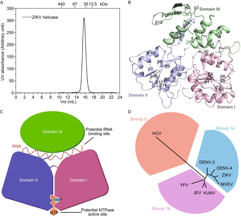
The monomeric structure of ZIKV helicase. (A) Size-exclusion chromatograms of ZIKV helicase. The molecular masses of protein standards are indicated at the top. (B) The overall structure of ZIKV helicase with the three domains colored and labeled respectively. (C) A cartoon diagram illustrating of the overall fold with potential RNA binding site and NTPase active site labelled. (D) Structure-based phylogenetic tree of 8 viral helicase structures from the Flaviviradae family using the program SHP (Stuart et al., 1979) and PHYLIP (Felsenstein, 1997). The following structures with PDB ID in parentheses are included: DENV-2 (2BMF), DENV-4 (2JLQ), JEV (2Z83), KUNV (2QEQ), YFV (1YKS), MVEV (2V8O), HCV (1HEI)
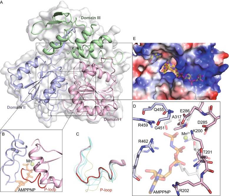
Structural insight into ZIKV helicase. (A) Cartoon and surface representation of the overall fold with the three domains of ZIKV helicase, colored and labeled respectively; (B) The electrostatic surface representation showing the tunnel for potential RNA binding. Positive potentials are colored blue and the negative are colored red. The putative position of the nucleic acid is marked as semi-transparent sticks. The model was obtained by superposition with the DENV-4 helicase in complex with ssRNA (PDB code 2JLV). (C) A clear view of the NTPase active site. The positions of putative nucleotide substrate (as sticks) and Mn2+ (as sphere) are marked semi-transparently by superposition with the DENV-4 helicase bound to AMPPNP and Mn2+ (PDB code 2JLR). P-loop is shown in red. (D) Isolated P-loops are shown by superimposing the structures of 7 flavivirus apo helicases. ZIKV helicase is in red ribbon and the others are shown in finer lines. The P-loop of DENV-4 helicase is colored green. The following structures of helicases with PDB ID in parentheses are included: DENV-2 (2BMF), DENV-4 (2JLQ), JEV (2Z83), KUNV (2QEQ), YFV (1YKS), MVEV (2V8O). (E) Interactions at NTPase active site by superposition of ZIKV helicase (solid) with DENV-4 helicase in complex with AMPPNP and Mn2+ (semitransparent, PDB code 2JLR). Conserved residues are shown as sticks and labeled
Similar articles
-
Structure of the NS3 helicase from Zika virus.Nat Struct Mol Biol. 2016 Aug;23(8):752-4. doi: 10.1038/nsmb.3258. Epub 2016 Jul 11. Nat Struct Mol Biol. 2016. PMID: 27399257 Free PMC article.
-
Discovery of Novel Druggable Sites on Zika Virus NS3 Helicase Using X-ray Crystallography-Based Fragment Screening.Int J Mol Sci. 2018 Nov 20;19(11):3664. doi: 10.3390/ijms19113664. Int J Mol Sci. 2018. PMID: 30463319 Free PMC article.
-
Crystal structures of the methyltransferase and helicase from the ZIKA 1947 MR766 Uganda strain.Acta Crystallogr D Struct Biol. 2017 Sep 1;73(Pt 9):767-774. doi: 10.1107/S2059798317010737. Epub 2017 Aug 15. Acta Crystallogr D Struct Biol. 2017. PMID: 28876240
-
Unzippers, resolvers and sensors: a structural and functional biochemistry tale of RNA helicases.Int J Mol Sci. 2015 Jan 22;16(2):2269-93. doi: 10.3390/ijms16022269. Int J Mol Sci. 2015. PMID: 25622248 Free PMC article. Review.
-
Binding and unwinding: SF3 viral helicases.Curr Opin Struct Biol. 2005 Feb;15(1):77-85. doi: 10.1016/j.sbi.2004.12.001. Curr Opin Struct Biol. 2005. PMID: 15718137 Review.
Cited by
-
Mechanistic Insights into Zika Virus NS3 Helicase Inhibition by Epigallocatechin-3-Gallate.ACS Omega. 2020 May 4;5(19):11217-11226. doi: 10.1021/acsomega.0c01353. eCollection 2020 May 19. ACS Omega. 2020. PMID: 32455246 Free PMC article.
-
Crystal structure of the Ilheus virus helicase: implications for enzyme function and drug design.Cell Biosci. 2022 Apr 15;12(1):44. doi: 10.1186/s13578-022-00777-8. Cell Biosci. 2022. PMID: 35428322 Free PMC article.
-
Discovery and Computational Analyses of Novel Small Molecule Zika Virus Inhibitors.Molecules. 2019 Apr 13;24(8):1465. doi: 10.3390/molecules24081465. Molecules. 2019. PMID: 31013906 Free PMC article.
-
Molecular docking and simulation of Zika virus NS3 helicase.BMC Chem. 2019 May 17;13(1):67. doi: 10.1186/s13065-019-0582-y. eCollection 2019 Dec. BMC Chem. 2019. PMID: 31384814 Free PMC article.
-
Identification of potential inhibitors of Zika virus targeting NS3 helicase using molecular dynamics simulations and DFT studies.Mol Divers. 2023 Aug;27(4):1689-1701. doi: 10.1007/s11030-022-10522-5. Epub 2022 Sep 5. Mol Divers. 2023. PMID: 36063275
References
Publication types
MeSH terms
Substances
LinkOut - more resources
Full Text Sources
Other Literature Sources
Medical
