Comparative Study
. 2016 Aug;132(2):175-196.
doi: 10.1007/s00401-016-1575-8. Epub 2016 May 10.

Comparative interactomics analysis of different ALS-associated proteins identifies converging molecular pathways

Affiliations
Comparative Study

Comparative interactomics analysis of different ALS-associated proteins identifies converging molecular pathways

Anna M Blokhuis et al. Acta Neuropathol. 2016 Aug.

Abstract

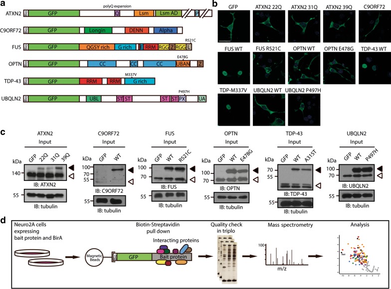
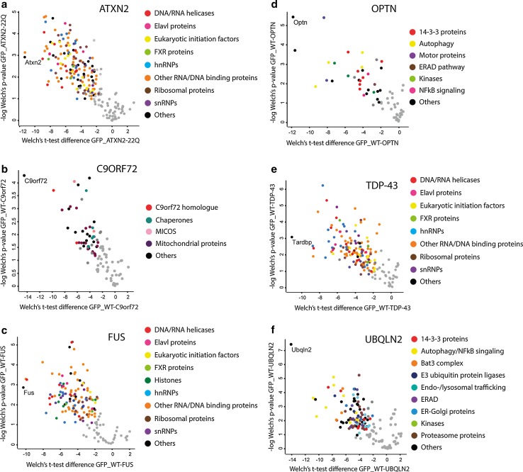
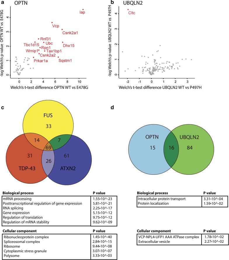
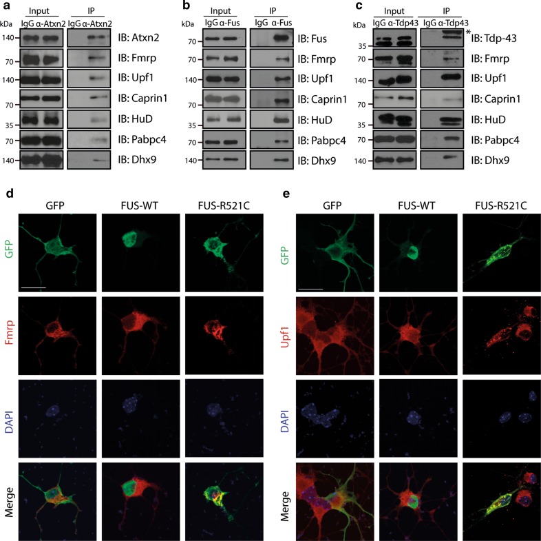
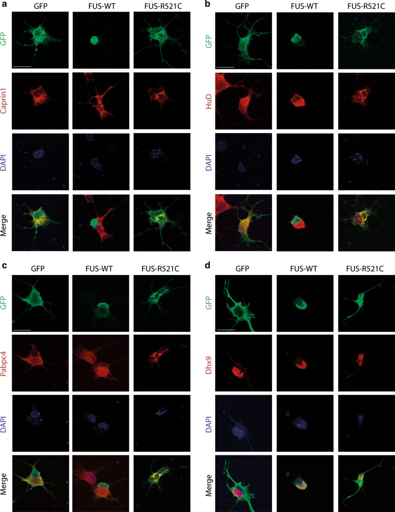
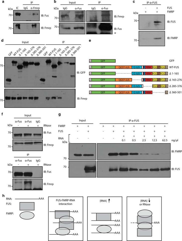
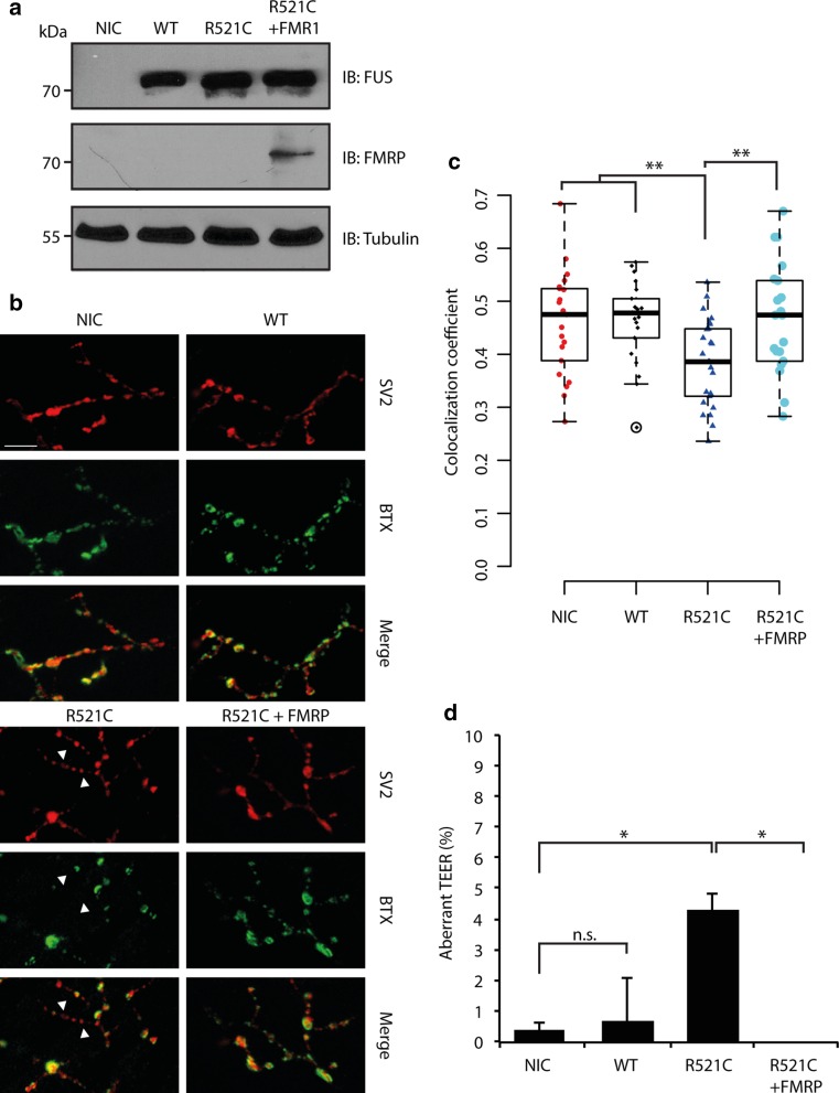
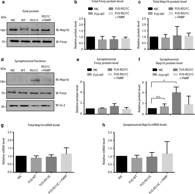
Amyotrophic lateral sclerosis (ALS) is a devastating neurological disease with no effective treatment available. An increasing number of genetic causes of ALS are being identified, but how these genetic defects lead to motor neuron degeneration and to which extent they affect common cellular pathways remains incompletely understood. To address these questions, we performed an interactomic analysis to identify binding partners of wild-type (WT) and ALS-associated mutant versions of ATXN2, C9orf72, FUS, OPTN, TDP-43 and UBQLN2 in neuronal cells. This analysis identified several known but also many novel binding partners of these proteins. Interactomes of WT and mutant ALS proteins were very similar except for OPTN and UBQLN2, in which mutations caused loss or gain of protein interactions. Several of the identified interactomes showed a high degree of overlap: shared binding partners of ATXN2, FUS and TDP-43 had roles in RNA metabolism; OPTN- and UBQLN2-interacting proteins were related to protein degradation and protein transport, and C9orf72 interactors function in mitochondria. To confirm that this overlap is important for ALS pathogenesis, we studied fragile X mental retardation protein (FMRP), one of the common interactors of ATXN2, FUS and TDP-43, in more detail in in vitro and in vivo model systems for FUS ALS. FMRP localized to mutant FUS-containing aggregates in spinal motor neurons and bound endogenous FUS in a direct and RNA-sensitive manner. Furthermore, defects in synaptic FMRP mRNA target expression, neuromuscular junction integrity, and motor behavior caused by mutant FUS in zebrafish embryos, could be rescued by exogenous FMRP expression. Together, these results show that interactomics analysis can provide crucial insight into ALS disease mechanisms and they link FMRP to motor neuron dysfunction caused by FUS mutations.

Keywords: Amyotrophic lateral sclerosis; C9orf72; FMRP; FUS; Motor neuron; TDP-43.


Figures

Fig. 1
Fig. 1
Experimental setup of interactomics analysis of six ALS-associated proteins. a Schematic representation of bioGFP-tagged ATNX2, C9orf72, FUS, OPTN, UBQLN2 and TDP-43. Numbers indicate common ALS-associated mutations. Bio biotin-tag, CC coiled-coil domain, DENN differentially expressed in normal and neoplasia, E nuclear export signal, G-rich glycine-rich region, L nuclear localization signal, Lsm like-Sm domain, Lsm AD Lsm associated domain, P Poly(A)-binding protein interacting motif, PX proline XX repeat region, Q poly-glutamine stretch, QGSY glutamine/glycine/serine/tyrosine-rich region, RRM RNA recognition motif, RGG arganine/glycine-rich region, ST heat shock chaperone binding motif, UBAN ubiquitin binding in ABIN and NEMO domain, UBL ubiquitin-like domain, UA ubiquitin-associated domain, WT wild-type, Z zinc finger motif. b Immunocytochemistry for GFP on Neuro2A (N2A) cells transfected with the indicated constructs (shown in a). Exogenous proteins show an endogenous distribution pattern. c Western blot analysis of lysates of N2A cells transfected with the indicated constructs. Tubulin is used a loading control. Exogenous proteins are indicated with closed arrowheads, open arrowheads indicate endogenous proteins. d Overview of experimental design. N2A cells were co-transfected with bioGFP-tagged constructs and the biotin ligase BirA. After 48 h, exogenous proteins were purified using magnetic streptavidin-coated beads. Purified protein complexes were separated using gel electrophoresis and subjected to silver staining to confirm equal protein loading and detection of bait and interacting proteins. Mass spectrometry analysis was performed, followed by MaxQuant analysis and statistical analysis using PERSEUS. IB immunoblot. Scale bar 30 μm
Fig. 2
Fig. 2
Quantitative analysis of the binding partners of six wild-type ALS-associated proteins. af Volcano plots showing interactors of wild-type, non-mutated forms of ATXN2 22Q (a), C9ORF72 (b), FUS (c), OPTN (d), TDP-43 (e) and UBQLN2 (f). Logarithmized ratios of relative protein intensities are plotted against the negative logarithmic p value from triplicate experiments (Welch’s t test; S0: 1.5, FDR: 0.01). PERSEUS was used for statistical analysis and visualization; colored dots represent significantly enriched proteins and the color refers to the corresponding functional category indicated at the right. FXR fragile-X related, MICOS mitochondrial contact site complex, ERAD endoplasmatic-reticulum-associated degradation
Fig. 3
Fig. 3
The effect of ALS-associated mutations on interactome composition and overlap between different interactomes. a, b Volcano plots showing a comparison of the interactors of OPTN E478G and OPTN wild-type (WT) (a) and UBQLN2 P487H and UBQLN WT (b). Logarithmized ratios of relative protein intensities are plotted against the negative logarithmic p value from triplicate experiments (Welch’s t test; S0: 1.5, FDR: 0.01). PERSEUS was used for statistical analysis and visualization; red dots and labels refer to proteins significantly altered compared to the WT interactome. c, d Venn diagrams showing the number of unique and overlapping interactors of ATXN2, FUS and TDP-43 (c) and of OPTN and UBQLN2 (d). Tables show GO analyses performed on the shared interactors. Csnk2a casein kinase II subunit alpha, Cltc clathrin heavy chain 1, Dhx15 Pre-mRNA-splicing factor ATP-dependent RNA helicase DHX15, Iap IgE-binding protein, Prkar1a cAMP-dependent protein kinase type I-alpha regulatory subunit, Rnf31 E3 ubiquitin-protein ligase RNF31, Rpn1, Dolichyl-diphospho-oligosaccharideprotein glycosyltransferase subunit 1, Sqstm1 Sequestosome1, Tax1bp1 Tax1-binding protein 1, Tbc1d15 TBC1 domain family member 15, Ub ubiquitin, Vcp valosin-containing protein, Wrnip1 ATPase WRNIP1
Fig. 4
Fig. 4
Interactors show endogenous binding to Atxn2, Fus and Tdp-43 and cytoplasmic mislocalization following transfection of mutant FUS. ac Western blot analysis of input and immunoprecipitation (IP) samples with the indicated antibodies following endogenous immunoprecipitation with antibodies against Atxn2 (a), Fus (b), or Tdp-43 (c) from Neuro2A (N2A) cell lysates. Asterisk indicates IgG band. IgG IgG control antibody. d, e Dissociated primary motor neuron cultures generated from E13.5 mouse embryos were transfected with DNA constructs expressing GFP, FUS wild-type (WT) or FUS R521C and, after 2 days in culture, fixed and immunostained using antibodies against FMRP (d) or Upf1 (e). DAPI staining was used to visualize the cell nucleus. IB immunoblot. Scale bar 30 μm (d, e)
Fig. 5
Fig. 5
Interactors show cytoplasmic mislocalization following transfection of mutant FUS (continued). ad Dissociated primary motor neuron cultures generated from E13.5 mouse embryos were transfected with DNA constructs expressing GFP, FUS wild-type (WT) or FUS R521C and, after 2 days in culture, fixed and immunostained using antibodies against Caprin1 (a), HuD (b), Pabpc4 (c) or Dhx9 (d). DAPI staining was used to visualize the cell nucleus. Scale bar 30 μm (ad)
Fig. 6
Fig. 6
Biochemical characterization of FMRP-FUS binding. a FMRP pull down samples (IP) from NSC-34 cells were immunoblotted with the indicated antibodies. IgG was used as a negative control. b IP of Fus from E14 primary mouse cortical neurons. Input and IP samples were analyzed with the indicated antibodies. Asterisk indicates IgG band. c Recombinant FUS and FMRP proteins were mixed and immunoprecipitated with anti-FUS antibodies. Samples were analyzed with the indicated antibodies. d Neuro2A (N2A) cells were transfected with bioGFP-tagged FUS deletion constructs (indicated in e) and immunoprecipitated using streptavidin beads (IP). Samples were immunoblotted and probed with the indicated antibodies. e Schematic overview of bioGFP-tagged FUS deletion constructs used in d. Bio biotin-tag, E nuclear export signal, G rich glycine-rich domain, L nuclear localization signal, RRM RNA recognition motif, RGG arganine/glycine-rich region, QGSY glutamine/glycine/serine/tyrosine-rich region, WT wild-type, Z zinc finger motif. f Total cell lysates were treated with RNase (+) and immunoprecipitated (IP) with IgG (control) or anti-Fus antibodies. Samples were analyzed with the indicated antibodies. g Recombinant FMRP and FUS proteins were mixed and RNA was added in increasing concentrations. Protein mixtures were incubated and immunoprecipitated with anti-FUS antibody and subjected to immunoblotting with the indicated antibodies. h Schematic summary illustrating how RNA modulates FUS-FMRP binding. FUS and FMRP bind to each other and are both able to bind RNA (left panel). The interaction between FUS and FMRP decreases with increasing RNA concentrations (middle panel). Vice versa, degradation of RNA enhances the interaction between FUS and FMRP (right panel). IB immunoblot, IC input control
Fig. 7
Fig. 7
Exogenous FMRP rescues NMJ and locomotion defects caused by mutant FUS. a Western blot analysis of 72 hpf zebrafish embryo lysates using antibodies against human FUS or FMRP following injection of FUS WT, FUS R521C and/or FMR1 RNAs. Tubulin is used as a loading control. NIC non-injected control. b Representative images of neuromuscular junctions (NMJ) in 72 hpf NIC zebrafish embryos or following injection of FUS WT, FUS R521C, and FUS R521C and FMR1 RNAs. Anti-synaptic vesicles 2 (SV2) and anti-bungarotoxin (BTX) were used to label the pre- and postsynaptic compartments. Arrowheads indicate absence of colocalization between SV2 and BTX staining. c Quantification of the Pearson’s correlation coefficient for overlap between immunolabeling for pre- and post-synaptic markers, as shown in b. n = 20 per condition. IB immunoblot. d Quantification of touch-evoked escape responses (TEER) in zebrafish embryos at 72 hpf following injection of the indicated RNAs. Data is obtained over five experiments with n = 267 for NIC, n = 182 for FUS WT, n = 163 for FUS R521C and n = 111 for FUS R521C + FMR1. *p < 0.05, **p < 0.01, Fisher’s exact test. Scale bar 20 μm
Fig. 8
Fig. 8
Synaptosomal expression of Map1b is increased by mutant FUS in vivo. a Total lysates were generated from 150 to 300 72 hpf zebrafish embryos injected with the indicated RNAs and subjected to immunoblotting with antibodies detecting endogenous Map1b and FMRP expression. Exogenously expressed human FMRP is not visible in this blot. NIC non-injected control, WT wild-type. b, c Quantification of band intensities as in a from at least 3 independent experiments. d Synaptosomal fractions were generated from 150 to 300 72 hpf zebrafish embryos injected with the indicated RNAs and subjected to immunoblotting with antibodies detecting endogenous Map1b, FMRP and Sv-2 expression. Exogenously expressed human FMRP is not visible in this blot. e, f Quantification of band intensities as in d from at least three independent experiments. *p < 0.05, n.s. = non significant, one-way ANOVA. g, h Quantitative RT-PCR for Map1b on total (g) or synaptosomal (h) RNA generated from 72 hpf zebrafish embryos injected with the indicated RNAs. Data are represented as mean ± SEM. IB immunoblot

Similar articles

Cited by

References

    1. Armstrong GA, Drapeau P. Loss and gain of FUS function impair neuromuscular synaptic transmission in a genetic model of ALS. Hum Mol Genet. 2013;22:4282–4292. doi: 10.1093/hmg/ddt278. - DOI - PubMed
    1. Barmada SJ, Ju S, Arjun A, et al. Amelioration of toxicity in neuronal models of amyotrophic lateral sclerosis by hUPF1. Proc Natl Acad Sci USA. 2015;112:7821–7826. doi: 10.1073/pnas.1509744112. - DOI - PMC - PubMed
    1. Bassell GJ, Warren ST. Fragile X syndrome: loss of local mRNA regulation alters synaptic development and function. Neuron. 2008;60:201–214. doi: 10.1016/j.neuron.2008.10.004. - DOI - PMC - PubMed
    1. Blauw HM, van Rheenen W, Koppers M, et al. NIPA1 polyalanine repeat expansions are associated with amyotrophic lateral sclerosis. Hum Mol Genet. 2012;21:2497–2502. doi: 10.1093/hmg/dds064. - DOI - PubMed
    1. Blokhuis AM, Groen EJ, Koppers M, van den Berg LH, Pasterkamp RJ. Protein aggregation in amyotrophic lateral sclerosis. Acta Neuropathol. 2013;125:777–794. doi: 10.1007/s00401-013-1125-6. - DOI - PMC - PubMed

Publication types

MeSH terms

Substances