Talin tension sensor reveals novel features of focal adhesion force transmission and mechanosensitivity
- PMID: 27161398
- PMCID: PMC4862330
- DOI: 10.1083/jcb.201510012
Talin tension sensor reveals novel features of focal adhesion force transmission and mechanosensitivity
Erratum in
-
Correction: Talin tension sensor reveals novel features of focal adhesion force transmission and mechanosensitivity.J Cell Biol. 2016 Jul 18;214(2):231. doi: 10.1083/jcb.20151001207062016c. J Cell Biol. 2016. PMID: 27432899 Free PMC article. No abstract available.
Abstract
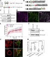
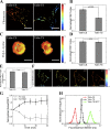
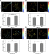
Integrin-dependent adhesions are mechanosensitive structures in which talin mediates a linkage to actin filaments either directly or indirectly by recruiting vinculin. Here, we report the development and validation of a talin tension sensor. We find that talin in focal adhesions is under tension, which is higher in peripheral than central adhesions. Tension on talin is increased by vinculin and depends mainly on actin-binding site 2 (ABS2) within the middle of the rod domain, rather than ABS3 at the far C terminus. Unlike vinculin, talin is under lower tension on soft substrates. The difference between central and peripheral adhesions requires ABS3 but not vinculin or ABS2. However, differential stiffness sensing by talin requires ABS2 but not vinculin or ABS3. These results indicate that central versus peripheral adhesions must be organized and regulated differently, and that ABS2 and ABS3 have distinct functions in spatial variations and stiffness sensing. Overall, these results shed new light on talin function and constrain models for cellular mechanosensing.
© 2016 Kumar et al.
Figures


Similar articles
-
Vinculin controls talin engagement with the actomyosin machinery.Nat Commun. 2015 Dec 4;6:10038. doi: 10.1038/ncomms10038. Nat Commun. 2015. PMID: 26634421 Free PMC article.
-
Actin flow-dependent and -independent force transmission through integrins.Proc Natl Acad Sci U S A. 2020 Dec 22;117(51):32413-32422. doi: 10.1073/pnas.2010292117. Epub 2020 Dec 1. Proc Natl Acad Sci U S A. 2020. PMID: 33262280 Free PMC article.
-
Force-dependent vinculin binding to talin in live cells: a crucial step in anchoring the actin cytoskeleton to focal adhesions.Am J Physiol Cell Physiol. 2014 Mar 15;306(6):C607-20. doi: 10.1152/ajpcell.00122.2013. Epub 2014 Jan 22. Am J Physiol Cell Physiol. 2014. PMID: 24452377
-
Integrating actin dynamics, mechanotransduction and integrin activation: the multiple functions of actin binding proteins in focal adhesions.Eur J Cell Biol. 2013 Oct-Nov;92(10-11):339-48. doi: 10.1016/j.ejcb.2013.10.009. Epub 2013 Nov 4. Eur J Cell Biol. 2013. PMID: 24252517 Review.
-
Integrin connections to the cytoskeleton through talin and vinculin.Biochem Soc Trans. 2008 Apr;36(Pt 2):235-9. doi: 10.1042/BST0360235. Biochem Soc Trans. 2008. PMID: 18363566 Review.
Cited by
-
β2 integrins impose a mechanical checkpoint on macrophage phagocytosis.Nat Commun. 2024 Sep 18;15(1):8182. doi: 10.1038/s41467-024-52453-9. Nat Commun. 2024. PMID: 39294148 Free PMC article.
-
The Importance of Mechanical Forces for in vitro Endothelial Cell Biology.Front Physiol. 2020 Jun 18;11:684. doi: 10.3389/fphys.2020.00684. eCollection 2020. Front Physiol. 2020. PMID: 32625119 Free PMC article. Review.
-
Organization, dynamics and mechanoregulation of integrin-mediated cell-ECM adhesions.Nat Rev Mol Cell Biol. 2023 Feb;24(2):142-161. doi: 10.1038/s41580-022-00531-5. Epub 2022 Sep 27. Nat Rev Mol Cell Biol. 2023. PMID: 36168065 Free PMC article. Review.
-
Adhesions Assemble!-Autoinhibition as a Major Regulatory Mechanism of Integrin-Mediated Adhesion.Front Mol Biosci. 2019 Dec 17;6:144. doi: 10.3389/fmolb.2019.00144. eCollection 2019. Front Mol Biosci. 2019. PMID: 31921890 Free PMC article. Review.
-
Adjustable viscoelasticity allows for efficient collective cell migration.Semin Cell Dev Biol. 2019 Sep;93:55-68. doi: 10.1016/j.semcdb.2018.05.027. Epub 2018 Jun 1. Semin Cell Dev Biol. 2019. PMID: 29859995 Free PMC article. Review.
References
-
- Balaban N.Q., Schwarz U.S., Riveline D., Goichberg P., Tzur G., Sabanay I., Mahalu D., Safran S., Bershadsky A., Addadi L., and Geiger B.. 2001. Force and focal adhesion assembly: a close relationship studied using elastic micropatterned substrates. Nat. Cell Biol. 3:466–472. 10.1038/35074532 - DOI - PubMed
Publication types
MeSH terms
Substances
Grants and funding
LinkOut - more resources
Full Text Sources
Other Literature Sources
Research Materials
Miscellaneous

