

Protein Folding-How and Why: By Hydrogen Exchange, Fragment Separation, and Mass Spectrometry
- PMID: 27145881
- PMCID: PMC5588872
- DOI: 10.1146/annurev-biophys-062215-011121
Protein Folding-How and Why: By Hydrogen Exchange, Fragment Separation, and Mass Spectrometry
Abstract
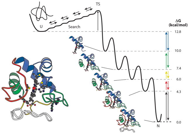
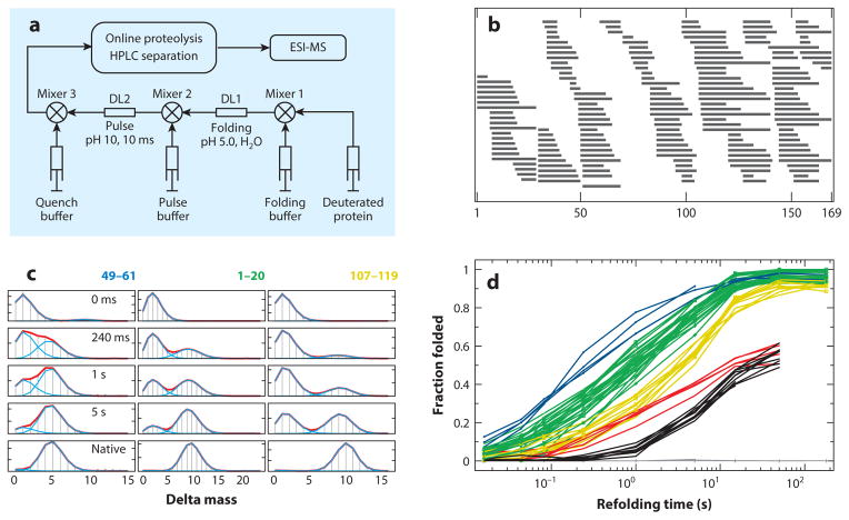
Advanced hydrogen exchange (HX) methodology can now determine the structure of protein folding intermediates and their progression in folding pathways. Key developments over time include the HX pulse labeling method with nuclear magnetic resonance analysis, the fragment separation method, the addition to it of mass spectrometric (MS) analysis, and recent improvements in the HX MS technique and data analysis. Also, the discovery of protein foldons and their role supplies an essential interpretive link. Recent work using HX pulse labeling with MS analysis finds that a number of proteins fold by stepping through a reproducible sequence of native-like intermediates in an ordered pathway. The stepwise nature of the pathway is dictated by the cooperative foldon unit construction of the protein. The pathway order is determined by a sequential stabilization principle; prior native-like structure guides the formation of adjacent native-like structure. This view does not match the funneled energy landscape paradigm of a very large number of folding tracks, which was framed before foldons were known and is more appropriate for the unguided residue-level search to surmount an initial kinetic barrier rather than for the overall unfolded-state to native-state folding pathway.
Keywords: HX MS; energy landscape theory; hydrogen exchange; protein folding.
Figures
Similar articles
-
Cytochrome c folds through foldon-dependent native-like intermediates in an ordered pathway.Proc Natl Acad Sci U S A. 2016 Apr 5;113(14):3809-14. doi: 10.1073/pnas.1522674113. Epub 2016 Mar 10. Proc Natl Acad Sci U S A. 2016. PMID: 26966231 Free PMC article.
-
Stepwise protein folding at near amino acid resolution by hydrogen exchange and mass spectrometry.Proc Natl Acad Sci U S A. 2013 May 7;110(19):7684-9. doi: 10.1073/pnas.1305887110. Epub 2013 Apr 19. Proc Natl Acad Sci U S A. 2013. PMID: 23603271 Free PMC article.
-
How cytochrome c folds, and why: submolecular foldon units and their stepwise sequential stabilization.J Mol Biol. 2004 Oct 8;343(1):223-33. doi: 10.1016/j.jmb.2004.08.005. J Mol Biol. 2004. PMID: 15381432
-
Protein folding and misfolding: mechanism and principles.Q Rev Biophys. 2007 Nov;40(4):287-326. doi: 10.1017/S0033583508004654. Epub 2008 Apr 14. Q Rev Biophys. 2007. PMID: 18405419 Free PMC article. Review.
-
The nature of protein folding pathways.Proc Natl Acad Sci U S A. 2014 Nov 11;111(45):15873-80. doi: 10.1073/pnas.1411798111. Epub 2014 Oct 17. Proc Natl Acad Sci U S A. 2014. PMID: 25326421 Free PMC article. Review.
Cited by
-
Evolutionary adaptation of the protein folding pathway for secretability.EMBO J. 2022 Dec 1;41(23):e111344. doi: 10.15252/embj.2022111344. Epub 2022 Aug 29. EMBO J. 2022. PMID: 36031863 Free PMC article.
-
Co-translational folding of nascent polypeptides: Multi-layered mechanisms for the efficient biogenesis of functional proteins.Bioessays. 2021 Jul;43(7):e2100042. doi: 10.1002/bies.202100042. Epub 2021 May 13. Bioessays. 2021. PMID: 33987870 Free PMC article.
-
A photoswitchable helical peptide with light-controllable interface/transmembrane topology in lipidic membranes.iScience. 2021 Jun 24;24(7):102771. doi: 10.1016/j.isci.2021.102771. eCollection 2021 Jul 23. iScience. 2021. PMID: 34286233 Free PMC article.
-
Hysteresis behavior in the unfolding/refolding processes of a protein trapped in metallo-cages.Chem Sci. 2023 Feb 22;14(11):2910-2914. doi: 10.1039/d2sc05879k. eCollection 2023 Mar 15. Chem Sci. 2023. PMID: 36937586 Free PMC article.
-
Computational Tools for Hydrogen-Deuterium Exchange Mass Spectrometry Data Analysis.Chem Rev. 2024 Nov 13;124(21):12242-12263. doi: 10.1021/acs.chemrev.4c00438. Epub 2024 Oct 31. Chem Rev. 2024. PMID: 39481095 Free PMC article. Review.
References
Publication types
MeSH terms
Substances
Grants and funding
LinkOut - more resources
Full Text Sources
Other Literature Sources
