

Zika virus damages the human placental barrier and presents marked fetal neurotropism
- PMID: 27143490
- PMCID: PMC4878297
- DOI: 10.1590/0074-02760160085
Zika virus damages the human placental barrier and presents marked fetal neurotropism
Abstract
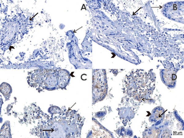
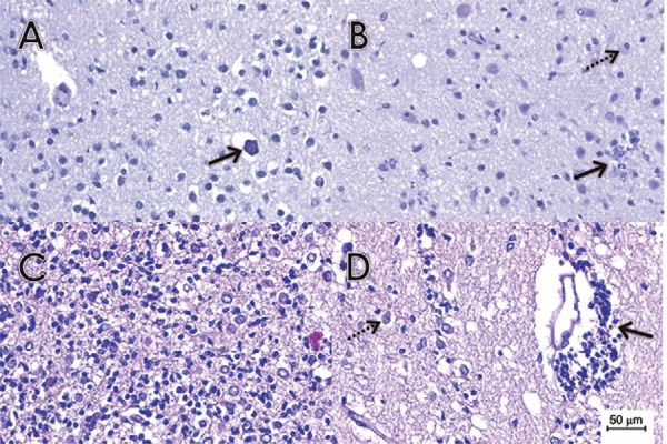
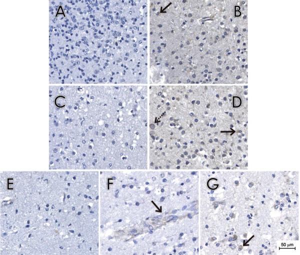
An unusually high incidence of microcephaly in newborns has recently been observed in Brazil. There is a temporal association between the increase in cases of microcephaly and the Zika virus (ZIKV) epidemic. Viral RNA has been detected in amniotic fluid samples, placental tissues and newborn and fetal brain tissues. However, much remains to be determined concerning the association between ZIKV infection and fetal malformations. In this study, we provide evidence of the transplacental transmission of ZIKV through the detection of viral proteins and viral RNA in placental tissue samples from expectant mothers infected at different stages of gestation. We observed chronic placentitis (TORCH type) with viral protein detection by immunohistochemistry in Hofbauer cells and some histiocytes in the intervillous spaces. We also demonstrated the neurotropism of the virus via the detection of viral proteins in glial cells and in some endothelial cells and the observation of scattered foci of microcalcifications in the brain tissues. Lesions were mainly located in the white matter. ZIKV RNA was also detected in these tissues by real-time-polymerase chain reaction. We believe that these findings will contribute to the body of knowledge of the mechanisms of ZIKV transmission, interactions between the virus and host cells and viral tropism.
Figures
Similar articles
-
Pathology of congenital Zika syndrome in Brazil: a case series.Lancet. 2016 Aug 27;388(10047):898-904. doi: 10.1016/S0140-6736(16)30883-2. Epub 2016 Jun 29. Lancet. 2016. PMID: 27372395
-
Congenital Zika syndrome: Pitfalls in the placental barrier.Rev Med Virol. 2018 Sep;28(5):e1985. doi: 10.1002/rmv.1985. Epub 2018 May 15. Rev Med Virol. 2018. PMID: 29761581 Review.
-
Congenital cerebral malformations and dysfunction in fetuses and newborns following the 2013 to 2014 Zika virus epidemic in French Polynesia.Euro Surveill. 2016;21(13). doi: 10.2807/1560-7917.ES.2016.21.13.30181. Euro Surveill. 2016. PMID: 27063794
-
Zika virus - reigniting the TORCH.Nat Rev Microbiol. 2016 Nov;14(11):707-715. doi: 10.1038/nrmicro.2016.125. Epub 2016 Aug 30. Nat Rev Microbiol. 2016. PMID: 27573577 Review.
-
Zika virus detection in amniotic fluid and Zika-associated birth defects.Am J Obstet Gynecol. 2020 Jun;222(6):610.e1-610.e13. doi: 10.1016/j.ajog.2020.01.009. Epub 2020 Jan 15. Am J Obstet Gynecol. 2020. PMID: 31954155 Free PMC article.
Cited by
-
Two Sides of a Coin: a Zika Virus Mutation Selected in Pregnant Rhesus Macaques Promotes Fetal Infection in Mice but at a Cost of Reduced Fitness in Nonpregnant Macaques and Diminished Transmissibility by Vectors.J Virol. 2020 Nov 23;94(24):e01605-20. doi: 10.1128/JVI.01605-20. Print 2020 Nov 23. J Virol. 2020. PMID: 32999034 Free PMC article.
-
Congenital Zika Syndrome Is Associated With Interferon Alfa Receptor 1.Front Immunol. 2021 Nov 25;12:764746. doi: 10.3389/fimmu.2021.764746. eCollection 2021. Front Immunol. 2021. PMID: 34899713 Free PMC article.
-
Intraamniotic Zika virus inoculation of pregnant rhesus macaques produces fetal neurologic disease.Nat Commun. 2018 Jun 20;9(1):2414. doi: 10.1038/s41467-018-04777-6. Nat Commun. 2018. PMID: 29925843 Free PMC article.
-
The Role of Secretory Autophagy in Zika Virus Transfer through the Placental Barrier.Front Cell Infect Microbiol. 2017 Jan 9;6:206. doi: 10.3389/fcimb.2016.00206. eCollection 2016. Front Cell Infect Microbiol. 2017. PMID: 28119857 Free PMC article. Review.
-
Maternal-Fetal Transmission of Zika Virus: Routes and Signals for Infection.J Interferon Cytokine Res. 2017 Jul;37(7):287-294. doi: 10.1089/jir.2017.0011. Epub 2017 Apr 12. J Interferon Cytokine Res. 2017. PMID: 28402153 Free PMC article. Review.
References
-
- Alpert S, Fergerson J, Noël L. Intrauterine West Nile virus: ocular and systemic findings. Am J Ophthalmol. 2003;136(4):733–735. - PubMed
-
- Benirchke K, Kaufmann P, Baergen RN. Benirchke K, Kaufmann P, Baergen RN.editors . Pathology of the human placenta. New York: Springer Science & Business Media; 2006b. Classifications of villous maldevelopment; pp. 502–507.
-
- Benirchke K, Kaufmann P, Baergen RN. Benirchke K, Kaufmann P, Baergen RN.editors . Pathology of the human placenta. New York: Springer Science & Business Media; 2006a. Infectious diseases; pp. 700–715.
-
- Boussemart T, Babe P, Sibille G, Neyret C, Berchel C. Prenatal transmission of dengue: two new cases. J Perinatol. 2001;21(4):255–257. - PubMed
Publication types
MeSH terms
Substances
LinkOut - more resources
Full Text Sources
Other Literature Sources
Medical
