. 2016 May 2:7:11383.
doi: 10.1038/ncomms11383.

The topography of mutational processes in breast cancer genomes

Affiliations

The topography of mutational processes in breast cancer genomes

Sandro Morganella et al. Nat Commun. .

Abstract

Somatic mutations in human cancers show unevenness in genomic distribution that correlate with aspects of genome structure and function. These mutations are, however, generated by multiple mutational processes operating through the cellular lineage between the fertilized egg and the cancer cell, each composed of specific DNA damage and repair components and leaving its own characteristic mutational signature on the genome. Using somatic mutation catalogues from 560 breast cancer whole-genome sequences, here we show that each of 12 base substitution, 2 insertion/deletion (indel) and 6 rearrangement mutational signatures present in breast tissue, exhibit distinct relationships with genomic features relating to transcription, DNA replication and chromatin organization. This signature-based approach permits visualization of the genomic distribution of mutational processes associated with APOBEC enzymes, mismatch repair deficiency and homologous recombinational repair deficiency, as well as mutational processes of unknown aetiology. Furthermore, it highlights mechanistic insights including a putative replication-dependent mechanism of APOBEC-related mutagenesis.


Figures

Figure 1
Figure 1. Distribution of all mutations across the cell cycle.
Replication domains were identified by using conservatively defined transition zones in DNA replication time data. Data were separated into deciles, with each segment containing exactly 10% of the observed replication time signal. Normalized mutation density per decile is presented for early (left) to late (right) replication domains. (a) Aggregated distribution of mutations (green), rearrangements (purple) and indels (orange) across the cell cycle. (b) Distribution of the 12 base substitution signatures across the cell cycle. Dashed grey lines represent the predicted distribution of mutations for each signature based on simulations that take into account mutation burden and sequence characteristics of individual mutations and of the signatures that were estimated to be present in each patient (Methods section). (c) Distribution of the six rearrangement signatures across the cell cycle. Dashed grey lines represent the predicted distribution of mutations for each signature based on simulations. (d) Distribution of the indel signatures across the cell cycle.
Figure 2
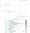
Figure 2. Replication and transcriptional strand bias and strand-coordinated mutagenesis of mutational signatures.
Forest plots showing replication (blue) and transcription (orange) strand bias for the 6 base substitution classes (a) and for the 12 base substitution signatures (b). Mutations were oriented in the pyrimidine context (the current convention for characterizing mutational signatures). Observed distribution between strands is shown as a diamond for replication and circle for transcriptional strands with 95% confidence intervals, against an expected probability of 0.5 (Supplementary Table 1 for values). (c) Relationship between processive group lengths (columns) and mutational signatures (rows). Processive groups were defined as sets of adjacent substitutions of the same mutational signature sharing the same reference allele, and the group length indicates the number of adjacent substitutions within each group. The size of each circle represents the number of groups (log10) observed for the specified group length (column) for each signature (row). The intensity of colour of each circle indicates significance of the likelihood of detection of a processive group of a defined length (−log10 of the P value obtained by comparing observed data to simulations, further details in Methods section).
Figure 3
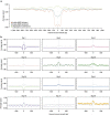
Figure 3. Relationship between mutational signatures and nucleosome occupancy.
The distribution of the signal of nucleosome density (y axis) is shown in a 2 kb window centred on each mutation (position 0 on the x axis), for each signature. The averaged signal was calculated as the total amount of signal observed at each point divided by total number of mutations contributing to that signal. (a) Nucleosome density for aggregated substitutions (green), and for deletions observed in MMR-proficient (blue) and MMR-deficient (orange) samples. (b) Nucleosome density for the twelve base substitution signatures (note the degree of variation between substitution signatures relative to aggregated substitutions in a). The grey line shows the distribution predicted by simulations if mutations from each signature were randomly distributed. The analysis reveals that most of the observed distributions showed similar trends to those expected from simulations, apart from signatures 17, 18 and 26 and to a lesser extent signatures 5 and 8.
Figure 4
Figure 4. A replication-related model of mutagenesis for putative APOBEC-related signatures 2 and 13.
1. During replication, transient moments of increased availability of single-stranded DNA (ssDNA) (for example, uncoupling between leading and lagging replicative strands or delays in elongation of the nascent lagging strand by Okazaki fragments) could occur, exposing ssDNA for APOBEC deamination, potentially for long genomic tracts. 2. Uracil-N-glycosylase (UNG) acts to remove undesirable uracils leaving a trail of abasic sites in its wake. Divergence of mutational processes occurs from this point. 3A Earlier in replication, error-prone translesion polymerases such as REV1 have been postulated to insert cytosines opposite abasic sites to avoid detrimental replication fork stalling or collapse. 4A The final outcome is stretches of successive C>G transversions at a TpC sequence context characteristic of signature 13. 3B Alternatively, uracils and abasic sites that are not fixed via REV1, undergo contingency processing, for example, the ‘A' rule. 4B The final outcome is of C>T mutations at a TpC sequence context.

Comment in

Similar articles

  • Landscape of somatic mutations in 560 breast cancer whole-genome sequences.
    Nik-Zainal S, Davies H, Staaf J, Ramakrishna M, Glodzik D, Zou X, Martincorena I, Alexandrov LB, Martin S, Wedge DC, Van Loo P, Ju YS, Smid M, Brinkman AB, Morganella S, Aure MR, Lingjærde OC, Langerød A, Ringnér M, Ahn SM, Boyault S, Brock JE, Broeks A, Butler A, Desmedt C, Dirix L, Dronov S, Fatima A, Foekens JA, Gerstung M, Hooijer GK, Jang SJ, Jones DR, Kim HY, King TA, Krishnamurthy S, Lee HJ, Lee JY, Li Y, McLaren S, Menzies A, Mustonen V, O'Meara S, Pauporté I, Pivot X, Purdie CA, Raine K, Ramakrishnan K, Rodríguez-González FG, Romieu G, Sieuwerts AM, Simpson PT, Shepherd R, Stebbings L, Stefansson OA, Teague J, Tommasi S, Treilleux I, Van den Eynden GG, Vermeulen P, Vincent-Salomon A, Yates L, Caldas C, van't Veer L, Tutt A, Knappskog S, Tan BK, Jonkers J, Borg Å, Ueno NT, Sotiriou C, Viari A, Futreal PA, Campbell PJ, Span PN, Van Laere S, Lakhani SR, Eyfjord JE, Thompson AM, Birney E, Stunnenberg HG, van de Vijver MJ, Martens JW, Børresen-Dale AL, Richardson AL, Kong G, Thomas G, Stratton MR. Nik-Zainal S, et al. Nature. 2016 Jun 2;534(7605):47-54. doi: 10.1038/nature17676. Epub 2016 May 2. Nature. 2016. PMID: 27135926 Free PMC article.
  • Mutational signatures reveal ternary relationships between homologous recombination repair, APOBEC, and mismatch repair in gynecological cancers.
    Farmanbar A, Firouzi S, Kneller R, Khiabanian H. Farmanbar A, et al. J Transl Med. 2022 Feb 2;20(1):65. doi: 10.1186/s12967-022-03259-0. J Transl Med. 2022. PMID: 35109853 Free PMC article.
  • Mutational spectra and mutational signatures: Insights into cancer aetiology and mechanisms of DNA damage and repair.
    Phillips DH. Phillips DH. DNA Repair (Amst). 2018 Nov;71:6-11. doi: 10.1016/j.dnarep.2018.08.003. Epub 2018 Aug 24. DNA Repair (Amst). 2018. PMID: 30236628 Free PMC article. Review.
  • The repertoire of mutational signatures in human cancer.
    Alexandrov LB, Kim J, Haradhvala NJ, Huang MN, Tian Ng AW, Wu Y, Boot A, Covington KR, Gordenin DA, Bergstrom EN, Islam SMA, Lopez-Bigas N, Klimczak LJ, McPherson JR, Morganella S, Sabarinathan R, Wheeler DA, Mustonen V; PCAWG Mutational Signatures Working Group; Getz G, Rozen SG, Stratton MR; PCAWG Consortium. Alexandrov LB, et al. Nature. 2020 Feb;578(7793):94-101. doi: 10.1038/s41586-020-1943-3. Epub 2020 Feb 5. Nature. 2020. PMID: 32025018 Free PMC article.
  • Mechanisms underlying mutational signatures in human cancers.
    Helleday T, Eshtad S, Nik-Zainal S. Helleday T, et al. Nat Rev Genet. 2014 Sep;15(9):585-98. doi: 10.1038/nrg3729. Epub 2014 Jul 1. Nat Rev Genet. 2014. PMID: 24981601 Free PMC article. Review.

Cited by

References

    1. De S. & Michor F. DNA replication timing and long-range DNA interactions predict mutational landscapes of cancer genomes. Nat. Biotechnol. 29, 1103–1108 (2011). - PMC - PubMed
    1. Drier Y. et al.. Somatic rearrangements across cancer reveal classes of samples with distinct patterns of DNA breakage and rearrangement-induced hypermutability. Genome Res. 23, 228–235 (2013). - PMC - PubMed
    1. Koren A. et al.. Differential relationship of DNA replication timing to different forms of human mutation and variation. Am. J. Hum. Genet. 91, 1033–1040 (2012). - PMC - PubMed
    1. Kundaje A. et al.. Ubiquitous heterogeneity and asymmetry of the chromatin environment at regulatory elements. Genome Res. 22, 1735–1747 (2012). - PMC - PubMed
    1. Liu L., De S. & Michor F. DNA replication timing and higher-order nuclear organization determine single-nucleotide substitution patterns in cancer genomes. Nat. Commun. 4, 1502 (2013). - PMC - PubMed

Publication types