Clinical Trial
. 2017 Jan 1;23(1):26-34.
doi: 10.1158/1078-0432.CCR-16-0134. Epub 2016 Apr 28.

A Phase Ib Study of Alpelisib (BYL719), a PI3Kα-Specific Inhibitor, with Letrozole in ER+/HER2- Metastatic Breast Cancer

Affiliations
Clinical Trial

A Phase Ib Study of Alpelisib (BYL719), a PI3Kα-Specific Inhibitor, with Letrozole in ER+/HER2- Metastatic Breast Cancer

Ingrid A Mayer et al. Clin Cancer Res. .

Abstract

Purpose: Alpelisib, a selective oral inhibitor of the class I PI3K catalytic subunit p110α, has shown synergistic antitumor activity with endocrine therapy against ER+/PIK3CA-mutated breast cancer cells. This phase Ib study evaluated alpelisib plus letrozole's safety, tolerability, and preliminary activity in patients with metastatic ER+ breast cancer refractory to endocrine therapy.

Experimental design: Twenty-six patients received letrozole and alpelisib daily. Outcomes were assessed by standard solid-tumor phase I methods. Tumor blocks were collected for DNA extraction and next-generation sequencing.

Results: Alpelisib's maximum-tolerated dose (MTD) in combination with letrozole was 300 mg/d. Common drug-related adverse events included hyperglycemia, nausea, fatigue, diarrhea, and rash with dose-limiting toxicity occurring at 350 mg/d of alpelisib. The clinical benefit rate (lack of progression ≥6 months) was 35% (44% in patients with PIK3CA-mutated and 20% in PIK3CA wild-type tumors; 95% CI, 17%-56%), including five objective responses. Of eight patients remaining on treatment ≥12 months, six had tumors with a PIK3CA mutation. Among evaluable tumors, those with FGFR1/2 amplification and KRAS and TP53 mutations did not derive clinical benefit. Overexpression of FGFR1 in ER+/PIK3CA mutant breast cancer cells attenuated the response to alpelisib in vitro CONCLUSIONS: The combination of letrozole and alpelisib was safe, with reversible toxicities. Clinical activity was observed independently of PIK3CA mutation status, although clinical benefit was seen in a higher proportion of patients with PIK3CA-mutated tumors. Phase II and III trials of alpelisib and endocrine therapy in patients with ER+ breast cancer are ongoing. Clin Cancer Res; 23(1); 26-34. ©2016 AACR.


Figures

Figure 1
Figure 1. Treatment duration (months) with corresponding radiologic response and tumor PIK3CA mutation status
Best response by Response Evaluation Criteria in Solid Tumors (RECIST v1.1) is depicted in yellow (partial response), blue (stable disease) and black (progression of disease) shading. Red shading indicates discontinuation of treatment due to toxicity or withdrawal. Textured bars reflect patients treated with 350 mg/d of alpelisib; solid bars reflect the ones on 300 mg/d. Arrows indicate patients that are still on study. PIK3CA mutations by NGS (in black) or SNaPShot (in pink) are shown on the vertical axis; Ø represents absence of PIK3CA mutation. Mutations on exons 9 and 20 (hot spots) were the most prevalent ones; exon 20 (H1047R) mutations appear to be associated with more clinical durable responses, in contrast to exon 9 mutations (E545K).
Figure 2
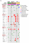
Figure 2. Next generation sequencing analysis of DNA from tumors of patients evaluable for clinical response
A, Of 341 cancer-associated genes, the somatic alterations (amplifications - red, deletions - blue, truncating - black, inframe - brown, and missense - green mutations) most commonly seen (i.e. >5% cases) are depicted here. Clinical benefit was defined as lack of progression for at least 6 months on letrozole/alpelisib. A total of 23 cases with adequate tissue for DNA extraction are depicted. Tissue origin in 9/23 (39%) tumors was a metastatic biopsy (pink), whereas 14/23 (61%) of tumors were from the primary diagnostic tumor specimen (purple). Tumors from patients with clinical benefit are depicted in green (top), and tumors from patients without clinical benefit are depicted in maroon (top). Patients that discontinued trial early (i.e., < 6 months) due to toxicity are depicted in gold as non-evaluable (top). A higher proportion of PIK3CA mutations were seen in patients with clinical benefit, and overall less genomic alterations were seen when compared to patients without clinical benefit. Two of the patients with clinical benefit were found to have both PIK3CA and ESR1 mutations in their tumor. FGFR1/2 amplification and mutation in TP53, BRCA1 and KRAS were only found in cancers from patients who did not benefit from treatment. PTEN mutations were found in both groups. B, Of patients with tumors harboring the genetic alterations depicted in the X axis, the blue bars indicate the proportion of patients with clinical benefit; red bars indicate the proportion of patients without clinical benefit. The graph reveals that most patients with PIK3CA and ESR1 mutated tumors had clinical benefit, in contrast to patients with alterations in KRAS, TP53 or FGFR1, none of which had clinical benefit. C, FGFR1 amplification was confirmed by FISH analysis in the three evaluable patients with FGFR1 amplification detected by NGS.
Figure 3
Figure 3. FGFR1 overexpression attenuates the antiproliferative action of alpelisib
A, FGFR1 amplification was confirmed by FISH analysis in the CAMA-1 ER+ breast cancer cells as described in Methods. B, CAMA-1 cells in estrogen free-media (IMEM + 10% dextran-charcoal-treated FBS) with 5 ng/mL FGF2 were treated with 2 μM alpelisib, 2 μM lucitanib or the combination. Representative images and quantification of integrated intensity are shown (**p<0.01 vs. control, t-test). Growth of CAMA-1 cells was completely insensitive to alpelisib but inhibited by lucitanib. C, Immunoblot analysis with the indicated antibodies of lysates from CAMA-1 cells treated for 6 h with vehicle, alpelisib, lucitanib and the combination of both drugs in estrogen free-media supplemented with 5 ng/mL FGF2. Alpelisib inhibits p-AKT but not p-ERK whereas lucitanib inhibits p-ERK but not p-AKT. D, Immunoblot analysis with the indicated antibodies of lysates of MCF7GFP and MCF7FGFR1 cells that were treated for 6 h with vehicle, alpelisib, lucitanib and the combination in estrogen free-media with 5 ng/mL FGF2. Alpelisib inhibits p-AKT but not p-ERK whereas lucitanib inhibits p-ERK but not p-AKT. E, MCF7GFP and MCF7FGFR1 cells were plated in estrogen free-media and FGF2 and treated with alpelisib alone or in combination with lucitanib for 72 h. Cell proliferation was assessed by CellTiter-Glo (Promega) assay; each data point represents the mean ± SD of triplicate wells. MCF7GFP cells (black) grown as monolayers had an IC50 to alpelisib <1 μM, whereas this was at least 10-fold higher in MCF7FGFR1 cells (green). Addition of lucitanib re-sensitized MCF7FGFR1 cells to alpelisib (red).

Similar articles

Cited by

References

    1. Samuels Y, Diaz LA, Jr., Schmidt-Kittler O, Cummins JM, Delong L, Cheong I, et al. Mutant PIK3CA promotes cell growth and invasion of human cancer cells. Cancer Cell. 2005;7(6):561–73. - PubMed
    1. Isakoff SJ, Engelman JA, Irie HY, Luo J, Brachmann SM, Pearline RV, et al. Breast cancer-associated PIK3CA mutations are oncogenic in mammary epithelial cells. Cancer Res. 2005;65(23):10992–1000. - PubMed
    1. Eichhorn PJ, Gili M, Scaltriti M, Serra V, Guzman M, Nijkamp W, et al. Phosphatidylinositol 3-kinase hyperactivation results in lapatinib resistance that is reversed by the mTOR/phosphatidylinositol 3-kinase inhibitor NVP-BEZ235. Cancer Res. 2008;68(22):9221–30. - PMC - PubMed
    1. Chakrabarty A, Rexer BN, Wang SE, Cook RS, Engelman JA, Arteaga CL. H1047R phosphatidylinositol 3-kinase mutant enhances HER2-mediated transformation by heregulin production and activation of HER3. Oncogene. 2010;29(37):5193–203. - PMC - PubMed
    1. Perez-Tenorio G, Alkhori L, Olsson B, Waltersson MA, Nordenskjold B, Rutqvist LE, et al. PIK3CA mutations and PTEN loss correlate with similar prognostic factors and are not mutually exclusive in breast cancer. Clin Cancer Res. 2007;13(12):3577–84. - PubMed

Publication types

MeSH terms