. 2016 Apr;97(2):97-106.
doi: 10.1111/iep.12172. Epub 2016 Apr 29.

Different effects of arginine vasopressin on high-mobility group box 1 expression in astrocytes isolated from stroke-prone spontaneously hypertensive rats and congenic SHRpch1_18 rats

Affiliations

Different effects of arginine vasopressin on high-mobility group box 1 expression in astrocytes isolated from stroke-prone spontaneously hypertensive rats and congenic SHRpch1_18 rats

Kazuo Yamagata et al. Int J Exp Pathol. 2016 Apr.

Abstract

Stroke-prone spontaneously hypertensive rats (SHRSP/Izm) develop severe hypertension and astrocytic oedema following ischaemic stimulation. During ischaemic stress high-mobility group box 1 (Hmgb1) expression in astrocytes is induced, and subsequently potentiates deterioration of the brain due to ischaemic injury, which manifests as both cerebral inflammation and astrocytic oedema. Arginine vasopressin (AVP) induces brain injury and increases astrocytic swelling. After stroke, Hmgb1 and peroxiredoxin (Prx) are released at different times and activate macrophages in the brain via Toll-like receptors (Tlr2s). The purpose of this study was to examine whether AVP and/or hypoxia and reoxygenation (H/R) contribute to Hmgb1 regulation following ischaemic stroke. Thus, Hmgb1, Prx2 and Tlr2 expression levels in astrocytes isolated from Wistar Kyoto rats (WKY/Izm), spontaneously hypertensive rats (SHR/Izm), SHRSP/Izm and congenic rat strain SHRpch1_18 treated with AVP and/or H/R were compared. Gene and protein expression levels were determined by reverse transcriptase-polymerase chain reaction (RT-PCR) and real-time quantitative PCR, and Western blot. mRNA expression of Hmgb1, Prx2 and Tlr2 induced by AVP was dose-dependent, and Hmgb1 and Prx2 expression was higher in SHR/Izm, SHRSP/Izm and SHRch1_18 than in WKY/Izm. Tlr2 expression with AVP was reduced in SHR/Izm compared to WKY/Izm. In SHRpch1_18, Hmgb1 expression increased after AVP plus H/R. AVP-modulated expression of Hmgb1 protein was reduced by the addition of the antioxidant N-acetylcysteine (NAC). These results suggest that oxidative stress by AVP enhanced expression of Hmgb1, Prx2 and Tlr2 in astrocytes. We hypothesize that regulation of Hmgb1 by AVP during H/R might be related to induction of inflammation and stroke in SHRSP/Izm and SHRpch1_18 rats.

Keywords: HMGB1; SHRSP; astrocytes.


Figures

Figure 1
Figure 1
Effect of AVP on Hmgb1 expression in astrocytes of WKY/Izm, SHR/Izm, SHRSP/Izm and SHRpch1_18 rats. Astrocytes isolated from WKY/Izm, SHR/Izm, SHRSP/Izm and SHRpch1_18 rats were exposed to AVP (0, 10, 50 and 100 nM) for 8 h, after which total cellular RNA was used for RTPCR analysis. Summation and comparison of Hmgb1 expression from astrocytes from all assayed rat strains. Gene expression levels were normalized to 18S ribosomal RNA. Columns show mean ± SE (n = 4). AVP, arginine vasopressin; 18s rRNA, 18S ribosomal RNA. *P < 0.05.
Figure 2
Figure 2
Effect of AVP on Prx2 expression in astrocytes isolated from WKY/Izm, SHR/Izm, SHRSP/Izm and SHRpch1_18 rats. Astrocytes isolated from WKY/Izm, SHR/Izm, SHRSP/Izm and SHRpch1_18 rats were exposed to AVP (0, 10, 50 and 100 nM) for 8 h, after which total cellular RNA was used for RTPCR analysis. Summation and comparison of Prx2 expression from astrocytes from all assayed rat strains. Gene expression levels were normalized to 18S ribosomal RNA. Columns show mean ± SE (n = 4). AVP, arginine vasopressin; 18s rRNA, 18S ribosomal RNA. *P < 0.05.
Figure 3
Figure 3
Effect of AVP on Tlr2 expression in astrocytes isolated from WKY/Izm, SHR/Izm, SHRSP/Izm and SHRpch1_18 rats. Astrocytes isolated from WKY/Izm, SHR/Izm, SHRSP/Izm and SHRpch1_18 rats were exposed to AVP (0, 10, 50 and 100 nM) for 8 h, after which total cellular RNA was used for RTPCR analysis. Summation and comparison of Hmgb1 expression from astrocytes from all assayed rat strains. Gene expression levels were normalized to 18S ribosomal RNA. Columns show mean ± SE (n = 4). AVP, arginine vasopressin; 18s rRNA, 18S ribosomal RNA. *P < 0.05.
Figure 4
Figure 4
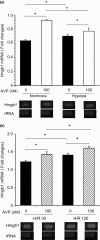
Effect of AVP during H/R on Hmgb1 expression in astrocytes isolated from SHRpch1_18 rats. Astrocytes isolated from SHRpch1_18 rats were exposed to (a) normoxic and hypoxic conditions for 24 h with or without AVP (100 nM) and (b) H/R conditions for 24 h with or without AVP (100 nM). After hypoxic culture, they were maintained under normoxic conditions for 30 min or for 2 h with or without AVP (100 nM). Total cellular RNA was isolated from the cultured astrocytes for RTPCR. Columns show means ± SE (n = 4). Normoxia, 21% O2; hypoxia: 1% O2; H/R30: after hypoxic culture (1% O2), cells were maintained in normoxic conditions (21% O2): for 30 min; H/R2 h: after hypoxic culture (1% O2), cells were maintained in normoxic conditions (21% O2) for 2 h. AVP, arginine vasopressin; rRNA, 18S ribosomal RNA. *P < 0.05.
Figure 5
Figure 5
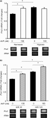
Effect of AVP during H/R on Prx2 expression in astrocytes isolated from SHRpch1_18 rats. Astrocytes isolated from SHRpch1_18 rats were exposed to (a) normoxic and hypoxic conditions for 24 h with or without AVP (100 nM) and (b) H/R conditions for 24 h with or without AVP (100 nM). After hypoxic culture, they were maintained under normoxic conditions for 30 min or for 2 h with or without AVP (100 nM). Total cellular RNA was isolated from the cultured astrocytes for RTPCR. Columns show means ± SE (n = 4). Normoxia, 21% O2; hypoxia: 1% O2; H/R30: after hypoxic culture (1% O2), cells were maintained in normoxic conditions (21% O2) for 30 min; H/R2 h: after hypoxic culture (1% O2), cells were maintained in normoxic conditions (21% O2) for 2 h. AVP, arginine vasopressin; rRNA, 18S ribosomal RNA. *P < 0.05.
Figure 6
Figure 6
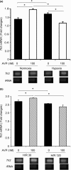
Effect of AVP during H/R on Tlr2 expression in astrocytes isolated from SHRpch1_18 rats. Astrocytes isolated from SHRpch1_18 rats were exposed to (a) normoxia and hypoxia and (b) H/R conditions for 24 h with or without AVP (100 nM). After hypoxic culture, they were maintained under normoxic conditions for 30 min or for 2 h with or without AVP (100 nM). Total cellular RNA was isolated from the cultured astrocytes for RTPCR. Columns show means ± SE (n = 4). Normoxia, 21% O2; hypoxia: 1% O2; H/R30: after hypoxic culture (1% O2), cells were maintained in normoxic conditions (21% O2): for 30 min; H/R2 h: after hypoxic culture (1% O2), cells were maintained in normoxic conditions (21% O2) for 2 h. AVP, arginine vasopressin; rRNA, 18S ribosomal RNA. *P < 0.05.
Figure 7
Figure 7
AVP‐induced expression of Hmgb1 protein from astrocytes isolated from WKY/Izm, SHR/Izm, SHRSP/Izm and SHRpch1_18 rats and effects of NAC. Astrocytes isolated from WKY/Izm, SHR/Izm, SHRSP/Izm and SHRpch1_18 rats were exposed to 100 nM AVP for 24 h, after which protein was used for WB analysis (a). In addition, astrocytes isolated from WKY/Izm and SHRpch1_18 were treated with or without AVP (100 nM) for 24 h. In addition, astrocytes isolated from SHRpch1_18 were treated with or without AVP (100 nM), with or without NAC for 24 h (b). Astrocytes were exposed to NAC for 30 min before the addition of AVP. Total protein (30 μg) was analysed by Western blot with anti‐Hmgb1 antibody. AVP, arginine vasopressin; WKY, WKY/Izm; SHRpch, SHRpch1_18; NAC, N‐acetylcysteine. *P < 0.05.

References

    1. Borysiewicz E., Fil D. & Konat G.W. (2009) Rho proteins are negative regulators of TLR2, TLR3, and TLR4 signaling in astrocytes. J. Neurosci. Res. 87, 1565–1572. - PubMed
    1. Caso J.R., Pradillo J.M., Hurtado O., Leza J.C., Moro M.A. & Lizasoain I. (2008) Toll‐like receptor 4 is involved in subacute stress‐induced neuroinflammation and in the worsening of experimental stroke. Stroke 39, 1314–1320. - PubMed
    1. Faraco G., Fossati S., Bianchi M.E. et al (2007) High mobility group box 1 protein is released by neural cells upon different stresses and worsens ischemic neurodegeneration in vitro and in vivo. J. Neurochem. 103, 590–603. - PubMed
    1. Freria C.M., Velloso L.A. & Oliveira A.L. (2012) Opposing effects of Toll‐like receptors 2 and 4 on synaptic stability in the spinal cord after peripheral nerve injury. J. Neuroinflammation 9, 240. - PMC - PubMed
    1. Gandolgor T.A., Ohara H., Cui Z.H. et al (2013) Two genomic regions of chromosomes 1 and 18 explain most of the stroke susceptibility under salt loading in stroke‐prone spontaneously hypertensive rat/Izm. Hypertension 62, 55–61. - PubMed

MeSH terms

Associated data