. 2016 Apr 19;12(4):e1005574.
doi: 10.1371/journal.ppat.1005574. eCollection 2016 Apr.

The Ancient Evolutionary History of Polyomaviruses

Affiliations

The Ancient Evolutionary History of Polyomaviruses

Christopher B Buck et al. PLoS Pathog. .

Abstract

Polyomaviruses are a family of DNA tumor viruses that are known to infect mammals and birds. To investigate the deeper evolutionary history of the family, we used a combination of viral metagenomics, bioinformatics, and structural modeling approaches to identify and characterize polyomavirus sequences associated with fish and arthropods. Analyses drawing upon the divergent new sequences indicate that polyomaviruses have been gradually co-evolving with their animal hosts for at least half a billion years. Phylogenetic analyses of individual polyomavirus genes suggest that some modern polyomavirus species arose after ancient recombination events involving distantly related polyomavirus lineages. The improved evolutionary model provides a useful platform for developing a more accurate taxonomic classification system for the viral family Polyomaviridae.


Conflict of interest statement

AJM is an employee of Georgia Aquarium, Inc., a 501(c)3 not-for-profit organization. This affiliation does not alter our adherence to all PLOS Pathogens policies on sharing data and materials.

Figures
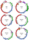
Fig 1
Fig 1. Predicted genetic organization of newly discovered polyomaviruses.
Merkel cell polyomavirus (MCV) is shown as a well-studied reference species. The size of each genome (in basepairs) is listed below the species name. Large T antigen (LT) is indicated in red. Dark gray lollipops indicate the signature HPDKGG motif of the LT “DNAJ” domain (which appears to be missing from the sea bass and notothen polyomaviruses). White lollipops indicate LXCXE motifs, which are hypothetically involved in binding pRb and related tumor suppressor proteins. Each virus encodes a potential myristoylation signal that defines the N-terminus of the minor capsid protein VP2 (green). The VP2 of the supermarket sheep meat-associated virus encodes an internal MALXXΦ motif [1] that defines the N-terminus of a predicted VP3 minor capsid protein, while the other viruses do not. Predicted VP1 major capsid protein genes are shaded blue. ORFs found in the same general arrangement as previously described accessory proteins are also shown. These include small T antigen (sT, pink) Agnoprotein (purple), and the recently described ALTO (orange), which is overprinted in the LT +1 frame. Un-named ORFs of potential interest are shaded light gray. Yellow bars indicate hypothetical metal-binding motifs (CXCXXC or related sequences) observed in some of the predicted accessory proteins. Aside from MCV, for which expressed proteins have been experimentally confirmed, the predicted proteins are hypothetical and do not necessarily account for possible spliced transcripts.
Fig 2
Fig 2. Structural modeling of LT proteins.
The solved OBD-Zn-ATPase SV40 LT structure (PDB identifier 4GDF) was used as template for all OBD and Zn-ATPase domain models. The model of the guitarfish polyomavirus J domain was generated using the solved structure of the SV40 LT DNAJ domain (PDB identifier 1GH6) as template. For the DNAJ domain of scorpion polyomavirus LT, the best modeling template match is a Thermus thermophilus DNAJ protein (PDB identifier 4J7Z). The solved structure of the bacterial DNAJ is highlighted in magenta in the pairwise superimposition (top left). The LT proteins of the indicated polyomavirus species are shown in black. The known structures of SV40 LT domains are superimposed in gold. The conserved HPD motif of the DNAJ domain is positioned on the top and highlighted in cyan. The N-terminal domain of the notothen polyomavirus has no discernible structural similarity to known DNAJ structures.
Fig 3
Fig 3. Midpoint-rooted phylogenetic tree for polyomavirus Large T antigen (LT) protein sequences.
Species with different clade affiliations in VP1 analyses (Fig 4) are indicated in colored bold oblique text. The script ƒ character indicates fragmentary (sub-genomic) sequences. A key to species nicknames, genetic characteristics, and accession numbers is provided in S1 File. Percent bootstrap values are indicated for selected nodes. A FigTree file containing detailed bootstrap values is provided as S2 File. Scale bar shows one substitution per site.
Fig 4
Fig 4. Midpoint-rooted phylogenetic tree for polyomavirus VP1 protein sequences.
Species with different clade affiliations in LT analyses (Fig 3) are indicated in colored bold oblique text. The script ƒ character indicates fragmentary (sub-genomic) sequences. Percent bootstrap values for selected nodes are indicated. A FigTree file containing detailed bootstrap values is provided as S3 File. Scale bar shows one substitution per site.
Fig 5
Fig 5. A hypothetical framework for ancient recombination events among major polyomavirus clades.
The model attempts to reconcile observed incongruities between LT and VP1 phylogenetic trees shown in Figs 3 and 4. In the model, a hypothetical ancient polyomavirus, designated Arche, is inferred to have infected the last common ancestor of bilaterian animals. The ancient Arche lineage then gave rise to separate polyomavirus lineages found in arthropods and fish, as well as the mammalian Ortho/Almi lineages. The figure depicts Avi and Wuki clades arising after recombination events involving an unknown vertebrate-Arche lineage and Ortho-like species. The figure does not depict the inferred evolution of the HPyV6/7 clade, which appears to have arisen after a separate recombination event involving the late region of a hypothetical vertebrate-Arche lineage and the early region of a basal Almi-like species. The TSV lineage, which shows evidence of recombination between the Ortho and Almi lineages, is also omitted. White lollipops represent predicted pRb-binding motifs (LXCXE or related sequences). Yellow bars represent hypothetical metal-binding motifs (CXCXXC or related sequences). The absence of metal-binding motifs in Avi small T antigen (sT) proteins suggests a different evolutionary origin than the classic metal-binding Ortho/Almi sT. Possible ALTO-like ORFs predicted for some Ortho species are shaded gray.
Fig 6
Fig 6. Standard virus/host co-divergence models.
The top panels depict the evolution of polyomaviruses within animal lineages. Idealized cartoon trees in the bottom panels represent the expected polyomavirus phylogeny. The silhouettes in the bottom panels represent the animal type in which the polyomavirus at the branch tip would be found.
Fig 7
Fig 7. Virus-host co-divergence plot.
SDT software was used to score individual pairs of polyomaviruses within various clades for percent divergence across the entire viral genome. The nucleotide divergence score was plotted against the estimated time (in millions of years ago, mya) of the last common ancestor of the host animals in which the polyomavirus pair was found. Apparent recent transmission of some Avi polyomaviruses between distantly related bird species is represented by points close to the x-axis. The absence of such points in the Almi and Ortho clades indicates a lack of evidence for recent transmission of polyomaviruses between distantly related mammal species. The arbitrary dashed reference line has a slope of about 0.5% polyomavirus divergence per million years after host divergence.

Similar articles

Cited by

References

    1. Schowalter RM, Buck CB. The Merkel cell polyomavirus minor capsid protein. PLoS pathogens. 2013;9(8):e1003558 Epub 2013/08/31. 10.1371/journal.ppat.1003558 - DOI - PMC - PubMed
    1. Gross L. A filterable agent, recovered from Ak leukemic extracts, causing salivary gland carcinomas in C3H mice. Proceedings of the Society for Experimental Biology and Medicine Society for Experimental Biology and Medicine (New York, NY. 1953;83(2):414–21. . - PubMed
    1. Stewart SE, Eddy BE, Gochenour AM, Borgese NG, Grubbs GE. The induction of neoplasms with a substance released from mouse tumors by tissue culture. Virology. 1957;3(2):380–400. . - PubMed
    1. Dang-Tan T, Mahmud SM, Puntoni R, Franco EL. Polio vaccines, Simian Virus 40, and human cancer: the epidemiologic evidence for a causal association. Oncogene. 2004;23(38):6535–40. Epub 2004/08/24. 10.1038/sj.onc.1207877 . - DOI - PubMed
    1. DeCaprio JA, Garcea RL. A cornucopia of human polyomaviruses. Nature reviews Microbiology. 2013;11(4):264–76. 10.1038/nrmicro2992 - DOI - PMC - PubMed

Publication types