. 2016 Apr 16:11:28.
doi: 10.1186/s13024-016-0094-3.

MicroRNA-7 targets Nod-like receptor protein 3 inflammasome to modulate neuroinflammation in the pathogenesis of Parkinson's disease

Affiliations

MicroRNA-7 targets Nod-like receptor protein 3 inflammasome to modulate neuroinflammation in the pathogenesis of Parkinson's disease

Yan Zhou et al. Mol Neurodegener. .

Abstract

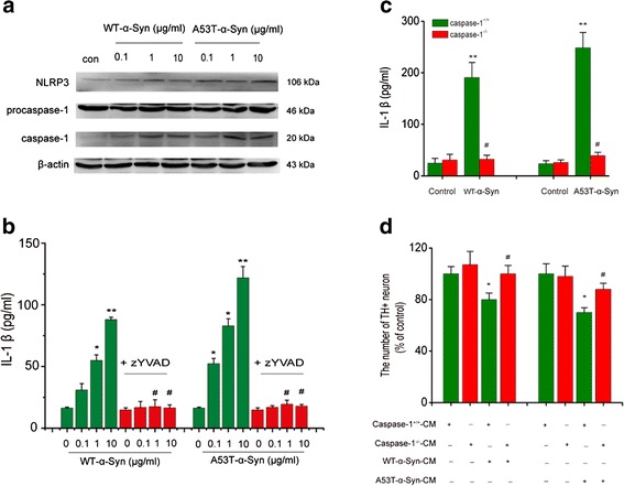
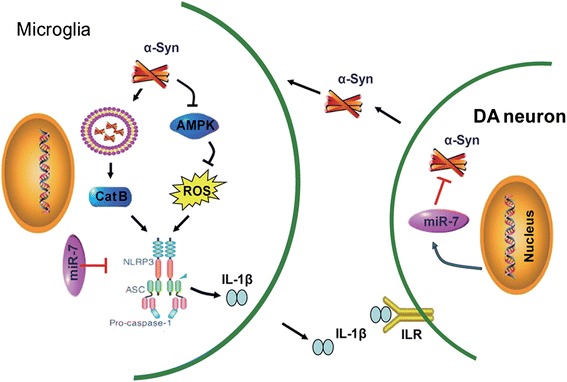
Background: α-Synuclein (α-Syn), a pathological hallmark of Parkinson's disease (PD), has been recognized to induce the production of interleukin-1β in a process that depends, at least in vitro, on nod-like receptor protein 3 (NLRP3) inflammasome in monocytes. However, the role of NLRP3 inflammasome activation in the onset of PD has not yet been fully established.

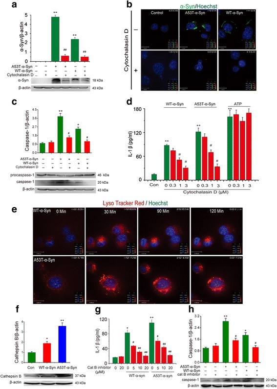
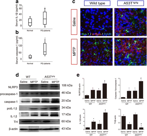
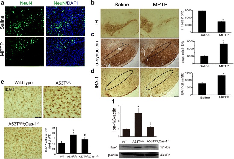
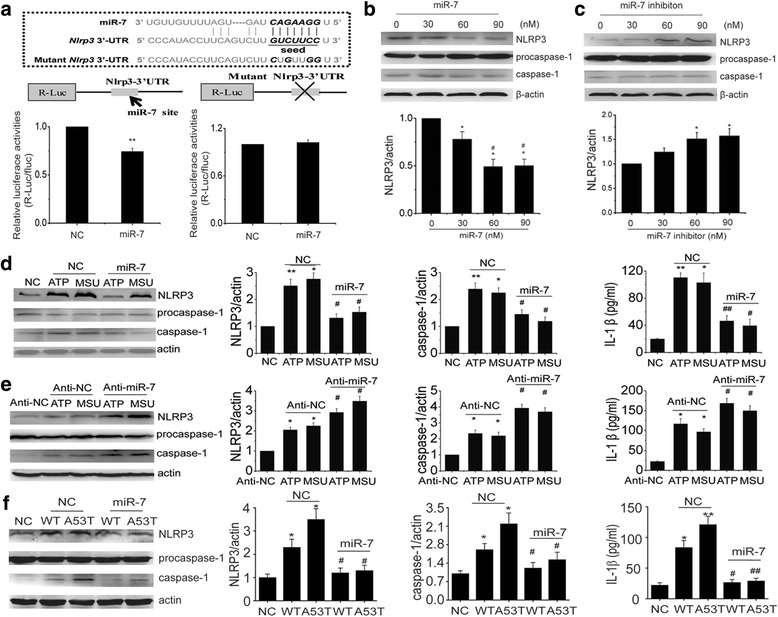
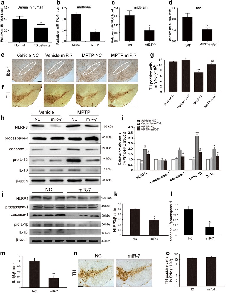
Results: In this study, we showed that NLRP3 inflammasomes were activated in the serum of PD patients and the midbrain of PD model mice. We further clarified that α-syn activated the NLRP3 inflammasome through microglial endocytosis and subsequent lysosomal cathepsin B release. Deficiency of caspase-1, an important component of NLRP3 inflammasome, significantly inhibited α-syn-induced microglia activation and interleukin-1β production, which in turn alleviated the reduction of mesencephalic dopaminergic neurons treated by microglia medium. Specifically, we demonstrated for the first time that Nlrp3 is a target gene of microRNA-7 (miR-7). Transfection of miR-7 inhibited microglial NLRP3 inflammasome activation whereas anti-miR-7 aggravated inflammasome activation in vitro. Notably, stereotactical injection of miR-7 mimics into mouse striatum attenuated dopaminergic neuron degeneration accompanied by the amelioration of microglial activation in MPTP-induced PD model mice.

Conclusions: Our study provides a direct link between miR-7 and NLRP3 inflammasome-mediated neuroinflammation in the pathogenesis of PD. These findings will give us an insight into the potential of miR-7 and NLRP3 inflammasome in terms of opening up novel therapeutic avenues for PD.

Keywords: NLRP3 inflammasome; Neuroinflammation; Parkinson’s disease; microRNA-7; α-Synuclein.


Figures

Fig. 1
Fig. 1
α-Synuclein activates the NLRP3 inflammasome in BV2 cells. a Wide-type and A53T mutants showing α-Syn induces the up-regulation of NLRP3 and Caspase-1 in BV2 cells. Data are presented as the mean ± S.E.M from four independent experiments. b Caspase-1-specific inhibitor (z-YVAD, 10 μM) suppresses IL-1β formation induced by both A53T mutant and wide-type α-Syn. LPS + ATP were used for a positive control drug. Data are presented as the mean ± S.E.M from four independent experiments. c Caspase-1 knockout abolishes WT or A53T α-Syn (10 μg/ml) induced elevation of IL-1β release in primary cultured microglia. d Midbrain TH neurons showed an attenuated impairment and a resistance to condition medium (CM) that was obtained from Caspase-/- microglia treated with WT or A53T α-Syn. Data are presented as the mean ± S.E.M from four independent experiments. * p < 0.05, ** p < 0.01 vs. control group, # p < 0.05, ## p < 0.01 vs. α-Syn treatment group
Fig. 2
Fig. 2
α-Syn activates NLRP3 inflammasome via endocytosis and lysosomal impairment. a Immunoblot analysis of α-Syn in BV2 cells stimulated with A53T mutant or wide-type α-Syn, cytochalasin D (3 μM) inhibits endocytosis of α-Syn into BV2 cells. Data are presented as the mean ± S.E.M from four independent experiments. b Confocal microscopy of immortalized microglia BV2 cells incubated for 4 h with FITC-labeled α-Syn (10 μM) and then processed for immunocytochemistry, cell nuclei were visualized with hoechst. Data are presented as the mean ± S.E.M from three independent experiments. c Immunoblot analysis of caspase-1 in BV2 cells stimulated with A53T mutant or wide-type α-Syn, cytochalasin D (3 μM) inhibits activation of caspase-1 in a concentration-dependent manner in BV2 cells. Data are presented as the mean ± S.E.M from three independent experiments. d ELISA of the release of IL-1β into supernatants of BV2 cells treated with cytochalasin D during stimulation with A53T mutant or wide-type α-Syn or ATP. Data are presented as the mean ± S.E.M from four independent experiments. e α-Syn-containing lysosomes adopt a swollen morphology and underwent structural damage once α-Syn was phagocytosed by BV2 cells. Data are presented as the mean ± S.E.M from three independent experiments. f WT or A53T α-Syn in turn triggers the release of lysosomal protease cathepsin B into the cytoplasm. g-h Cathepsin B inhibitor significantly suppresses α-Syn evoked increase of caspase-1 expression and IL-1β production. Data are presented as the mean ± S.E.M from four independent experiments. * p < 0.05, ** p < 0.01 vs. control group, # p < 0.05, ## p < 0.01 vs. α-Syn treatment group
Fig. 3
Fig. 3
Inflammasome is activated in serum of PD patients and in the midbrain of α-Syn-overexpressed mice. a Serum levels of IL-1β and b caspase-1 activity are upregulated in PD patients. * p < 0.05, ** p < 0.01 vs. normal group. Data are presented as the mean ± S.E.M, n = 12. c Immunofluorescence exhibits enhanced α-Syn accumulation and aggravated Iba-1+ microglial immunofluorescence intensity in the SNc of A53T transgenic mice. Green: Iba-1, red: α-Syn, blue: DAPI, n = 5–6. d-e A53T mutant mice exhibit aggravated activation of NLRP3 inflammasome and the lower levels of TH expression in the midbrain in the presence or absence of MPTP injections, n = 4. * p < 0.05 vs. wild type group, # p < 0.05 vs. A53Ttg/tg mice
Fig. 4
Fig. 4
Caspase-1 deficiency attenuates α-Syn-induced microglial activation in mouse SNc. a NeuN immunostaining exhibits a decrease of total neurons in the SNc of MPTP/p mice. Stereological counts reveal that MPTP treatment for 5-week results in a loss of TH+ DA neurons in SNc (b) and a dramatic increase of α-Syn-ir cells accumulation (c). d Concomitantly, MPTP induces the overactivation of microglia which was characterized by the increased Iba-1-ir cells in SNc. Scale bar: 150 μm, data are presented as the mean ± S.E.M, n = 5. e IBA-1+ microglial activation and f IBA-1 expressions in WT, A53T alone transgenic mice and A53Ttg/tg;Caspase-1-/- double transgenic mice. Scale bar: 40 μm, data are presented as the mean ± S.E.M, n = 4–6. * p < 0.05 vs. saline group
Fig. 5
Fig. 5
Nlrp3 is a target gene of microRNA-7. a Luciferase reporter assays confirm that Nlrp3 is a direct target gene of miR-7. Data are presented as the mean ± S.E.M from three independent experiments. b Transfection of miR-7 into BV2 cells significantly reduces NLRP3 expression but fails to affect procaspase-1 and caspase-1 expressions. c Anti-miR-7 upregulates NLRP3 expression but has no effect on caspase-1 maturation. Data are presented as the mean ± S.E.M from four independent experiments. d Transfection of miR-7 into BV2 cells markedly suppresses ATP or MSU triggerd upregulation of NLRP3 and caspase-1 as well as inhibits IL-1β production. e In contrast, miR-7 inhibitor exacerbates inflammasome activation characterized by aggravated NLRP3, caspase-1 expression and IL-1β release upon the stimulus of ATP or MSU. f Transfection of miR-7 suppresses WT or A53T α-Syn-induced upregulation of NLRP3 and caspase-1 as well as reduces IL-1β production. Data are presented as the mean ± S.E.M from four independent experiments. * p < 0.05, ** p < 0.01 vs. control or NC group, # p < 0.05, ## p < 0.01 vs. 30 nM of miR-7, anti-miR-7 treatment or corresponding NC plus ATP, MSU or α-Syn group
Fig. 6
Fig. 6
miR-7 inhibits NLRP3 inflammasome activation and attenuates DA neuronal loss in vivo. Relative miR-7 levels in the serum samples of PD patients (a, n = 12), the midbrain of MPTP-treated mice (b) and A53Ttg/tg mice (c) as well as α-Syn-treated BV2 cells (d). Data are presented as the mean ± S.E.M from four independent experiments. e miR-7 dramatically decreases the activation of IBA-1+ microglia in WT mice treated with MPTP. Scale bar: 150 μm (f-g) miR-7 mimics remarkedly increase the number of TH+ neurons in the SNc of WT mice treated with MPTP. Data are presented as the mean ± S.E.M, n = 6. h-i Injection of miR-7 mimics obviously inhibited NLRP3 inflammasome activation in MPTP-treated mice. j-m Stereotactic injection of miR-7 mimics into the striatum of A53T mice significantly downregulates NLRP3 expression and inhibits production of caspase-1 and IL-1β in vivo. n-o immunostaining showed miR-7 mimics had no significant effect on TH+ cell number in A53Ttg/tg mice at basal state. Data are presented as the mean ± S.E.M, n = 4. * p < 0.05, ** p < 0.01 vs. NC or vehicle group, # p < 0.05, ## p < 0.01 vs MPTP-NC group
Fig. 7
Fig. 7
Schematic illustration demonstrates that microglial NLRP3 inflammasome mediates α-Syn-induced neuroinflammation and DA neuron degeneration in MPTP/p PD mouse model. miR-7 targets Nlrp3 expression and modulates neuroinflammation in the pathogenesis of PD

Similar articles

Cited by

References

    1. Gasser T. Molecular pathogenesis of Parkinson disease: insights from genetic studies. Expert Rev Mol Med. 2009;11:e22. doi: 10.1017/S1462399409001148. - DOI - PubMed
    1. Schapira AH, Bezard E, Brotchie J, Calon F, Collingridge GL, Ferger B, Hengerer B, Hirsch E, Jenner P, Le Novere N, et al. Novel pharmacological targets for the treatment of Parkinson’s disease. Nat Rev Drug Discov. 2006;5:845–854. doi: 10.1038/nrd2087. - DOI - PubMed
    1. Devine MJ, Gwinn K, Singleton A, Hardy J. Parkinson’s disease and alpha-synuclein expression. Mov Disord. 2011;26:2160–2168. doi: 10.1002/mds.23948. - DOI - PMC - PubMed
    1. Zhang W, Wang T, Pei Z, Miller DS, Wu X, Block ML, Wilson B, Zhou Y, Hong JS, Zhang J. Aggregated alpha-synuclein activates microglia: a process leading to disease progression in Parkinson’s disease. FASEB J. 2005;19:533–542. doi: 10.1096/fj.04-2751com. - DOI - PubMed
    1. Gao HM, Jiang J, Wilson B, Zhang W, Hong JS, Liu B. Microglial activation-mediated delayed and progressive degeneration of rat nigral dopaminergic neurons: relevance to Parkinson’s disease. J Neurochem. 2002;81:1285–1297. doi: 10.1046/j.1471-4159.2002.00928.x. - DOI - PubMed

Publication types

MeSH terms