. 2016 Apr 6:7:11284.
doi: 10.1038/ncomms11284.

Calcium-sensing receptors signal constitutive macropinocytosis and facilitate the uptake of NOD2 ligands in macrophages

Affiliations

Calcium-sensing receptors signal constitutive macropinocytosis and facilitate the uptake of NOD2 ligands in macrophages

Johnathan Canton et al. Nat Commun. .

Abstract

Macropinocytosis can be induced in several cell types by stimulation with growth factors. In selected cell types, notably macrophages and dendritic cells, macropinocytosis occurs constitutively, supporting the uptake of antigens for subsequent presentation. Despite their different mode of initiation and contrasting physiological roles, it is tacitly assumed that both types of macropinocytosis are mechanistically identical. We report that constitutive macropinocytosis is stringently calcium dependent, while stimulus-induced macropinocytosis is not. Extracellular calcium is sensed by G-protein-coupled calcium-sensing receptors (CaSR) that signal macropinocytosis through Gα-, phosphatidylinositol 3-kinase and phospholipase C. These pathways promote the recruitment of exchange factors that stimulate Rac and/or Cdc42, driving actin-dependent formation of ruffles and macropinosomes. In addition, the heterologous expression of CaSR in HEK293 cells confers on them the ability to perform constitutive macropinocytosis. Finally, we show that CaSR-induced constitutive macropinocytosis facilitates the sentinel function of macrophages, promoting the efficient delivery of ligands to cytosolic pattern-recognition receptors.


Figures
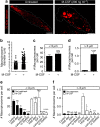
Figure 1
Figure 1. Two distinct modes of macropinocytosis exist in hMDMs.
(a) hMDMs were incubated with fluorescently labelled 70 kDa dextran (0.025 mg ml−1) for 15 min at 37 °C, in either the presence or absence of M-CSF (200 ng ml−1). Cells were then washed and imaged immediately by spinning disc confocal microscopy. (b) Macropinosomes were then counted using ImageJ software and the diameter of each macropinosome was recorded and plotted. A size cutoff (dashed line) was set to distinguish between constitutive and growth factor-induced macropinosomes. The number of macropinosomes under (c) or above (d) 3 μm in diameter per cell was plotted. hMDMs were pretreated with HOE-694 (10 μM), EIPA (10 μM), latrunculin A (2 μM) or LY294002 (10 μM) for 1 h and then incubated with fluorescently labelled 70 kDa dextran (0.025 mg ml−1) for 15 min at 37 °C, in either the presence or absence of M-CSF (200 ng ml−1). The number of macropinosomes under (e) or above (f) 3 μm in diameter is plotted. Data represent the means±s.e.m. of at least three independent experiments using cells from at least 2 separate healthy donors. Scale bar, 10 μm. *P≤0.05, **P≤0.01, ***P≤0.001, ****P≤0.0001, NS, not significantly different.
Figure 2
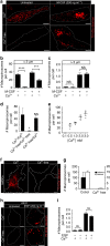
Figure 2. Removal of extracellular calcium abolishes constitutive, but not growth factor-induced macropinocytosis.
(a) hMDMs were incubated with fluorescently labelled 70 kDa dextran (0.025 mg ml−1) for 15 min at 37 °C, in the presence or absence of M-CSF (200 ng ml−1), in either calcium-containing or calcium-free medium (see Methods). The number of macropinosomes under (b) or above (c) 3 μm in diameter is plotted. (d) hMDMs were preincubated in either calcium-containing or calcium-free medium and then allowed to undergo macropinocytosis for 15 min at 37 °C. The total number of macropinosomes per cell is plotted. (e) hMDMs were incubated with fluorescently labelled 70 kDa dextran (0.025 mg ml−1) for 15 min at 37 °C in Na+-rich buffer containing the indicated concentrations of CaCl2. The total number of macropinosomes per cell is plotted. (f) hMDDCs were incubated with fluorescently labelled 70 kDa dextran (0.025 mg ml−1) for 15 min at 37 °C, in either calcium-containing or calcium-free medium. (g) hMDDCs and murine BMDMs were incubated with fluorescently labelled 70 kDa dextran (0.025 mg ml−1) for 15 min at 37 °C, in either calcium-containing or calcium-free medium. The total number of macropinosomes per cell is plotted. (h) A431 cells were incubated with fluorescently labelled 70 kDa dextran (0.025 mg ml−1) for 15 min at 37 °C, in the presence or absence of EGF (200 ng ml−1), in either calcium-containing or calcium-free medium. (i) The total number of macropinosomes per cell is plotted for each condition, as indicated. Data represent the means±s.e.m. of at least three independent experiments using cells from at least two separate healthy donors. Scale bar, 10 μm. *P≤0.05, ***P≤0.001, ****P≤0.0001, NS, not significantly different.
Figure 3
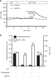
Figure 3. Role of intracellular calcium in constitutive macropinocytosis.
(a) hMDMs were loaded with 3 μM Fura2-AM and, where indicated, with 10 μM BAPTA-AM (see Methods), and imaged for 3 min in calcium-containing medium, acquiring an image every minute. The medium was then replaced with calcium-free medium and the cells imaged for an additional 4 min. Thapsigargin (100 nM) was then added and images were acquired for an additional 5 min. Data are representative of at least 30 independent determinations±s.e.m. (b) hMDMs were preloaded with Fura2-AM and treated with or without thapsigargin (100 nM), BAPTA-AM (10 μM), and extracellular calcium as indicated. The total number of macropinosomes per cell and the corresponding [Ca2+]I were then plotted. Data represent the means±s.e.m. of at least three independent experiments using cells from at least two separate healthy donors. **P≤0.01, ****P≤0.0001, NS, not significantly different.
Figure 4
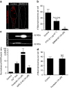
Figure 4. CaSR is necessary for constitutive macropinocytosis in hMDMs and is sufficient to induce macropinocytosis when heterologously expressed in HEK293 cells.
(a) The expression of CaSR mRNA in HEK293 cells and hMDMs was determined by RT–PCR. The expression of GAPDH mRNA was determined as a control. (b) HEK293 cells were transfected with SeP-CaSR and, 24 h after transfection, were incubated on ice with an anti-GFP monoclonal antibody for 15 min to label SeP-CaSR expressed at the cell surface. Cells were then washed, fixed and labelled with a fluorescent secondary antibody (red). Cell nuclei were labelled with DAPI (cyan). (c) HEK293 cells were transfected with SeP-CaSR and, 24 h after transfection, were labelled with fluorescent phalloidin (red). (d) Untransfected (upper panel) and HEK293 cells transfected with SeP-CaSR (lower panel) were incubated with labelled 70 kDa dextran (0.025 mg ml−1) for 15 min at 37 °C. Cells were then imaged to quantify the uptake of dextran from the fluid phase. (e,f) Untransfected and HEK293 cells transfected with SeP-CaSR were incubated with DyLight 650-labelled 70 kDa dextran (0.025 mg ml−1) for 15 min at 37 °C. Cells were then gently lifted and dextran uptake assessed by flow cytometry. Gates were drawn on GFP(+) (SeP-positive) and GFP(-) (SeP-negative) populations as shown in (e) and the relative dextran uptake in the respective populations in shown in (f). (g) hMDMs were pretreated with the indicated dose of the specific CaSR antagonist NPS2143 and then incubated with labelled 70 kDa dextran (0.025 mg ml−1) for 15 min at 37 °C. (h) The total number of macropinosomes per cell is plotted at the indicated concentration of NPS2143 for hMDMs (main panel) and hMDDCs (inset, where 10 μM was used). (i) hMDMs were pretreated with NPS2143 (10 μM), and then incubated with 70 kDa dextran (0.025 mg/ml) and M-CSF (200 ng/mL) for 15 min at 37 °C. (j) CaSR expression was knocked down in hMDMs using CaSR-specific siRNA oligonucleotides. The cells were then incubated with labelled 70 kDa dextran (0.025 mg ml−1) for 15 min at 37 °C. The expression level of CaSR mRNA, normalized to actin mRNA, was determined by quantitative PCR (qPCR); the total number of macropinosomes per cell is also plotted. Scale bar, 10 μm. *P≤0.05, **P≤0.01, NS, not significantly different.
Figure 5
Figure 5. Gαi-dependent signalling is required for constitutive macropinocytosis.
(a) hMDMs were pretreated in the absence or presence of BIM46187 (10 μM) for 1 h and then incubated with fluorescent 70 kDa dextran (0.025 mg ml−1) for 15 min at 37 °C. (b) hMDMs were pretreated with BIM46187 (10 μM) or PTX (0.1 μg ml−1) for 1 h and then incubated with fluorescent 70 kDa dextran (0.025 mg ml−1) for 15 min at 37 °C. The total number of macropinosomes per cell is plotted. (c) hMDMs were incubated with forskolin (10 μM) or NPS2143 (10 μM) in either calcium-containing or calcium-free medium for 30 min, then lysed, fractionated by 12% SDS–PAGE and subjected to immunoblotting with antibodies to phosphorylated VASP (pVASP) and α-tubulin (as loading control). A representative immunoblot is shown on top and quantification of the pVASP/α-tubulin ratio from three independent experiments is shown below. (d) hMDMs were pretreated with forskolin (10 μM) for 30 min and then incubated with labelled 70 kDa dextran (0.025 mg ml−1) for 15 min at 37 °C. The total number of macropinosomes per cell was then plotted. Scale bar, 10 μm. **P≤0.01, NS, not significantly different.
Figure 6
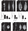
Figure 6. CaSR signals through a Gα-, PtdIns3Kγ- and PLC-dependent pathway.
(a) hMDMs were transfected with the PtdIns(3,4,5)P3 probe (PH)Akt-GFP and imaged live in either calcium-containing or calcium-free medium. Where indicated, the cells were treated with NPS2143 (10 μM) for 1 h before imaging. (b) hMDMs were transfected with (PH)Akt-GFP and imaged live. Where indicated, the cells were treated with BIM46187 (10 μM) for 1 h before imaging. (c) hMDMs were pretreated with the PtdIns3Kγ inhibitor AS605240 at the indicated concentrations for 1 h and then incubated with fluorescent 70 kDa dextran (0.025 mg ml−1) for 15 min at 37 °C. The total number of macropinosomes per cell is plotted. (d) mBMDMs from wild-type and PtdIns3Kγ knockout mice were incubated with fluorescently labelled 70 kDa dextran (0.025 mg ml−1) for 15 min at 37 °C. The total number of macropinosomes per cell is plotted. (e) hMDMs were pretreated with the PLC inhibitor U73122 (1 μM) for 1 h and then incubated with fluorescently labelled 70 kDa dextran (0.025 mg ml−1) for 15 min at 37 °C. The total number of macropinosomes per cell is plotted. (f) hMDMs were transfected with the PtdOH probe GFP-2PABD and were treated with U73122 (1 μM) for 1 h before imaging. (g) hMDMs were transfected with GFP-2PABD and imaged live in either calcium-containing or calcium-free medium. Where indicated, the cells were treated with NPS2143 (10 μM) for 1 h before imaging. Scale bar, 10 μm. **P≤0.01, ***P≤0.001, NS, not significantly different.
Figure 7
Figure 7. Inhibition of CaSR results in cytoskeletal changes in MDMs.
(a) hMDMs were treated with NPS2143 (10 μM) and then fixed and labelled with fluorescently labelled phalloidin (red) and DAPI (cyan). (b) hMDMs were transfected with the active Rac1/Cdc42 biosensor PBD(Pak)-YFP and imaged live in either calcium-containing or calcium-free medium. Where indicated, the cells were treated with NPS2143 (10 μM) before imaging. Scale bar, 10 μm. (c) hMDMs were pretreated with either the C. difficile toxin B (50 ng ml−1) or Tat-C3 (10 μg ml−1; see Methods) and then incubated with labelled 70 kDa dextran (0.025 mg ml−1) for 15 min at 37 °C. The total number of macropinosomes per cell was then plotted. ****P≤0.0001, NS, not significantly different.
Figure 8
Figure 8. CaSR-dependent macropinocytosis delivers NOD2 ligands to the cytosol.
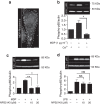
(a) hMDMs were incubated with the fluorescently labelled NOD2 ligand muramyl dipeptide (MDP-rhodamine; 1 μg ml−1) for 15 min at 37 °C. Cells were then washed and imaged immediately by spinning disc confocal microscopy. Scale bar, 10 μm. (b) hMDMs were incubated in calcium-containing or calcium-free medium in the presence or absence of MDP (1 μg ml−1) for 15 min and then lysed, separated by 12% SDS–PAGE and subjected to immunoblotting. Image shows representative immunoblot for phosphorylated p65 (p65) and actin (used as loading control); quantification of the normalized phospho-p65/actin ratio from three independent experiments is shown below. (c) hMDMs were incubated with NPS2143 at the indicated concentrations in either the presence or absence of MDP (1 μg ml−1) for 30 min and then lysed, separated by 12% SDS–PAGE and subjected to immunoblotting. Image shows representative immunoblot for phosphorylated p65 (p65) and α-tubulin (tub.; used as loading control); quantification of the phospho-p65/α-tubulin ratio from three independent experiments is shown below. (d) hMDMs were incubated with NPS2143 at the indicated concentrations in either the presence or absence of LPS (0.5 μg ml−1) for 30 min and then lysed, separated by 12% SDS–PAGE and subjected to immunoblotting. Image shows representative immunoblot for phosphorylated p65 (p65) and α-tubulin; quantification of the phospho-p65/α-tubulin ratio from three independent experiments is shown below. *P≤0.05, **P≤0.01, NS, not significantly different.

Similar articles

Cited by

References

    1. Commisso C. et al.. Macropinocytosis of protein is an amino acid supply route in Ras-transformed cells. Nature 497, 633–637 (2013). - PMC - PubMed
    1. Gu Z., Noss E. H., Hsu V. W. & Brenner M. B. Integrins traffic rapidly via circular dorsal ruffles and macropinocytosis during stimulated cell migration. J. Cell Biol. 193, 61–70 (2011). - PMC - PubMed
    1. Holt M., Cooke A., Wu M. M. & Lagnado L. Bulk membrane retrieval in the synaptic terminal of retinal bipolar cells. J. Neurosci. 23, 1329–1339 (2003). - PMC - PubMed
    1. Kabayama H. et al.. Ca2+ induces macropinocytosis via F-actin depolymerization during growth cone collapse. Mol. Cell Neurosci. 40, 27–38 (2009). - PubMed
    1. Welliver T. P. & Swanson J. A. A growth factor signaling cascade confined to circular ruffles in macrophages. Biol. Open 1, 754–760 (2012). - PMC - PubMed

Publication types

MeSH terms

LinkOut - more resources