

doi: 10.1126/scisignal.aaf6029.
IP3 receptors: Take four IP3 to open
Affiliations
- PMID: 27048564
- PMCID: PMC5019202
- DOI: 10.1126/scisignal.aaf6029
Item in Clipboard
IP3 receptors: Take four IP3 to open
Sci Signal.
.
Abstract
Inositol 1,4,5-trisphosphate receptors (IP3Rs) and ryanodine receptors are the channels responsible for Ca(2+)release from the endoplasmic and sarcoplasmic reticulum. Research inScience Signalingby Alzayadyet al show that all four IP3-binding sites within the tetrameric IP3R must bind IP3before the channel can open, which has important consequences for the distribution of both IP3and IP3R activity within cells.
Copyright © 2016, American Association for the Advancement of Science.
Conflict of interest statement
The authors declare that they have no competing interests.
Figures

(A) The essential 4- and 5-phosphate groups of IP3 bind to opposite sides (β- and α-domains) of the clam-like IP3-binding core (IBC) causing the clam to close and re-position the suppressor domain (SD), which moves with the α-domain of the IBC (Protein Data Bank, PDB, 3U4J) (5). Locations of key domains within the primary sequence are shown below. (B) View from the cytosol showing the four N-terminal regions (SD and IBC) and the C-terminal domains (CTD) from each subunit (the central splayed α-helices). The coloured subunits (orange and green) show how the CTD from one subunit contacts the N-terminal region (the β-IBC) of another. The N- (blue) and C-termini (red) of each subunit, through which Alzayady et al. linked subunits to form concatamers (16), are highlighted; the termini of one subunit are enclosed in the yellow box. Within each IBC, the position where IP3 would bind is shown by a yellow circle (PDB, 3JAV) (11). (C) View across the ER membrane, with 2 subunits highlighted in shades of orange and green. The transmembrane region with its 24 α-helices, six from each subunit, are shown (PDB, 3JAV) (11). (D) One of the likely inter-subunit interactions through which IP3R binding to the IBC is communicated to the pore. The β-domain of the IBC of one subunit contacts the C-terminal domain (CTD) of another. The CTD extends as an almost unbroken α-helix through the centre of the protein to helix S6, which lines the pore. For the pore to open and allow Ca2+ to pass, S6 must move to disrupt the hydrophobic constriction that occludes the closed pore (red arrow).
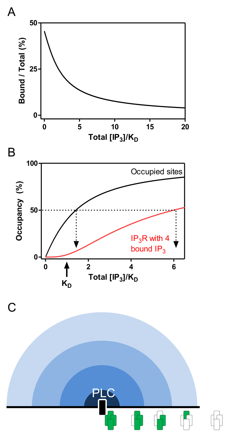
(A) Within a typical cell, the concentration of IP3-binding sites and their affinity (KD) for IP3 are similar (~100nM). An inescapable consequence is that within the range of IP3 concentrations that achieve graded occupancy of the binding sites, a substantial fraction of the IP3 is bound, thereby depleting the cytosol of free IP3 (18). (B) Buffering of cytosolic IP3 by IP3R shifts the relationship between IP3 concentration and IP3 binding to higher concentrations of IP3: a total concentration that exceeds the KD is now required to occupy 50% of the binding sites. Because 4 sites must be occupied to activate an IP3R, the IP3 concentrations needed to activate 50% of IP3Rs are much higher than those needed to occupy 50% of the binding sites. In this simple scheme, a concentration of IP3 that occupied 50% of all binding sites, would allow only 6% of all IP3Rs, about 450 IP3Rs in a typical cell, to bind the 4 IP3 needed for activity. (C) IP3 produced by PLC is captured by IP3Rs as it diffuses into the cytosol creating a radial concentration gradient. Within the gradient, IP3Rs very close to PLC may acquire the 4 IP3 molecules needed for activity (green), but beyond that there is an extensive penumbra of IP3Rs that bind IP3 to fewer of their subunits.
Similar articles
-
Defining the stoichiometry of inositol 1,4,5-trisphosphate binding required to initiate Ca2+ release.Sci Signal. 2016 Apr 5;9(422):ra35. doi: 10.1126/scisignal.aad6281. Sci Signal. 2016. PMID: 27048566 Free PMC article.
-
IP3-mediated gating mechanism of the IP3 receptor revealed by mutagenesis and X-ray crystallography.Proc Natl Acad Sci U S A. 2017 May 2;114(18):4661-4666. doi: 10.1073/pnas.1701420114. Epub 2017 Apr 17. Proc Natl Acad Sci U S A. 2017. PMID: 28416699 Free PMC article.
-
IP3 receptors - lessons from analyses ex cellula.J Cell Sci. 2018 Dec 14;132(4):jcs222463. doi: 10.1242/jcs.222463. J Cell Sci. 2018. PMID: 30552138 Review.
-
Structural and functional conservation of the activating Ca2+ binding site in inositol 1,4.5-trisphosphate and ryanodine receptors.Cell Calcium. 2022 Dec;108:102671. doi: 10.1016/j.ceca.2022.102671. Epub 2022 Nov 5. Cell Calcium. 2022. PMID: 36370621
-
Structure and Function of IP3 Receptors.Cold Spring Harb Perspect Biol. 2019 Apr 1;11(4):a035063. doi: 10.1101/cshperspect.a035063. Cold Spring Harb Perspect Biol. 2019. PMID: 30745293 Free PMC article. Review.
Cited by
-
Advances in Intracellular Calcium Signaling Reveal Untapped Targets for Cancer Therapy.Biomedicines. 2021 Aug 24;9(9):1077. doi: 10.3390/biomedicines9091077. Biomedicines. 2021. PMID: 34572262 Free PMC article. Review.
-
Dynamic Ca2+ imaging with a simplified lattice light-sheet microscope: A sideways view of subcellular Ca2+ puffs.Cell Calcium. 2018 May;71:34-44. doi: 10.1016/j.ceca.2017.11.005. Epub 2017 Dec 1. Cell Calcium. 2018. PMID: 29604962 Free PMC article.
-
Inside the Insulin Secretory Granule.Metabolites. 2021 Aug 5;11(8):515. doi: 10.3390/metabo11080515. Metabolites. 2021. PMID: 34436456 Free PMC article. Review.
-
TNFα induces Ca2+ influx to accelerate extrinsic apoptosis in hepatocellular carcinoma cells.J Exp Clin Cancer Res. 2018 Mar 5;37(1):43. doi: 10.1186/s13046-018-0714-6. J Exp Clin Cancer Res. 2018. PMID: 29506556 Free PMC article.
-
Computational investigation of IP3 diffusion.Sci Rep. 2023 Feb 20;13(1):2922. doi: 10.1038/s41598-023-29876-3. Sci Rep. 2023. PMID: 36808161 Free PMC article.
References
-
- Bosanac I, Alattia J-R, Mal TK, Chan J, Talarico S, Tong FK, Tong KI, Yoshikawa F, Furuichi T, Iwai M, Michikawa T, et al. Structure of the inositol 1,4,5-trisphosphate receptor binding core in complex with its ligand. Nature. 2002;420:696–700. - PubMed
Publication types
MeSH terms
Substances
Grants and funding
LinkOut - more resources
Full Text Sources
Other Literature Sources
Miscellaneous
