. 2016 May;157(5):1956-66.
doi: 10.1210/en.2015-1846. Epub 2016 Mar 25.

CCDC141 Mutation Identified in Anosmic Hypogonadotropic Hypogonadism (Kallmann Syndrome) Alters GnRH Neuronal Migration

Affiliations

CCDC141 Mutation Identified in Anosmic Hypogonadotropic Hypogonadism (Kallmann Syndrome) Alters GnRH Neuronal Migration

B Ian Hutchins et al. Endocrinology. 2016 May.

Abstract

The first mutation in a gene associated with a neuronal migration disorder was identified in patients with Kallmann Syndrome, characterized by hypogonadotropic hypogonadism and anosmia. This pathophysiological association results from a defect in the development of the GnRH and the olfactory system. A recent genetic screening of Kallmann Syndrome patients revealed a novel mutation in CCDC141. Little is known about CCDC141, which encodes a coiled-coil domain containing protein. Here, we show that Ccdc141 is expressed in GnRH neurons and olfactory fibers and that knockdown of Ccdc141 reduces GnRH neuronal migration. Our findings in human patients and mouse models predict that CCDC141 takes part in embryonic migration of GnRH neurons enabling them to form a hypothalamic neuronal network to initiate pulsatile GnRH secretion and reproductive function.


Figures

Figure 1.
Figure 1.
Structural alterations in the affected family vs healthy control. A and B, Magnetic resonance imaging of the brain in coronal sections. Subjects were also described in Ref. . A, Healthy adult individual, arrow indicating normal olfactory bulb. B, Aplastic olfactory bulbs in the proband. C, Genotype sequencing of CCDC141 gene shows CCDC141 mutation (p.R724X) (black box indicates mutation site). The top line shows the homozygous wild-type genotype (healthy control individual), the middle line shows the homozygous mutant genotype (proband), and the bottom line shows the heterozygous genotype (mother).
Figure 2.
Figure 2.
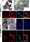
Ccdc141 is expressed in migrating GnRH neurons in the developing mouse. A1, Schematic of an E11.5 parasagittal mouse head; *, developing VNO; OE, olfactory epithelium, arrow points to nasal forebrain junction; OB, olfactory bulb. Peripherin-positive olfactory axons (in brown) are shown extending to the olfactory bulb. GnRH neurons (shown in black) migrate on these axon bundles until they cross the nasal forebrain junction (black arrow) and enter the brain. A2, E11.5 embryo stained for peripherin (brown) and GnRH (black), with high-magnification image of boxed area on right. Arrowhead indicates GnRH neurons streaming out from the VNO (*). B1–B4, Fluorescent staining for Ccdc141 (red), GnRH (green), and nuclear staining with DAPI (blue) at E11.5. B1, Ccdc141 staining is expressed in GnRH neurons and in mesenchymal cells. Robust staining is evident in the axonal tracts leaving the VNO where GnRH neurons are located (arrows). B2–B4, Higher magnification showing GnRH/Ccdc141 neurons in the VNO. C1, Schematic of a coronal section of an E14.5 embryo nose; VNO (*), respiratory epithelium (RE), OE, nasal forebrain junction (arrow), and OB. C2, Magnified coronal sections from an E14.5 embryo stained for GnRH (black) and peripherin (brown), showing GnRH neurons at the nasal/forebrain junction (upper box in schematic) and leaving the VNO (*, bottom box in schematic). D, Fluorescent staining of Ccdc141 red at E14.5 showing staining in VNO (*) and olfactory axons (OX). E, Serial section stained with antibody + blocking peptide (10 μg/mL). After primary antibody/primary antibody + blocking peptide, D and E were stained together. No specific staining was detected after blocking peptide was added to the primary antibody. F1–F4, Fluorescent staining for Ccdc141 (red), GnRH (green), and nuclear staining with DAPI (blue) at E14.5. By E14.5, Ccdc141 levels appeared to decrease in mesenchyme cells (F1) yet remained clearly present in sensory axons and GnRH neurons (F2–F4). Scale bars: 100 μm (A), 50 μm (B1 and F1), and 10 μm (B2–B4 and F2–F4).
Figure 3.
Figure 3.
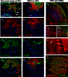
Olfactory neurons and fibers express Ccdc141. A1–A4, Fluorescent staining for Ccdc141 (red) neural markers TUJ1/Hu (green), and nuclear staining with DAPI (blue) in an E14.5 section, highlights expression of Ccdc141 in olfactory sensory axons (arrows) in nasal region, leaving the olfactory epithelium (OE), at the nasal forebrain junction (NFJ) and entering the olfactory bulb (OB); higher magnification of boxed region is shown in A2–A4. B1–B4, In contrast to olfactory axons, Ccdc141 expression (red) was not detected in BLBP-Cre/rosaYFP-positive OECs (green). Ccdc141 (red) was also detected in areas of the developing central (C) and peripheral nervous systems (D and E) and overlapped with early neuronal markers (TUJ1/Hu, green). C1–C3, At E14.5, Ccdc141 was expressed in multiple layers of the developing cortex and in the outer layer (higher magnification) (C2–C3). D and E, At E12.5–E14.5, Ccdc141 expression was detected in multiple sensory ganglia, such as the dorsal root ganglia shown here (cell soma, asterisk; arrow, fibers). Scale bars, 50 μm (A1, B1, C1, and F) and 100 μm (D).
Figure 4.
Figure 4.
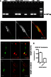
Ccdc141 expression and knockdown in GnRH neurons. A, PCR product of approximately 207 bp indicated presence of Ccdc141 in cDNA made from transcript in 6- and 3-div whole nasal explants (Exp, lanes 1 and 2), and in 4 of 7 cDNA made from individual GnRH cells isolated at 4.5 div (single cells, lanes 3–9). Control brain (Br) cDNAs from embryonic (lane 10) and adult brain (lane 11), and negative control water (lane 12); M, DNA marker. B, Confocal fluorescence immunostaining showing GnRH expression (B1), Ccdc141 expression (B2), and merged image confirming coexpression (B3). C–E, GnRH (C1 and D1) and Ccdc141 (C2 and D2) coexpression (merge, C3 and D3) in explants treated with control (C) and Ccdc141-targeting siRNA (D). E, Measurements of background-subtracted fluorescence indicated levels of cytoplasmic Ccdc141 in GnRH neurons decreased after treatment with Ccdc141-targeting siRNA compared with control siRNA (P < .001, n ≥ 85 GnRH neurons, 2 explants/group). Mann-Whitney U test used for this comparison. Scale bars, 10 μm.
Figure 5.
Figure 5.
CCDC141 knockdown reduces GnRH neuronal migration. A and B, Frequency distributions of the linear (A) and total (B) migration rates fit with lognormal curves showing a shift in the distribution in the CCDC141 siRNA-treated cultures vs the controls (P < .05, Mann-Whitney U test, n = 73 control, and n = 40 CCDC141 siRNA-treated cells; error bars, SEM). C and D, Examples of measurement of linear migration (C) and total migration (D) of GnRH neurons over a 40-minute imaging session. C, Linear migration rates represent the displacement of the neuron (black arrow) over the imaging period. D, Total migration rates represent the average of the frame-to-frame movement rates (represented by the simplified time series and trajectory trace). Scale bars, 10 μm.
Figure 6.
Figure 6.
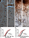
CCDC141 knockdown reduces GnRH neuronal migration but not olfactory outgrowth. A and B, Immunopositive GnRH neurons in control (A) or Ccdc141-siRNA-treated explants (B). Distance GnRH neurons (arrowheads, examples) migrated from center of nasal pit (asterisk) outward was measured 3 days after electroporation. Scale bars, 100 μm (A and B). C and F, Cumulative frequency of 2 independent siRNA experiments showing distributions of distances GnRH neurons migrated. In both experiments, a shift to the left after Ccdc141 knockdown was detected (P < .001, two-way ANOVA, n = 4 explants/condition). D and E, Olfactory axon growth was unaffected by electroporation with Ccdc141 siRNA. Images of explants electroporated with control (D) or Ccdc141 siRNA (E) and stained for peripherin (marker of olfactory axons, brown fibers) and GnRH (blue cell bodies). Farthest GnRH neurons visible in the field of view are marked with arrows/arrowheads. Scale bars, 100 μm (D and E). Higher magnification of GnRH neurons (blue) closely apposed to olfactory axons (brown) from explants in A and B, respectively (arrows), are shown in boxes. Scale bars, 10 μm (insets in D and E).

Similar articles

Cited by

  • Genome-Wide Association Study of Accessory Atrioventricular Pathways.
    Aegisdottir HM, Andreasen L, Thorolfsdottir RB, Sveinbjornsson G, Jonsdottir AB, Stefansdottir L, Thorleifsson G, Sigurdsson A, Halldorsson GH, Barc J, Simonet F, Tragante V, Oddsson A, Ferkingstad E, Svendsen JH, Ghouse J, Ahlberg G, Paludan-Müller C, Hadji-Turdeghal K, Bustamante M, Ulfarsson MO, Helgadottir A, Gretarsdottir S, Saevarsdottir S, Jonsdottir I, Erikstrup C, Ullum H, Sørensen E, Brunak S, Jøns C, Zheng C, Bezzina CR, Knowlton KU, Nadauld LD, Sulem P, Ostrowski SR, Pedersen OB, Arnar DO, Gudbjartsson DF, Olesen MS, Bundgaard H, Holm H, Stefansson K; DBDS consortium. Aegisdottir HM, et al. JAMA Cardiol. 2024 Nov 1;9(11):1053-1058. doi: 10.1001/jamacardio.2024.2684. JAMA Cardiol. 2024. PMID: 39230897
  • Increased Burden of Rare Sequence Variants in GnRH-Associated Genes in Women With Hypothalamic Amenorrhea.
    Delaney A, Burkholder AB, Lavender CA, Plummer L, Mericq V, Merino PM, Quinton R, Lewis KL, Meader BN, Albano A, Shaw ND, Welt CK, Martin KA, Seminara SB, Biesecker LG, Bailey-Wilson JE, Hall JE. Delaney A, et al. J Clin Endocrinol Metab. 2021 Mar 8;106(3):e1441-e1452. doi: 10.1210/clinem/dgaa609. J Clin Endocrinol Metab. 2021. PMID: 32870266 Free PMC article.
  • New findings in oligogenic inheritance of congenital hypogonadotropic hypogonadism.
    Gach A, Pinkier I, Wysocka U, Sałacińska K, Salachna D, Szarras-Czapnik M, Pietrzyk A, Sakowicz A, Nykel A, Rutkowska L, Rybak-Krzyszkowska M, Socha M, Jamsheer A, Jakubowski L. Gach A, et al. Arch Med Sci. 2020 Sep 18;18(2):353-364. doi: 10.5114/aoms.2020.98909. eCollection 2022. Arch Med Sci. 2020. PMID: 35316923 Free PMC article.
  • Exome sequencing of families from Ghana reveals known and candidate hearing impairment genes.
    Wonkam A, Adadey SM, Schrauwen I, Aboagye ET, Wonkam-Tingang E, Esoh K, Popel K, Manyisa N, Jonas M, deKock C, Nembaware V, Cornejo Sanchez DM, Bharadwaj T, Nasir A, Everard JL, Kadlubowska MK, Nouel-Saied LM, Acharya A, Quaye O, Amedofu GK, Awandare GA, Leal SM. Wonkam A, et al. Commun Biol. 2022 Apr 19;5(1):369. doi: 10.1038/s42003-022-03326-8. Commun Biol. 2022. PMID: 35440622 Free PMC article.
  • Whole exome sequencing and trio analysis to broaden the variant spectrum of genes in idiopathic hypogonadotropic hypogonadism.
    Zhang J, Tang SY, Zhu XB, Li P, Lu JQ, Cong JS, Wang LB, Zhang F, Li Z. Zhang J, et al. Asian J Androl. 2021 May-Jun;23(3):288-293. doi: 10.4103/aja.aja_65_20. Asian J Androl. 2021. PMID: 33208564 Free PMC article.

References

    1. Schwanzel-Fukuda M, Pfaff DW. Origin of luteinizing hormone-releasing hormone neurons. Nature. 1989;338:161–164. - PubMed
    1. Wray S, Grant P, Gainer H. Evidence that cells expressing luteinizing hormone-releasing hormone mRNA in the mouse are derived from progenitor cells in the olfactory placode. Proc Natl Acad Sci USA. 1989;86:8132–8136. - PMC - PubMed
    1. Forni PE, Wray S. GnRH, anosmia and hypogonadotropic hypogonadism–where are we? Front Neuroendocrinol. 2015;36:165–177. - PMC - PubMed
    1. Franco B, Guioli S, Pragliola A, et al. A gene deleted in Kallmann's syndrome shares homology with neural cell adhesion and axonal path-finding molecules. Nature. 1991;353:529–536. - PubMed
    1. Ballabio A, Camerino G. The gene for X-linked Kallmann syndrome: a human neuronal migration defect. Curr Opin Genet Dev. 1992;2:417–421. - PubMed

Publication types

Substances