. 2016 Apr 28;59(8):4019-25.
doi: 10.1021/acs.jmedchem.5b01536. Epub 2016 Apr 11.

Affinity-Guided Design of Caveolin-1 Ligands for Deoligomerization

Affiliations

Affinity-Guided Design of Caveolin-1 Ligands for Deoligomerization

Amanda J H Gilliam et al. J Med Chem. .

Abstract

Caveolin-1 is a target for academic and pharmaceutical research due to its many cellular roles and associated diseases. We report peptide WL47 (1), a small, high-affinity, selective disrupter of caveolin-1 oligomers. Developed and optimized through screening and analysis of synthetic peptide libraries, ligand 1 has 7500-fold improved affinity compared to its T20 parent ligand and an 80% decrease in sequence length. Ligand 1 will permit targeted study of caveolin-1 function.


Figures

Figure 1
Figure 1
Evolution of CAV ligands. Sequence alignment showing the progression of peptide ligands from the native gp41 segment sequence (2) to T20 (3), to ligands 4, 5, and 6, and ultimately to ligand 1, which binds CAV(1-104) with 7500-fold higher affinity despite an 80% decrease in length compared to 3. Residues mutated from 3 are in red. Numbers above 2 indicate the residue numbers from the original protein.
Figure 2
Figure 2
Initial identification of CAV interacting region. The sequence of ligand 4 was truncated to generate 22 unique peptide sequences of 15 amino acids each. Only two truncation sequences, consisting of residues 21–35 and residues 22–36, respectively, bound CAV(1-104) more than 2-fold above the level of the negative control. The negative control was a scrambled sequence of the randomly selected 11th truncation. An additional, blank negative control had a linker region but lacked a peptide. The sequence corresponding to residues 22–36 (indicated by asterisk) was chosen as the template for subsequent libraries. and designated ligand 5.
Figure 3
Figure 3
Identification of key residues, and removal of detrimental amino acids. a) In the library of similar and dissimilar substitutions of amino acids in ligand 5, at Arg7, ligand binding was retained for Lys and His substituents but was abolished by neutral Gln or Ala substituents. At Lys13, ligand binding was eliminated for all substituents except Arg. We conclude that both these residues contribute to binding primarily via positive charge. b) For Glu1, ligand binding was retained for negatively charged Asp and neutral Gln, and was slightly elevated for Ala. For Glu4 and Glu6, ligand binding increased moderately for neutral Gln and Ala. In short, positively charged residues are optimal, followed by neutral residues. Ligand binding was reduced by substitution with negatively charged Asp, and all Lys substitutions at these three sites increased ligand binding. These trends suggest that these Glu are not optimal for binding. c) Truncation of the six N-terminal residues, which included all three Glu, without removing any of the positively charged residues, yielded a peptide (indicated by asterisk) designated ligand 6 that became the template for subsequent library design. All libraries include a scrambled ligand 5 sequence as a negative control.
Figure 4
Figure 4
Unnecessary residues removed, and sequences shuffled. Direct removal of Gly9 and Phe15 from the ligand 6 sequence to create ligand 6(ΔGF) decreased binding. When the sequence lacking Gly and Phe is shuffled, however, many library members retain or increase binding ability. The 7-mer shuffled sequence (indicated by asterisk) with the highest binding was used for subsequent studies.
Figure 5
Figure 5
Demonstration of selectivity. CAV(1-104) and several control proteins were labeled with rhodamine and screened against duplicate SPOT sheets containing ligands 5, 6, and 1 to demonstrate selectivity of the ligand. Bovine serum albumin (BSA), casein, and hen egg white lysozyme (HEWL) all bound ligand 1 with reduced apparent affinity relative to CAV(1-104) as measured by fluorescence. Blank SPOTs containing only the double β-Ala linker were included as negative controls, and these baseline signals were subtracted from the corresponding signal for ligands 5, 6 and 1 SPOTs on each sheet.
Figure 6
Figure 6
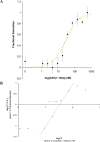
Binding affinity and cooperativity. a) Fluorescence anisotropy was measured for Mantyl-1 dimer incubated with the indicated concentrations of CAV(1-104). Using equation 1, a best fit binding curve was fit to the raw experimental data using a weighted method of least squares, and assuming each ligand 1 dimer functions as a single ligand. The KD for this binding interaction was calculated to 23 nM. b) A Hill plot yields a Hill coefficient (nH) of 1.97. This is the slope of the linear region of the plot as it crosses the x-axis (R2=0.992). Based on the two-site binding model, this coefficient indicates virtually complete positive cooperativity. Thus, upon binding one molecule of 1 (one half of the dimer), a second molecule (the other half of the dimer) binds essentially instantaneously the second site. This observation supports our decision to treat the 1 dimer as a single ligand for the purpose of KD calculations.
Figure 7
Figure 7
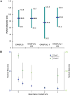
Disruption of oligomers. a) CAV(FLV) spontaneously forms oligomers which can be measured by dynamic light scattering (DLS). In this experiment, these oligomers had an apparent average diameter near 90 nm with polydispersity (indicated in teal) corresponding to approximately a 55 nm range. Incubating these oligomers with ligand 1 resulted in reduced average diameter and increased polydispersity, which are the expected outcomes of deoligomerization. With the disulfide dimerization of the ligand disrupted by reducing conditions, no deoligomerization effect was observed. b) In a separate DLS experiment, CAV(FLV) oligomeric peaks at 32 nm (blue, left axis) and 240 nm (green, right axis) each showed a dose-dependent reduction in diameter when incubated with varied concentrations of ligand 1. A maximum size reduction of 30% and 40% was observed for the respective peaks.

Similar articles

Cited by

References

    1. Cohen AW, Hnasko R, Schubert W, Lisanti MP. Role of Caveolae and Caveolins in Health and Disease. Physiol Rev. 2004;84:1341–1379. - PubMed
    1. Shvets E, Ludwig A, Nichols BJ. News from the Caves: Update on the Structure and Function of Caveolae. Curr Opin Cell Biol. 2014;29:99–106. - PubMed
    1. Strålfors P. Caveolins and Caveolae, Roles in Insulin Signalling and Diabetes. Adv Exp Med Biol. 2012;729:111–126. - PubMed
    1. Zou H, Stoppani E, Volonte D, Galbiati F. Caveolin-1, Cellular Senescence and Age-Related Diseases. Mech Ageing Dev. 2011;132:533–542. - PMC - PubMed
    1. Machado FS, Rodriguez NE, Adesse D, Garzoni LR, Esper L, Lisanti MP, Burk RD, Albanese C, Van Doorslaer K, Weiss LM, Nagajyothi F, Nosanchuk JD, Wilson ME, Tanowitz HB. Recent Developments in the Interactions between Caveolin and Pathogens. Adv Exp Med Biol. 2012;729:65–82. - PMC - PubMed

Publication types