

Role of interleukin-1 and its antagonism of hepatic stellate cell proliferation and liver fibrosis in the Abcb4(-/-) mouse model
- PMID: 27004088
- PMCID: PMC4794530
- DOI: 10.4254/wjh.v8.i8.401
Role of interleukin-1 and its antagonism of hepatic stellate cell proliferation and liver fibrosis in the Abcb4(-/-) mouse model
Abstract
Aim: To study the interleukin-1 (IL-1) pathway as a therapeutic target for liver fibrosis in vitro and in vivo using the ATP-binding cassette transporter b4(-/-) (Abcb4(-/-)) mouse model.
Methods: Female and male Abcb4(-/-) mice from 6 to 13 mo of age were analysed for the degree of cholestasis (liver serum tests), extent of liver fibrosis (hydroxyproline content and Sirius red staining) and tissue-specific activation of signalling pathways such as the IL-1 pathway [quantitative polymerase chain reaction (qPCR)]. For in vivo experiments, murine hepatic stellate cells (HSCs) were isolated via pronase-collagenase perfusion followed by density gradient centrifugation using female mice. Murine HSCs were stimulated with up to 1 ng/mL IL-1β with or without 2.5 μg/mL Anakinra, an IL-1 receptor antagonist, respectively. The proliferation of murine HSCs was assessed via the BrdU assay. The toxicity of Anakinra was evaluated via the fluorescein diacetate hydrolysis (FDH) assay. In vivo 8-wk-old Abcb4(-/-) mice with an already fully established hepatic phenotype were treated with Anakinra (1 mg/kg body-weight daily intraperitoneally) or vehicle and liver injury and liver fibrosis were evaluated via serum tests, qPCR, hydroxyproline content and Sirius red staining.
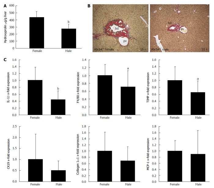
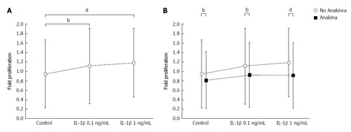
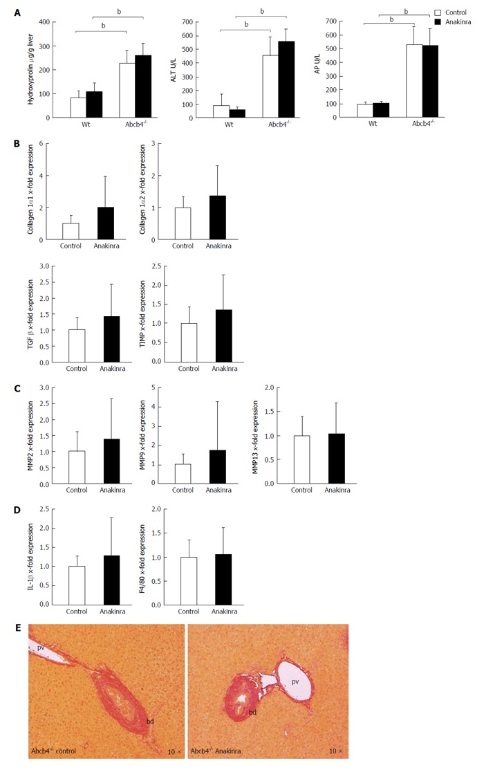
Results: Liver fibrosis was less pronounced in males than in female Abcb4(-/-) animals as defined by a lower hydroxyproline content (274 ± 64 μg/g vs 436 ± 80 μg/g liver, respectively; n = 13-15; P < 0.001; Mann-Whitney U-test) and lower mRNA expression of the profibrogenic tissue inhibitor of metalloproteinase-1 (TIMP) (1 ± 0.41 vs 0.66 ± 0.33 fold, respectively; n = 13-15; P < 0.05; Mann-Whitney U-test). Reduced liver fibrosis was associated with significantly lower levels of F4/80 mRNA expression (1 ± 0.28 vs 0.71 ± 0.41 fold, respectively; n = 12-15; P < 0.05; Mann-Whitney U-test) and significantly lower IL-1β mRNA expression levels (1 ± 0.38 vs 0.44 ± 0.26 fold, respectively; n = 13-15; P < 0.001; Mann-Whitney U-test). No gender differences in the serum liver parameters [bilirubin; alanine aminotransferase (ALT); aspartate aminotransferase and alkaline phosphatase (AP)] were found. In vitro, the administration of IL-1β resulted in a significant increase in HSC proliferation [0.94 ± 0.72 arbitrary units (A.U.) in untreated controls, 1.12 ± 0.80 A.U. at an IL-1β concentration of 0.1 ng/mL and 1.18 ± 0.73 A.U. at an IL-1β concentration of 1 ng/mL in samples from n = 6 donor animals; P < 0.001; analyses of variance (ANOVA)]. Proliferation was reduced significantly by the addition of 2.5 μg/mL Anakinra (0.81 ± 0.60 A.U. in untreated controls, 0.92 ± 0.68 A.U. at an IL-1β concentration of 0.1 ng/mL, and 0.91 ± 0.69 A.U. at an IL-1β concentration of 1 ng/mL; in samples from n = 6 donor animals; P < 0.001; ANOVA) suggesting an anti-proliferative effect of this clinically approved IL-1 receptor antagonist. The FDH assay showed this dose to be non-toxic in HSCs. In vivo, Anakinra had no effect on the hepatic hydroxyproline content, liver serum tests (ALT and AP) and pro-fibrotic (collagen 1α1, collagen 1α2, transforming growth factor-β, and TIMP-1) and anti-fibrotic [matrix metalloproteinase 2 (MMP2), MMP9 and MMP13] gene expression after 4 wk of treatment. Furthermore, the hepatic IL-1β and F4/80 mRNA expression levels were unaffected by Anakinra treatment.
Conclusion: IL-1β expression is associated with the degree of liver fibrosis in Abcb4(-/-) mice and promotes HSC proliferation. IL-1 antagonism shows antifibrotic effects in vitro but not in Abcb4(-/-) mice.
Keywords: Cholestasis; Interleukin-1; Liver fibrosis; Primary sclerosing cholangitis; The ATP-binding cassette transporter b4.
Figures
Similar articles
-
1,25-(OH)₂-vitamin D₃ prevents activation of hepatic stellate cells in vitro and ameliorates inflammatory liver damage but not fibrosis in the Abcb4(-/-) model.Biochem Biophys Res Commun. 2015 Apr 3;459(2):227-233. doi: 10.1016/j.bbrc.2015.02.074. Epub 2015 Feb 21. Biochem Biophys Res Commun. 2015. PMID: 25712522
-
Liuweiwuling tablets attenuate BDL-induced hepatic fibrosis via modulation of TGF-β/Smad and NF-κB signaling pathways.J Ethnopharmacol. 2018 Jan 10;210:232-241. doi: 10.1016/j.jep.2017.08.029. Epub 2017 Aug 31. J Ethnopharmacol. 2018. PMID: 28864168
-
[Dynamic expressions of IL-22 and hepatic stellate cells senescence in mice infected with Schistosoma japonicum].Zhongguo Xue Xi Chong Bing Fang Zhi Za Zhi. 2014 Apr;26(2):169-74. Zhongguo Xue Xi Chong Bing Fang Zhi Za Zhi. 2014. PMID: 25051830 Chinese.
-
Anakinra.2020 Apr 20. LiverTox: Clinical and Research Information on Drug-Induced Liver Injury [Internet]. Bethesda (MD): National Institute of Diabetes and Digestive and Kidney Diseases; 2012–. 2020 Apr 20. LiverTox: Clinical and Research Information on Drug-Induced Liver Injury [Internet]. Bethesda (MD): National Institute of Diabetes and Digestive and Kidney Diseases; 2012–. PMID: 31643927 Free Books & Documents. Review.
-
Targeting Interleukin-17 as a Novel Treatment Option for Fibrotic Diseases.J Clin Med. 2023 Dec 27;13(1):164. doi: 10.3390/jcm13010164. J Clin Med. 2023. PMID: 38202170 Free PMC article. Review.
Cited by
-
Modulation of Liver Inflammation and Fibrosis by Interleukin-37.Front Immunol. 2021 Mar 4;12:603649. doi: 10.3389/fimmu.2021.603649. eCollection 2021. Front Immunol. 2021. PMID: 33746950 Free PMC article.
-
New Concepts on Reversibility and Targeting of Liver Fibrosis; A Review Article.Middle East J Dig Dis. 2018 Jul;10(3):133-148. doi: 10.15171/mejdd.2018.103. Epub 2018 Jun 14. Middle East J Dig Dis. 2018. PMID: 30186577 Free PMC article.
-
Neupogen and mesenchymal stem cells are the novel therapeutic agents in regeneration of induced endometrial fibrosis in experimental rats.Biosci Rep. 2017 Oct 11;37(5):BSR20170794. doi: 10.1042/BSR20170794. Print 2017 Oct 31. Biosci Rep. 2017. PMID: 28883083 Free PMC article.
-
Fibrosis in IBD: from pathogenesis to therapeutic targets.Gut. 2024 Apr 5;73(5):854-866. doi: 10.1136/gutjnl-2023-329963. Gut. 2024. PMID: 38233198 Review.
-
Development of antifibrotic therapy for stricturing Crohn's disease: lessons from randomized trials in other fibrotic diseases.Physiol Rev. 2022 Apr 1;102(2):605-652. doi: 10.1152/physrev.00005.2021. Epub 2021 Sep 27. Physiol Rev. 2022. PMID: 34569264 Free PMC article. Review.
References
-
- Farrant JM, Hayllar KM, Wilkinson ML, Karani J, Portmann BC, Westaby D, Williams R. Natural history and prognostic variables in primary sclerosing cholangitis. Gastroenterology. 1991;100:1710–1717. - PubMed
-
- Chapman R, Fevery J, Kalloo A, Nagorney DM, Boberg KM, Shneider B, Gores GJ. Diagnosis and management of primary sclerosing cholangitis. Hepatology. 2010;51:660–678. - PubMed
-
- Boonstra K, Weersma RK, van Erpecum KJ, Rauws EA, Spanier BW, Poen AC, van Nieuwkerk KM, Drenth JP, Witteman BJ, Tuynman HA, et al. Population-based epidemiology, malignancy risk, and outcome of primary sclerosing cholangitis. Hepatology. 2013;58:2045–2055. - PubMed
-
- Washington MK. Autoimmune liver disease: overlap and outliers. Mod Pathol. 2007;20 Suppl 1:S15–S30. - PubMed
-
- Dilger K, Hohenester S, Winkler-Budenhofer U, Bastiaansen BA, Schaap FG, Rust C, Beuers U. Effect of ursodeoxycholic acid on bile acid profiles and intestinal detoxification machinery in primary biliary cirrhosis and health. J Hepatol. 2012;57:133–140. - PubMed
LinkOut - more resources
Full Text Sources
Other Literature Sources
Research Materials
Miscellaneous
