. 2016 Feb 5;2(2):e1501257.
doi: 10.1126/sciadv.1501257. eCollection 2016 Feb.

Citrullination-acetylation interplay guides E2F-1 activity during the inflammatory response

Affiliations

Citrullination-acetylation interplay guides E2F-1 activity during the inflammatory response

Fatemeh Ghari et al. Sci Adv. .

Abstract

Peptidyl arginine deiminase 4 (PAD4) is a nuclear enzyme that converts arginine residues to citrulline. Although increasingly implicated in inflammatory disease and cancer, the mechanism of action of PAD4 and its functionally relevant pathways remains unclear. E2F transcription factors are a family of master regulators that coordinate gene expression during cellular proliferation and diverse cell fates. We show that E2F-1 is citrullinated by PAD4 in inflammatory cells. Citrullination of E2F-1 assists its chromatin association, specifically to cytokine genes in granulocyte cells. Mechanistically, citrullination augments binding of the BET (bromodomain and extra-terminal domain) family bromodomain reader BRD4 (bromodomain-containing protein 4) to an acetylated domain in E2F-1, and PAD4 and BRD4 coexist with E2F-1 on cytokine gene promoters. Accordingly, the combined inhibition of PAD4 and BRD4 disrupts the chromatin-bound complex and suppresses cytokine gene expression. In the murine collagen-induced arthritis model, chromatin-bound E2F-1 in inflammatory cells and consequent cytokine expression are diminished upon small-molecule inhibition of PAD4 and BRD4, and the combined treatment is clinically efficacious in preventing disease progression. Our results shed light on a new transcription-based mechanism that mediates the inflammatory effect of PAD4 and establish the interplay between citrullination and acetylation in the control of E2F-1 as a regulatory interface for driving inflammatory gene expression.

Keywords: BRD4; E2F-1; PAD4; cancer; citrullination; immune response; inflammation.


Figures
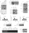
Fig. 1
Fig. 1. PAD4-mediated citrullination of E2F-1 augments its transcriptional activity.
(A) In vitro citrullination of GST (glutathione S-transferase)–E2F-1by Flag-PAD4, where recombinant GST–E2F-1 (1 μg) was incubated with Flag-PAD4 (1 μg). Lanes 1 to 7 were in the presence of CaCl2, and lane 8 in the absence of CaCl2. The arrow points to citrullinated GST-E2F-1. AMC, anti-modified citrulline (Millipore); IB, immunoblot. (B) Citrullination of E2F-1 in U2OS cells, transfected with HA (hemagglutinin)–PAD4 (2 μg). The lysate was incubated with 4 mM CaCl2 and 2 mM dithiothreitol (DTT). E2F-1 (C20) and rabbit immunoglobulin G (IgG) antibodies were used for immunoprecipitation (IP). (C) Citrullunation of N-terminal domain of E2F-1 in U2OS cells, transfected with Flag–E2F-1 (1 μg) and HA-PAD4 (2 μg) and treated with A23187 (5 μM, 30 min). Flag-agarose beads were used for immunoprecipitation. ΔC E2F-1: 1 to 194, ΔN E2F-1: 194 to 437. (D) Interaction between HA–E2F-1 and Flag-PAD4 in Flag-PAD4.pTRE and pTRE empty vector cell lines transfected with pcDNA or HA–E2F-1 (1 μg). Flag-agarose beads were used for immunoprecipitation. (E) Relative luciferase reporter comparing HA–E2F-1 wild-type (WT) and R4K activity in U2OS cells transfected with HA–E2F-1 WT or R4K mutant (R109K/R111K/R113K/R127K) (100 ng), Flag-PAD4 (300 ng), p73 luciferase reporter (100 ng), and β-gal (150 ng) plasmid. The luciferase reporter signal was normalized to β-gal reading. (F) Accompanying immunoblot ± SD. *P < 0.05. (G) Quantitative polymerase chain reaction (qPCR) analysis comparing Flag ΔC E2F-1 WT and R4K relative promoter occupancy in U2OS cells transfected with Flag ΔC E2F-1 WT or R4K mutant (R109/R111/R113/R127) (1 μg) and HA-PAD4 (2 μg). Flag antibody was used for ChIP. (H) Accompanying immunoblot ± SD. *P < 0.05. (I) ChIP analysis in doxycycline-inducible Flag-PAD4.pTRE cells {treated with or without doxycycline (1 μg/ml) [P4(+) and P4(−), respectively]}, measuring PAD4 promoter occupancy in nontargeting (NT) versus E2F-1 siRNA (30 nM)–transfected cells, and presented as visualized on ethidium bromide–stained gel. NS, nonspecific. (J) Accompanying immunoblot.
Fig. 2
Fig. 2. PAD4 regulates the recruitment of E2F-1 to cytokine gene promoters.
(A) U2OS cells were transfected with nontargeting, E2F-1, or PAD4 siRNA (50 nM). The transcript profile was analyzed using Illumina HumanHT12 v4.0 gene expression array. Transcripts for E2F-1 and PAD4 siRNA (E2F-1si and PAD4si) groups were normalized to nontargeting group and analyzed using GSEA. GSEA revealed down-regulation of immune system (green line) with both E2F-1 and PAD4 siRNAs. The black bars on the far right represent the genes that contribute to the plot. NES, normalized enrichment score. P value <0.01 and false discovery rate <0.05, *P < 0.05, **P < 0.01. GAPDH, glyceraldehyde phosphate dehydrogenase. (B) Expression of PAD4 in granulocyte-differentiated HL60 cells treated with 1% DMSO, ATRA (1 μM), or TPA (12-O-tetradecanoylphorbol 13-acetate) (10 ng/ml) for 48 hours. (C) Interaction between E2F-1 and PAD4 in DMSO-differentiated HL60 cells treated with 1% DMSO for 48 hours and immunoprecipitated using PAD4 or rabbit IgG antibodies. (D) Citrullination of E2F-1 in DMSO-differentiated HL60 cells. Lysates were incubated with CaCl2 (4 mM) and DTT (2 mM) and immunoprecipitated using E2F-1 (C20) or rabbit IgG antibodies. (E) ChIP analysis in undifferentiated (-) or DMSO-differentiated (D) HL60 cells. E2F-1 (C20), pRb (IF8), PAD4, and rabbit/mouse IgG antibodies were used for ChIP and presented as visualized on ethidium bromide–stained gel. (F to I) qPCR analysis in DMSO-differentiated HL60 cells measuring E2F-1 binding to inflammatory gene promoters, treated with LPS (lipopolysaccharide; L) (100 ng/ml, 3 hours) and the pan-PAD inhibitor BB-Cl-amidine (10 μM, 6 hours) (F) or the selective PAD4 inhibitor GSK484 (10 μM, 16 hours) (H), with accompanying immunoblots ± SD (G and I). *P < 0.05, **P < 0.01. Relative E2F-1 protein levels decrease twofold upon BB-Cl-amidine treatment (as quantified by ImageJ), whereas relative E2F-1 promoter occupancy on TNFα promoter decreases fivefold upon BB-Cl-amidine treatment.
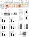
Fig. 3
Fig. 3. E2F-1 interacts with BRD4 on cytokine gene promoters.
(A) Heat map of bromodomain binding to acetylated E2F-1 peptides (sequence: 113-RHPGKGVKSPGEKSR-127). The bromodomains are structurally subgrouped into eight families (I to VIII) (21), and the multiple domains of a given bromodomain protein are represented by numbers in brackets. (B) Interaction between HA–E2F-1 and Flag-BD1/BD2 in human embryonic kidney (HEK) 293T cells transfected with HA–E2F-1 (1 μg) and Flag-BD1 or Flag-BD2 (2 μg) (BD 1 and BD 2 of BRD4, respectively). Flag-agarose beads were used for immunoprecipitation. (C) Interaction between E2F-1 and BRD4 in DMSO-differentiated HL60 cells. E2F-1 (C20) or rabbit IgG antibodies were used for immunoprecipitation. (D to I) qPCR double ChIP analysis of BRD4/E2F-1 interaction in DMSO-differentiated HL60 cells challenged with LPS (100 ng/ml, 3 hours) (D), treated with JQ1 (5 μM, 4 hours) (E and F), or BB-Cl-amidine (2.5 μM, 16 hours) (G), with accompanying immunoblot ± SD (H and I). *P < 0.05, **P < 0.01. (J) Interaction between His-BD1 and E2F-1 from U2OS cells treated with JQ1 (5 μM) or BB-Cl-amidine (5 μM) for 16 hours. The cell lysates were incubated with His-tagged BD1 of BRD4 and subject to Ni-agarose immunoprecipitation.
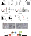
Fig. 4
Fig. 4. Additive immunosuppressive effects of PAD4 and BRD4 inhibition.
(A and B) Quantitative reverse transcription–PCR (qRT-PCR) analysis of cytokine transcript levels in DMSO-differentiated HL60 cells, treated with LPS (100 ng/ml, 3 hours), JQ1 (100 nM, 16 hours), and BB-Cl-amidine (2.5 μM, 16 hours) where indicated (J, JQ1; B, BB-Cl-amidine) (A), with accompanying immunoblot ± SD (B). *P < 0.05, **P < 0.01, ***P < 0.001. (C and D) Clinical scores for arthritic paw swelling (n = 7 for each treatment) in DBA/1 mice treated with vehicle, BB-Cl-amidine (1 mg/kg), JQ1 (1, 5, or 10 mg/kg), or combinations of, for 10 days from the onset of symptoms. ±SEM. *P < 0.05, **P < 0.01, ***P < 0.001. (E) qPCR ChIP analysis of E2F-1 promoter occupancy in murine spleen cells, collected from DBA/1 mice with CIA comparing vehicle and combination treatments. Enrichment of E2F-1 on mouse TNFα gene promoter expressed relative to IgG control. ±SD. *P < 0.05. (F) qRT-PCR analysis of TNFα transcript levels in murine spleen cells ± SD. **P < 0.01, ***P < 0.001. (G) Secreted levels of TNFα and IL-6 in murine inguinal lymph nodes ± SEM. *P < 0.05. (H) E2F-1 and IgG control immunohistochemistry staining of vehicle- and drug-treated paws. The arrows point to cells in the biopsy positively stained for E2F-1. (I) Model: PAD4-mediated citrullination targets E2F-1 to inflammatory genes, where E2F-1 interacts with BRD4 to drive inflammatory gene expression.

Similar articles

Cited by

References

    1. Wang S., Wang Y., Peptidylarginine deiminases in citrullination, gene regulation, health and pathogenesis. Biochim. Biophys. Acta 1829, 1126–1135 (2013). - PMC - PubMed
    1. Christophorou M. A., Castelo-Branco G., Halley-Stott R. P., Oliveira C. S., Loos R., Radzisheuskaya A., Mowen K. A., Bertone P., Silva J. C. R., Zernicka-Goetz M., Nielsen M. L., Gurdon J. B., Kouzarides T., Citrullination regulates pluripotency and histone H1 binding to chromatin. Nature 507, 104–108 (2014). - PMC - PubMed
    1. Sharma P., Azebi S., England P., Christensen T., Møller-Larsen A., Petersen T., Batsché E., Muchardt C., Citrullination of histone H3 interferes with HP1-mediated transcriptional repression. PLOS Genet. 8, e1002934 (2012). - PMC - PubMed
    1. Zhang X., Gamble M. J., Stadler S., Cherrington B. D., Causey C. P., Thompson P. R., Roberson M. S., Kraus W. L., Coonrod S. A., Genome-wide analysis reveals PADI4 cooperates with Elk-1 to activate c-Fos expression in breast cancer cells. PLOS Genet. 7, e1002112 (2011). - PMC - PubMed
    1. Roworth A. P., Ghari F., La Thangue N. B., To live or let die—Complexity within the E2F1 pathway. Mol. Cell. Oncol. 2, e970480 (2015). - PMC - PubMed

Publication types

MeSH terms

LinkOut - more resources