. 2016 May 13;291(20):10726-35.
doi: 10.1074/jbc.M116.722298. Epub 2016 Mar 11.

Rab13 Traffics on Vesicles Independent of Prenylation

Affiliations

Rab13 Traffics on Vesicles Independent of Prenylation

Maria S Ioannou et al. J Biol Chem. .

Abstract

Rab GTPases are critical regulators of membrane trafficking. The canonical view is that Rabs are soluble in their inactive GDP-bound form, and only upon activation and conversion to their GTP-bound state are they anchored to membranes through membrane insertion of a C-terminal prenyl group. Here we demonstrate that C-terminal prenylation is not required for Rab13 to associate with and traffic on vesicles. Instead, inactive Rab13 appears to associate with vesicles via protein-protein interactions. Only following activation does Rab13 associate with the plasma membrane, presumably with insertion of the C-terminal prenyl group into the membrane.

Keywords: DENN domain; DENND2B; GDI; GDP dissociation inhibitor; Rab; TI-VAMP; endosome; guanine nucleotide exchange factor (GEF); protein isoprenylation; vesicles.


Figures

FIGURE 1.
FIGURE 1.
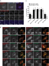
Inactive Rab13 traffics on vesicles. A, MCF10A cells expressing mCh-Rab13 Q67L or mCh-Rab13 T22N were imaged live. The arrows illustrate localization on the plasma membrane. Scale bars = 10 μm. B, the GST-Rab-binding domain (RBD) or GST alone, bound to glutathione-Sepharose, was incubated with HEK-293T cell lysates expressing mCh-Rab13 Q67L or T22N. Ponceau S staining revealed the level of GST-Rab-binding domain, whereas specifically bound Rab13 was detected by blot. Starting material (SM) equals 10% of the lysate used per condition. C, the percentage of transfected cells with mCh-Rab13 constructs on the plasma membrane. Data are mean ± S.D., measuring a minimum of 20 cells/experiment from a minimum of three independent experiments (Student's t test; ***, p < 0.001). D, PC12 cells differentiated for 24 h by addition of 50 ng/ml NGF and expressing mCh-Rab13 T22N were imaged live. The boxed region is magnified. The arrows follow a vesicle trafficking along a neurite. Scale bar = 10 μm (top panel) and 5 μm (magnified panel).
FIGURE 2.
FIGURE 2.
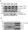
Rab13 is found on vesicles with multiple endosomal origins. A, MCF10A cells expressing mCh-Rab13 T22N and internalized Alexa Fluor (AF) 647-labeled transferrin were imaged live (i). The arrows illustrate co-localization. The boxed region from i is magnified below over a time-lapse imaging series (ii–iv). Scale bars = 10 μm (top panel) and 1 μm (magnified panels). B, MCF10A cells expressing mCh-Rab13 T22N or mCh-Rab13 Q67L co-expressed with GFP-cellubrevin (Cbv), GFP-TI-VAMP, GFP-Rab7, GFP-Rab9, or GFP-Rab5 were fixed and imaged. The boxed region in each image is magnified in the insets. The arrows illustrate co-localization. Scale bars = 10 μm. C, quantification of percent co-localization as determined in experiments such as those in B. Data are mean ± S.D., measuring a minimum of 8 cells/experiment from a minimum of two independent experiments.
FIGURE 3.
FIGURE 3.
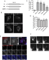
Rab13 resists membrane extraction by GDI. A, HEK-293T cell homogenates were spun for 30 min at 200,000 × g, and the resulting pellet was resuspended in HEPES buffer and incubated without or with increasing concentrations of purified myc-His-GDI as indicated. After 30 min of incubation at 37 °C, samples were spun for 30 min at 200,000 × g, and the resulting supernatant (S) and pellet (P) fractions were analyzed by Western blotting using the indicated antibodies. The Ponceau S-stained transfer revealed the presence of the purified GDI. B, HEK-293T cells were lysed in buffer containing 1% Triton X-100. Cell lysates were placed at the bottom of a discontinuous sucrose gradient and centrifuged at 230,000 × g for 16 h at 4 °C. Twelve fractions of 1 ml each were collected with fraction 1 from the top and fraction 12 from the bottom of the gradient. The fractions were analyzed by Western blotting with the indicated antibodies.
FIGURE 4.
FIGURE 4.
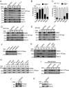
Inactive Rab13 traffics on vesicles following deletion of the C-terminal prenylation motif. A, schematic of the constructs used in the figure. FL, full-length; ΔC, C-terminal deletion of the last four amino acids CSLG (Cys-Ser-Leu-Gly); ΔHVD, hypervariable domain deletion. B, MCF10A cells expressing mCh-Rab13 Q67L FL, T22N FL, or T22N deletion constructs, as indicated, were imaged live. Scale bars = 10 μm. C, the percentage of transfected cells with mCh-Rab13 constructs on vesicles. Data are mean ± S.D., measuring a minimum of 20 cells/experiment from a minimum of three independent experiments. ***, p < 0.001. D, percentage of cells from experiments as in B with mCh-Rab13 on vesicles that exhibit perinuclear localization. Data are mean ± S.D., measuring a minimum of 20 cells/experiment from a minimum of three independent experiments. E, MCF10A cells expressing mCh-Rab13 T22N ΔC and internalized Alexa Fluor (AF) 647-labeled transferrin were imaged live. The arrows illustrate co-localization. The boxed region is magnified below over a time-lapse imaging series. Scale bars = 10 μm (top panel) and 1 μm (magnified panels). F, PC12 cells differentiated for 24 h with 50 ng/ml NGF and expressing mCh-Rab13 T22N-ΔC were imaged live. The boxed region is magnified. The arrows follow vesicle trafficking along a neurite. Scale bars = 10 μm (top panel) and 2.5 μm (magnified panel).
FIGURE 5.
FIGURE 5.
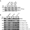
Rab13 associates with membranes via protein-protein interactions in cells. A, HEK-293T cell homogenates in HEPES buffer were spun for 30 min at 200,000 × g, and equal protein aliquots of the resulting pellet (P) were resuspended in ice-cold HEPES buffer with or without 1% Triton X-100 (TX100) or NaCO3 at pH 11. 0. After 15 min of incubation, samples were spun for 30 min at 200,000 × g, and the resulting supernatant (S2) and pellet (P2) fractions were analyzed by Western blotting using the indicated antibodies. B and C, quantification of the distribution of Rab proteins resuspended in Triton X-100 (B) and at pH 11.0 (C). Data are mean ± S.D. (n = 7 for Rab13, n = 5 for Rab5, n = 4 for Rab8 and Rab35, and n = 3 for Rab9). D--E, lysates of MCF10A cells (D) and MCH46 cells (E) were processed and analyzed as in A. F, HEK-293T cells were stably transduced with a lentivirus driving the expression of control shRNA or three different shRNAs targeting Rab13, and the indicated proteins were detected by blotting. G, HEK-293T cell homogenates in HEPES buffer were spun for 30 min at 200,000 × g, and equal protein aliquots of the resulting pellet were resuspended in ice-cold HEPES buffer with digitonin (Digit.), Nonidet P-40, CHAPS, or SDS/deoxycholate (DOC). After 15 min of incubation, samples were spun for 30 min at 200,000 × g, and the resulting supernatant and pellet fractions were analyzed by Western blotting using the indicated antibodies. H, HEK-293T cell homogenates in HEPES buffer were spun for 30 min at 200,000 × g, and equal protein aliquots of the resulting pellet were resuspended in ice-cold HEPES buffer with or without NaCl or KCl at the indicated concentrations. After 15 min of incubation, the samples were spun for 30 min at 200,000 × g, and the resulting supernatant and pellets were analyzed by Western blotting using the indicated antibodies. I, HEK-293T cell homogenates in HEPES buffer were spun for 30 min at 200,000 × g, and equal protein aliquots of the resulting pellet were resuspended in ice-cold HEPES buffer containing 100 mm NaCl with or without 1% Triton X-100 or NaCO3 at pH 11.0. After 15 min of incubation, samples were spun for 30 min at 200,000 × g, and the resulting supernatant and pellet fractions were analyzed by Western blotting using the indicated antibodies. J and K, HEK-293T cell homogenates in HEPES buffer were spun for 30 min at 200,000 × g, and equal protein aliquots of the resulting pellet were resuspended in ice-cold HEPES buffer. After 15 min of incubation, samples were spun for 30 min at 200,000 × g, and the resulting supernatant and pellets were analyzed by Western blotting using the indicated antibodies.
FIGURE 6.
FIGURE 6.
Rab13 associates with membranes via protein-protein interactions in tissue. A and B, equal protein aliquots of rat brain (A) and rat liver (B) homogenate in HEPES buffer were spun for 30 min at 200,000 × g, and the resulting pellet fraction (P) was resuspended in ice-cold HEPES buffer with or without 1% Triton X-100 (TX100) or NaCO3 at pH 11.0. After 15 min of incubation, the samples were spun for 30 min at 200,000 × g, and the resulting supernatant (S2) and pellets (P2) were analyzed by Western blotting using the indicated antibodies. C, quantification of the distribution of Rab proteins from extraction experiments as in A. Data are mean ± S.D. (n = 3 for Rab13 and Rab5). D, quantification of the distribution of Rab proteins from extraction experiments as in B. Data are mean ± S.D. (n = 5 for Rab5 and n = 3 Rab13).
FIGURE 7.
FIGURE 7.
Rab13 associates in a protein complex independent of nucleotide status. A, HEK-293T cell homogenates in HEPES buffer were spun for 30 min at 200,000 × g, and equal protein aliquots of the resulting pellet (P) were resuspended in ice-cold HEPES buffer with or without EDTA at the indicated concentrations. After 15 min of incubation, the samples were spun for 30 min at 200,000 × g, and the resulting supernatant (S2) and pellets (P2) were analyzed by Western blotting using the indicated antibodies. B, HEK-293T cells were left untransfected or were transfected with various mCh-Rab13 constructs as indicated. Cell homogenates in HEPES buffer were spun for 30 min at 200,000 × g, and equal protein aliquots of the resulting pellet were resuspended in ice-cold HEPES buffer with or without 1% Triton X-100 (TX100) or NaCO3 at pH 11.0. After 15 min of incubation, the samples were spun for 30 min at 200,000 × g, and the resulting supernatant (S2) and pellets (P2) were analyzed by Western blotting using the indicated antibodies for the first and second panels and an antibody against Rab13 to detect the various Rab13 constructs.
FIGURE 8.
FIGURE 8.
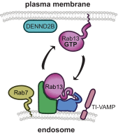
Model of Rab13 trafficking. Rab13 is targeted to and traffics on vesicles derived from late endosomes (depicted here) or recycling endosomes as part of a protein complex. When Rab13-positive vesicles reach the plasma membrane, Rab13 is activated locally by its GEF DENND2B, allowing active Rab13 to anchor to the plasma membrane via its C-terminal hydrophobic prenyl group.

Similar articles

Cited by

References

    1. Stenmark H. (2009) Rab GTPases as coordinators of vesicle traffic. Nat. Rev. Mol. Cell Biol. 10, 513–525 - PubMed
    1. Yang P. S., Yin P. H., Tseng L. M., Yang C. H., Hsu C. Y., Lee M. Y., Horng C. F., and Chi C. W. (2011) Rab5A is associated with axillary lymph node metastasis in breast cancer patients. Cancer Sci. 102, 2172–2178 - PubMed
    1. Allaire P. D., Seyed Sadr M., Chaineau M., Seyed Sadr E., Konefal S., Fotouhi M., Maret D., Ritter B., Del Maestro R. F., and McPherson P. S. (2013) Interplay between Rab35 and Arf6 controls cargo recycling to coordinate cell adhesion and migration. J. Cell Sci. 126, 722–731 - PubMed
    1. Bravo-Cordero J. J., Marrero-Diaz R., Megías D., Genís L., García-Grande A., García M. A., Arroyo A. G., and Montoya M. C. (2007) MT1-MMP proinvasive activity is regulated by a novel Rab8-dependent exocytic pathway. EMBO J. 26, 1499–1510 - PMC - PubMed
    1. Ioannou M. S., Bell E. S., Girard M., Chaineau M., Hamlin J. N., Daubaras M., Monast A., Park M., Hodgson L., and McPherson P. S. (2015) DENND2B activates Rab13 at the leading edge of migrating cells and promotes metastatic behavior. J. Cell Biol. 208, 629–648 - PMC - PubMed

Publication types

MeSH terms

Substances

LinkOut - more resources