

Necroptosis: an alternative cell death program defending against cancer
- PMID: 26968619
- PMCID: PMC4860077
- DOI: 10.1016/j.bbcan.2016.03.003
Necroptosis: an alternative cell death program defending against cancer
Abstract
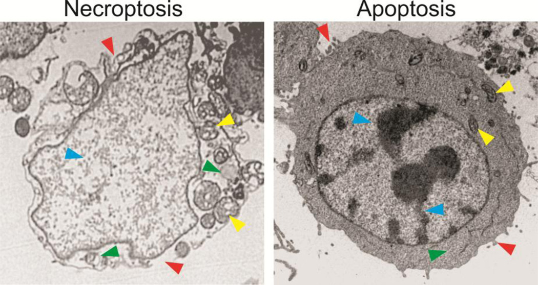
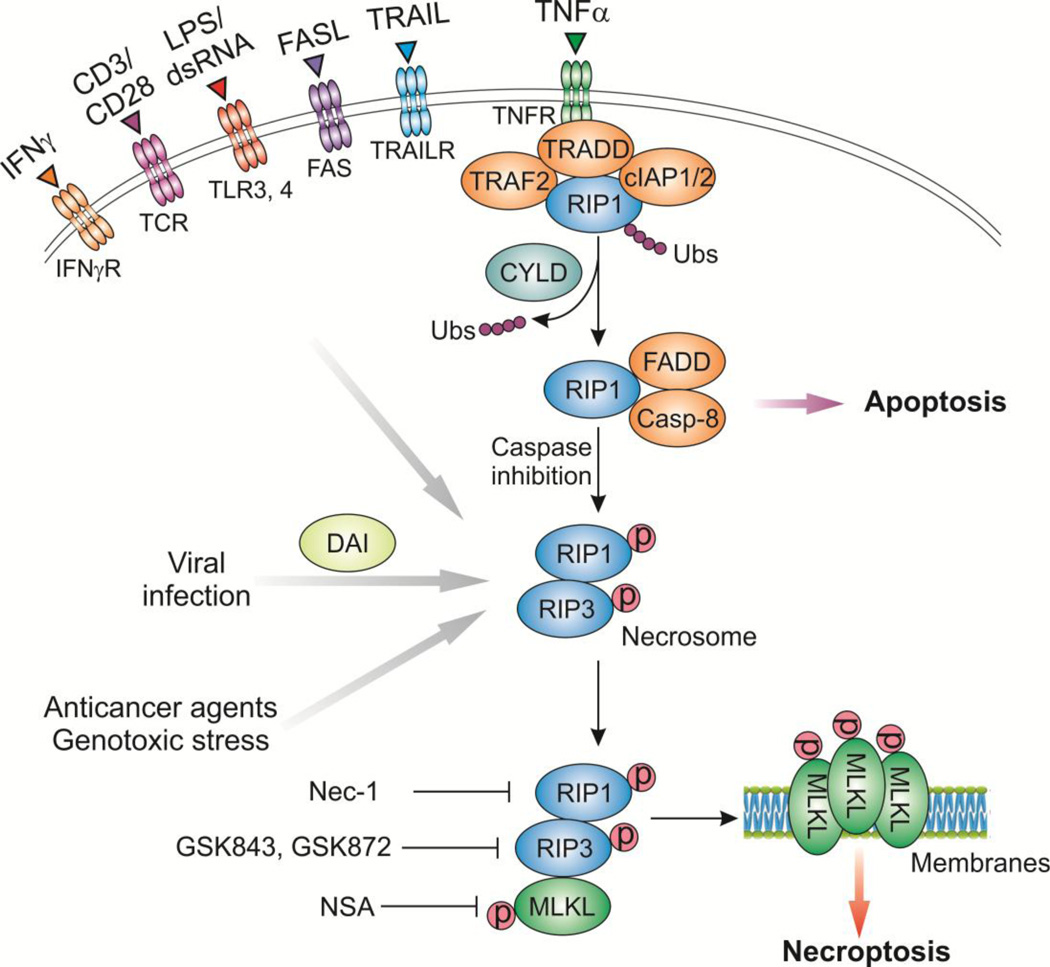
One of the hallmarks of cancer is resistance to programmed cell death, which maintains the survival of cells en route to oncogenic transformation and underlies therapeutic resistance. Recent studies demonstrate that programmed cell death is not confined to caspase-dependent apoptosis, but includes necroptosis, a form of necrotic death governed by Receptor-Interacting Protein 1 (RIP1), RIP3, and Mixed Lineage Kinase Domain-Like (MLKL) protein. Necroptosis serves as a critical cell-killing mechanism in response to severe stress and blocked apoptosis, and can be induced by inflammatory cytokines or chemotherapeutic drugs. Genetic or epigenetic alterations of necroptosis regulators such as RIP3 and cylindromatosis (CYLD), are frequently found in human tumors. Unlike apoptosis, necroptosis elicits a more robust immune response that may function as a defensive mechanism by eliminating tumor-causing mutations and viruses. Furthermore, several classes of anticancer agents currently under clinical development, such as SMAC and BH3 mimetics, can promote necroptosis in addition to apoptosis. A more complete understanding of the interplay among necroptosis, apoptosis, and other cell death modalities is critical for developing new therapeutic strategies to enhance killing of tumor cells.
Keywords: Cancer; MLKL; Necroptosis; RIP1; RIP3.
Copyright © 2016 Elsevier B.V. All rights reserved.
Figures
Similar articles
-
RIP1, RIP3, and MLKL Contribute to Cell Death Caused by Clostridium perfringens Enterotoxin.mBio. 2019 Dec 17;10(6):e02985-19. doi: 10.1128/mBio.02985-19. mBio. 2019. PMID: 31848291 Free PMC article.
-
Smac mimetic triggers necroptosis in pancreatic carcinoma cells when caspase activation is blocked.Cancer Lett. 2016 Sep 28;380(1):31-8. doi: 10.1016/j.canlet.2016.05.036. Epub 2016 Jun 3. Cancer Lett. 2016. PMID: 27267809
-
PUMA/RIP3 Mediates Chemotherapy Response via Necroptosis and Local Immune Activation in Colorectal Cancer.Mol Cancer Ther. 2024 Mar 4;23(3):354-367. doi: 10.1158/1535-7163.MCT-23-0162. Mol Cancer Ther. 2024. PMID: 37992761 Free PMC article.
-
Necroptosis in health and diseases.Semin Cell Dev Biol. 2014 Nov;35:14-23. doi: 10.1016/j.semcdb.2014.07.013. Epub 2014 Aug 1. Semin Cell Dev Biol. 2014. PMID: 25087983 Review.
-
Cancer therapy in the necroptosis era.Cell Death Differ. 2016 May;23(5):748-56. doi: 10.1038/cdd.2016.8. Epub 2016 Feb 26. Cell Death Differ. 2016. PMID: 26915291 Free PMC article. Review.
Cited by
-
Role of necroptosis in traumatic brain and spinal cord injuries.J Adv Res. 2022 Sep;40:125-134. doi: 10.1016/j.jare.2021.12.002. Epub 2021 Dec 22. J Adv Res. 2022. PMID: 36100321 Free PMC article. Review.
-
Therapeutic effects of exendin-4 on spinal cord injury via restoring autophagy function and decreasing necroptosis in neuron.CNS Neurosci Ther. 2024 Jul;30(7):e14835. doi: 10.1111/cns.14835. CNS Neurosci Ther. 2024. PMID: 39004783 Free PMC article.
-
Targeting necroptosis in anticancer therapy: mechanisms and modulators.Acta Pharm Sin B. 2020 Sep;10(9):1601-1618. doi: 10.1016/j.apsb.2020.01.007. Epub 2020 Jan 21. Acta Pharm Sin B. 2020. PMID: 33088682 Free PMC article. Review.
-
RIP1 upregulation promoted tumor progression by activating AKT/Bcl-2/BAX signaling and predicted poor postsurgical prognosis in HCC.Tumour Biol. 2016 Nov;37(11):15305-15313. doi: 10.1007/s13277-016-5342-1. Epub 2016 Oct 4. Tumour Biol. 2016. PMID: 27699664
-
p53 Up-regulated Modulator of Apoptosis Induction Mediates Acetaminophen-Induced Necrosis and Liver Injury in Mice.Hepatology. 2019 May;69(5):2164-2179. doi: 10.1002/hep.30422. Epub 2019 Mar 11. Hepatology. 2019. PMID: 30552702 Free PMC article.
References
-
- Hanahan D, Weinberg RA. Hallmarks of cancer: the next generation. Cell. 2011;144:646–674. - PubMed
-
- Galluzzi L, Vitale I, Abrams JM, Alnemri ES, Baehrecke EH, Blagosklonny MV, Dawson TM, Dawson VL, El-Deiry WS, Fulda S, Gottlieb E, Green DR, Hengartner MO, Kepp O, Knight RA, Kumar S, Lipton SA, Lu X, Madeo F, Malorni W, Mehlen P, Nunez G, Peter ME, Piacentini M, Rubinsztein DC, Shi Y, Simon HU, Vandenabeele P, White E, Yuan J, Zhivotovsky B, Melino G, Kroemer G. Molecular definitions of cell death subroutines: recommendations of the Nomenclature Committee on Cell Death 2012. Cell Death Differ. 2012;19:107–120. - PMC - PubMed
-
- Holler N, Zaru R, Micheau O, Thome M, Attinger A, Valitutti S, Bodmer JL, Schneider P, Seed B, Tschopp J. Fas triggers an alternative, caspase-8-independent cell death pathway using the kinase RIP as effector molecule. Nature immunology. 2000;1:489–495. - PubMed
-
- Degterev A, Huang Z, Boyce M, Li Y, Jagtap P, Mizushima N, Cuny GD, Mitchison TJ, Moskowitz MA, Yuan J. Chemical inhibitor of nonapoptotic cell death with therapeutic potential for ischemic brain injury. Nature chemical biology. 2005;1:112–119. - PubMed
Publication types
MeSH terms
Substances
Grants and funding
- R01CA172136/CA/NCI NIH HHS/United States
- U19 AI068021/AI/NIAID NIH HHS/United States
- R01CA106348/CA/NCI NIH HHS/United States
- P30 CA047904/CA/NCI NIH HHS/United States
- U19AI068021/AI/NIAID NIH HHS/United States
- R01 CA172136/CA/NCI NIH HHS/United States
- U01 DK085532/DK/NIDDK NIH HHS/United States
- R01CA203028/CA/NCI NIH HHS/United States
- U01 DK085570/DK/NIDDK NIH HHS/United States
- R01 CA106348/CA/NCI NIH HHS/United States
- R01 CA203028/CA/NCI NIH HHS/United States
- U01DK085570/DK/NIDDK NIH HHS/United States
- P30CA047904/CA/NCI NIH HHS/United States
- U24 DK085532/DK/NIDDK NIH HHS/United States
LinkOut - more resources
Full Text Sources
Other Literature Sources
Research Materials
Miscellaneous
