. 2016 Mar 1:7:10833.
doi: 10.1038/ncomms10833.

Stereocilia-staircase spacing is influenced by myosin III motors and their cargos espin-1 and espin-like

Affiliations

Stereocilia-staircase spacing is influenced by myosin III motors and their cargos espin-1 and espin-like

Seham Ebrahim et al. Nat Commun. .

Erratum in

Abstract

Hair cells tightly control the dimensions of their stereocilia, which are actin-rich protrusions with graded heights that mediate mechanotransduction in the inner ear. Two members of the myosin-III family, MYO3A and MYO3B, are thought to regulate stereocilia length by transporting cargos that control actin polymerization at stereocilia tips. We show that eliminating espin-1 (ESPN-1), an isoform of ESPN and a myosin-III cargo, dramatically alters the slope of the stereocilia staircase in a subset of hair cells. Furthermore, we show that espin-like (ESPNL), primarily present in developing stereocilia, is also a myosin-III cargo and is essential for normal hearing. ESPN-1 and ESPNL each bind MYO3A and MYO3B, but differentially influence how the two motors function. Consequently, functional properties of different motor-cargo combinations differentially affect molecular transport and the length of actin protrusions. This mechanism is used by hair cells to establish the required range of stereocilia lengths within a single cell.


Conflict of interest statement

The authors declare no competing financial interests.

Figures
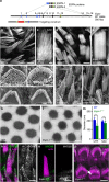
Figure 1
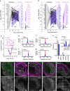
Figure 1. Altered length and width regulation in Espn-1−/− mice.
(a) Targeting strategy. PGK-Neo cassette targeted exon 1, which includes the translation start for ESPN-1; translation start for short ESPN isoforms is in exon 6. (b,c) Phalloidin-labelled stereocilia in the striola of Espn-1−/− utricles, imaged with confocal fluorescence microscopy, appear normal. (d,e) Phalloidin-labelled stereocilia in an extrastriolar region of Espn-1−/− utricles are of nearly uniform lengths. (f) SEM image of WT cochlear outer cell hair bundles. (g) SEM of P4 Espn-1−/− outer hair cells shows elongated protrusions opposite the bundle (arrow). (h) Extraneous protrusions disappear from Espn-1−/− outer hair cells by P10. (i) SEM image of WT utricle extrastriolar bundles. (j) SEM of extrastriolar bundles of Espn-1−/− utricle; stereocilia are of nearly uniform length. (k,l) Transmission electron micrographs show that extrastriolar stereocilia of Espn-1−/− utricle (l) have a smaller diameter than those of WT utricles (k). (m) Diameter of utricle (UTR) and saccule (SAC) stereocilia. Mean±s.d.; n=43 (WT utricle), 163 (Espn-1−/− utricle), 134 (WT saccule) and 168 (Espn-1−/− saccule). Differences are significant at P<0.001 (Student's t-test). (n) MYO3B targets to stereocilia tips of utricle bundles of WT mice. (o) MYO3B no longer targets to tips of Espn-1−/− stereocilia. (p) PB538 anti-ESPN-1 antibody detects antigen at some stereocilia tips (asterisks). Elongated protrusions (arrow) are visible in inner hair cells. Scale bars (b,d,mo), 5 μm; (c,ej), 2 μm; and (k,l), 100 nm.
Figure 2
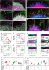
Figure 2. Proteomics of developing mouse hair bundles.
(a) Mole fractions of proteins in utricular epithelium (left) and hair bundles (right) of P5 mice. Proteins most highly enriched in epithelium are indicated at left and those highly enriched in bundles at right. Hue represents enrichment for each protein. Far right, proteins detected only in bundles. (b) Mole fractions of proteins in epithelium (left) and bundles (right) of P23 mice. (a,b) Derived from four biological replicates of 100 ear-equivalents of hair bundles and 10 whole utricles. (c) Volcano plot showing relationship between P23/P5 enrichment (x axis) and FDR-adjusted P value (y axis). Proteins that are enriched fivefold or greater between bundles and epithelium are labelled with purple. (d) Targeted MS2 signal for ESPN peptide LASLPAWR (m/z=457.2663, 2+ charge state) detected from P23 bundles; 10 fmol of the heavy-isotope-labelled peptide (462.2705, 2+) was included as a standard. (e) Targeted MS2 signal for ESPN-1 peptide YLVEEVALPAVSR (723.4036, 2+) detected from P23 bundles; 1 fmol of standard (728.4077, 2+). (f) Targeted MS2 signal for ESPNL peptide CQEYESELGR (635.7721, 2+) detected from P23 bundles; 1 fmol of standard (640.7762, 2+). (g) Targeted MS2 signal for MYO3B peptide NRDTLPADVVVVLR (522.9703, 3+) detected from P23 bundles; 1 fmol of standard (526.3063, 3+). (h) Summarized PRM signal for each protein, normalized to actin; two to three peptides were used for each protein, and four biological replicates were measured for each age (reported as mean±s.d.). While MYO3A was detectable in one sample at P5, its signal was below the assay limit of detection at P23. (im) Low-power images of mouse utricle stained with phalloidin (top, magenta) for actin and specific antibodies (top, green; below, grey). Approximate location of the striola, estimated from examination of the phalloidin channel, is indicated. ESPN-1 (j) and MYO3B (m) are enriched in extrastriolar regions; ESPNL (k) and MYO3A (l) are enriched in the striola. LES, lateral extrastriola; MES, medial extrastriola; S, striola. Scale bars, 100 μm. FDR, false discovery rate.
Figure 3
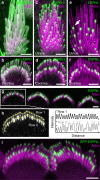
Figure 3. Immunolocalization of ESPN-1 and ESPNL.
(a,b) Pan-ESPN antibody (green) and actin (magenta) labels utricle (a) and cochlear outer hair cell (b) stereocilia throughout. (c,d) ESPN-1 antibody labels the tips of utricular (c) and cochlear (d) stereocilia. (e) ESPNL antibody (ab170747) labels some stereocilia tips strongly (arrow), some weakly (asterisk), but does not label tallest stereocilia of utricle hair cell. (f) ESPNL antibody labels most but not all stereocilia tips of row 2 in bundle of outer hair cell. (g) Structured illumination microscopic image of ESPNL labelling (BG35961) of inner hair-cell bundles. (h) Quantification of ESPNL labelling of one hair bundle from (G). (left) Yellow lines indicate quantification trajectories; and (right) uniform labelling in row 1 but saturation of some tip signals in row 2 (asterisks). (i) GFP-ESPNL labelling of P0.5 inner hair cells following in utero electroporation at E12. Scale bars, 2 μm.
Figure 4
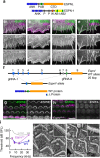
Figure 4. ESPNL functional characterization.
(a) Domain structure of ESPNL and ESPN-1. Blue, ankyrin repeats (ANK); black, proline-rich domain (P); red, WH2 domain (W); green, common actin-binding region (C); yellow, ESPN actin-binding domain (AB); and orange, ESPNL C-terminal domain (CTD). (b) COS7 cells transfected with mCherry-ESPN1 (green), also labelled with phalloidin (magenta). Individual channels shown below. ESPN-1 forms filopodia. (c) mCherry-ESPNL localizes with actin structures but does not induce filopodia. (d) ESPNL localizes to filopodia induced by the actin-bundling protein GFP-PLS1. (e) ESPNL localizes to filopodia induced by GFP-ESPN-3. (f) CRISPR targeting strategy. gRNA-1 targeted exon 1 and gRNA-2 targeted exon 8; one of 16 alleles was a deletion between those two exons, eliminating ∼20 kbp of genomic DNA. The Δ protein is hypothetical; we do not have evidence that this truncated product is even made. (g,h) BG35961 anti-ESPNL antibody labels WT cochlear hair bundles (g) but not those of EspnlΔ/Δ mice (h). (i) ABRs showing high-frequency hearing loss in 30-day mice. Mice with EspnlΔ/Δ, EspnlΔ/A and EspnlA/A genotypes had similar ABRs and were grouped together as Espnl−/− (n=13; WT, n=19; displayed as mean±s.d.). ***P<10−5 (Student’s t-test). (j) SEM of basal cochlear outer hair cells from WT mice. Note three rows of stereocilia in each bundle. (k) SEM of basal cochlear outer hair cells from EspnlΔ/Δ mice. Bundles only have two rows of stereocilia except for scattered row three stereocilia (asterisks). Scale bars, (be,j) 2 μm; and (g,h) 10 μm.
Figure 5
Figure 5. ESPNL binds myosin-III tail-homology domain 1.
(a) Domain structure of MYO3A and MYO3B. Light blue, motor domain. Magenta, calmodulin-binding IQ domains. Red, tail homology domain 1 (THD1). Grey, tail homology domain 2 (THD2). MYO3B lacks THD2. (b,c) ESPNL ARD binds THD1 of MYO3A (b) and MYO3B (c) indicated by red arrows. GFP-tagged MYO3A-3THDI and MYO3B-THDI protein from the cell lysates bound to glutathione-agarose beads preloaded with GST-ESPNL-ARD but not beads with GST only. Pre- and post-THDI MYO3A and MYO3B constructs did not bind to GST-ESPNL-ARD beads. (d) (left) GFP-ESPNL-ARD is transported to filopodia tips by MYO3A (red channel, phalloidin). (centre) Magnified view of one filopodium (red channel, MYO3A). (right) Intensity profile of filopodium. ESPNL-ARD and MYO3A puncta (asterisks) may represent transport aggregates. (e) GFP-ESPL-ARD is not transported into filopodia by MYO3B. Scale bars, 2 μm.
Figure 6
Figure 6. MYO3A and MYO3B separately localize in bundles but replace each other in the ear.
(a) Immunolabelling of MYO3A (green) in rat auditory hair cells (P3); actin (magenta) labelled with phalloidin. (right) MYO3A label only. (b) MYO3A and ESPNL-GFP do not completely overlap in hair bundle of outer hair cell (see inset). Elongated ESPNL-GFP labelling in row 2. (c) SIM image of outer hair-cell stereocilia showing MYO3A cap and elongated ESPNL-GFP. (d) MYO3A labelling in utricle bundle. (left) MYO3A image with transect indicated. (right) Intensity profile along transect. (e) (left) MYO3B labelling is greatest in tallest stereocilia (asterisks). (right) Intensity profile along transect. (f) Representation of exons 1–7 (blue boxes) of WT mouse Myo3a locus. The targeting vector replaced exons 4 and 5 with a floxed neomycin-resistance gene cassette, introducing a premature stop codon in exon 6. (g) Exons 1–8 of the Myo3b locus; the targeting vector (middle) was designed to eliminate exons 3 and 4, replacing them with a neomycin-resistance gene cassette conjugated to LacZ; a premature stop codon was introduced in exon 5. (h) RT–PCR on inner-ear mRNA showing 578 bp amplicon from WT mouse and expected 205 bp truncated band in Myo3a−/−. (i) Mean ABR measurements (±s.d.) from Myo3a−/− (n=29) and Myo3a+/− (n=27) mice at 6 months. (j) RT–PCR on inner-ear mRNA showing 695 bp amplicon from WT mouse and expected 319 bp truncated band from Myo3b−/−. (k) ABR from Myo3b−/− (n=9) and Myo3b+/− (n=9) mice at 6 months. (l) WT extrastriolar bundle (phalloidin stained). (m) Myo3a−/− extrastriolar bundle. (n) Myo3b−/− extrastriolar bundle. (o) Heights of short and tall stereocilia from indicated genotypes. Mean±s.d.; n=148 (WT), 123 (Myo3a−/−) and 199 (Myo3b−/−). Student’s t-test indicated Myo3b−/− short stereocilia lengths are different from WT at P=10−21; tall lengths are different at P=0.007. (p) Distribution of short stereocilia lengths. (q) Distribution of tall stereocilia lengths. Scale bars, (a,b,d,e) 5 μm; (c) 300 nm; and (ln) 2 μm.
Figure 7
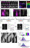
Figure 7. ESPN-1 and ESPNL cooperate with MYO3A and MYO3B to differentially control filopodia growth.
(a,b) COS7 cells co-expressing mCherry-MYO3A (magenta) with relatively low (a) or high (b) expression of mEmerald-tagged full-length ESPNL (green). (c,d) COS7 cells co-expressing mCherry-MYO3B (magenta) with relatively low (c) or high (d) expression of mEmerald-ESPNL (green). (e) MYO3A is unable to target filopodia tips in the presence of ESPNL, even when co-expressed with ESPN-1. (f) MYO3B consistently targets filopodia tips in cells expressing both ESPNL and untagged ESPN-1. (gj) Quantification of relationship between ESPNL fluorescence level (ESPNL Fl) and filopodia density (g,i) or length (h,j). There was an inverse relationship of filopodial number and length with ESPNL fluorescence for MYO3A (R2=0.4 and 0.5) but not MYO3B (R2<0.1 and <0.01). (k) COS7 cell expressing GFP-MYO3A (green) and mCherry-MYO3B (red), with actin labelled phalloidin (blue). (l) Filopodia are longer in the presence of MYO3A, GFP-MYO3B and ESPN1 (blue). (m) Filopodia are long with MYO3A, GFP-MYO3B, TagBFP2-ESPNL (blue) and untagged ESPN1. (n) Confocal images and relative pixel intensity profiles of single filopodia from cells transfected with the indicated constructs. Colours are identical in images and corresponding profiles. Scale in the profiles applies to the images. (o,p) Filopodia number per 10 μm of cell perimeter (o) and lengths (p) for each motor-cargo combination are presented as box plots, with upper and lower whiskers representing the range, top and bottom of the boxes representing the upper and lower 25th percentile, and the bars bisecting the boxes representing the median values (values from ∼30 COS7 cells used for each combination). Two-way analysis of variance analysis of data in these panels is reported in Supplementary Table 1. Scale bars, 2 μm.

Similar articles

Cited by

References

    1. Mattila P. K. & Lappalainen P. Filopodia: molecular architecture and cellular functions. Nat. Rev. Mol. Cell Biol. 9, 446–454 (2008). - PubMed
    1. Nambiar R., McConnell R. E. & Tyska M. J. Myosin motor function: the ins and outs of actin-based membrane protrusions. Cell. Mol. Life Sci. 67, 1239–1254 (2010). - PMC - PubMed
    1. Tilney L. G., Tilney M. S. & DeRosier D. J. Actin filaments, stereocilia, and hair cells: how cells count and measure. Annu. Rev. Cell Biol. 8, 257–274 (1992). - PubMed
    1. Manor U. & Kachar B. Dynamic length regulation of sensory stereocilia. Semin. Cell Dev. Biol. 19, 502–510 (2008). - PMC - PubMed
    1. Jacobo A. & Hudspeth A. J. Reaction-diffusion model of hair-bundle morphogenesis. Proc. Natl Acad. Sci. USA 111, 15444–15449 (2014). - PMC - PubMed

Publication types