Akermanite bioceramics promote osteogenesis, angiogenesis and suppress osteoclastogenesis for osteoporotic bone regeneration
- PMID: 26911441
- PMCID: PMC4766478
- DOI: 10.1038/srep22005
Akermanite bioceramics promote osteogenesis, angiogenesis and suppress osteoclastogenesis for osteoporotic bone regeneration
Abstract
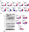
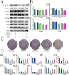
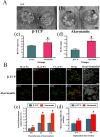
It is a big challenge for bone healing under osteoporotic pathological condition with impaired angiogenesis, osteogenesis and remodeling. In the present study, the effect of Ca, Mg, Si containing akermanite bioceramics (Ca2MgSi2O7) extract on cell proliferation, osteogenic differentiation and angiogenic factor expression of BMSCs derived from ovariectomized rats (BMSCs-OVX) as well as the expression of osteoclastogenic factors was evaluated. The results showed that akermanite could enhance cell proliferation, ALP activity, expression of Runx2, BMP-2, BSP, OPN, OCN, OPG and angiogenic factors including VEGF and ANG-1. Meanwhile, akermanite could repress expression of osteoclastogenic factors including RANKL and TNF-α. Moreover, akermanite could activate ERK, P38, AKT and STAT3 signaling pathways, while crosstalk among these signaling pathways was evident. More importantly, the effect of akermanite extract on RANKL-induced osteoclastogenesis was evaluated by TRAP staining and real-time PCR assay. The results showed that akermanite could suppress osteoclast formation and expression of TRAP, cathepsin K and NFATc1. The in vivo experiments revealed that akermanite bioceramics dramatically stimulated osteogenesis and angiogenesis in an OVX rat critical-sized calvarial defect model. All these results suggest that akermanite bioceramics with the effects of Mg and Si ions on osteogenesis, angiogenesis and osteoclastogenesis are promising biomaterials for osteoporotic bone regeneration.
Figures


Similar articles
-
The synergistic effects of Sr and Si bioactive ions on osteogenesis, osteoclastogenesis and angiogenesis for osteoporotic bone regeneration.Acta Biomater. 2017 Oct 1;61:217-232. doi: 10.1016/j.actbio.2017.08.015. Epub 2017 Aug 12. Acta Biomater. 2017. PMID: 28807800
-
Enhanced osteoporotic bone regeneration by strontium-substituted calcium silicate bioactive ceramics.Biomaterials. 2013 Dec;34(38):10028-42. doi: 10.1016/j.biomaterials.2013.09.056. Epub 2013 Oct 2. Biomaterials. 2013. PMID: 24095251
-
Stimulatory effects of the ionic products from Ca-Mg-Si bioceramics on both osteogenesis and angiogenesis in vitro.Acta Biomater. 2013 Aug;9(8):8004-14. doi: 10.1016/j.actbio.2013.04.024. Epub 2013 Apr 22. Acta Biomater. 2013. PMID: 23619289
-
Stem Cell and Advanced Nano Bioceramic Interactions.Adv Exp Med Biol. 2018;1077:317-342. doi: 10.1007/978-981-13-0947-2_17. Adv Exp Med Biol. 2018. PMID: 30357696 Review.
-
Exosome: A Novel Approach to Stimulate Bone Regeneration through Regulation of Osteogenesis and Angiogenesis.Int J Mol Sci. 2016 May 19;17(5):712. doi: 10.3390/ijms17050712. Int J Mol Sci. 2016. PMID: 27213355 Free PMC article. Review.
Cited by
-
Akermanite used as an alkaline biodegradable implants for the treatment of osteoporotic bone defect.Bioact Mater. 2016 Dec 7;1(2):151-159. doi: 10.1016/j.bioactmat.2016.11.004. eCollection 2016 Dec. Bioact Mater. 2016. PMID: 29744404 Free PMC article.
-
The MAZ transcription factor is a downstream target of the oncoprotein Cyr61/CCN1 and promotes pancreatic cancer cell invasion via CRAF-ERK signaling.J Biol Chem. 2018 Mar 23;293(12):4334-4349. doi: 10.1074/jbc.RA117.000333. Epub 2018 Feb 6. J Biol Chem. 2018. PMID: 29414775 Free PMC article.
-
Doped Calcium Silicate Ceramics: A New Class of Candidates for Synthetic Bone Substitutes.Materials (Basel). 2017 Feb 10;10(2):153. doi: 10.3390/ma10020153. Materials (Basel). 2017. PMID: 28772513 Free PMC article. Review.
-
Human amnion-derived mesenchymal stem cells promote osteogenic and angiogenic differentiation of human adipose-derived stem cells.PLoS One. 2017 Oct 11;12(10):e0186253. doi: 10.1371/journal.pone.0186253. eCollection 2017. PLoS One. 2017. PMID: 29020045 Free PMC article.
-
Magnesium-containing bioceramics stimulate exosomal miR-196a-5p secretion to promote senescent osteogenesis through targeting Hoxa7/MAPK signaling axis.Bioact Mater. 2023 Nov 4;33:14-29. doi: 10.1016/j.bioactmat.2023.10.024. eCollection 2024 Mar. Bioact Mater. 2023. PMID: 38024235 Free PMC article.
References
-
- Liu H. Y. et al. The balance between adipogenesis and osteogenesis in bone regeneration by platelet-rich plasma for age-related osteoporosis. Biomaterials 32, 6773–6780 (2011). - PubMed
-
- Genant H. K. et al. Interim report and recommendations of the World Health Organization Task-Force for Osteoporosis. Osteoporos Int 10, 259–264 (1999). - PubMed
-
- Jee W. S. & Yao W. Overview: animal models of osteopenia and osteoporosis. J Musculoskelet Neuronal Interact 1, 193–207 (2001). - PubMed
-
- Lerner U. H. Bone remodeling in post-menopausal osteoporosis. J Dent Res 85, 584–595 (2006). - PubMed
-
- Lin K. et al. Enhanced osteoporotic bone regeneration by strontium-substituted calcium silicate bioactive ceramics. Biomaterials 34, 10028–10042 (2013). - PubMed
Publication types
MeSH terms
Substances
LinkOut - more resources
Full Text Sources
Other Literature Sources
Research Materials
Miscellaneous

