. 2016 Feb 22;12(2):e1004774.
doi: 10.1371/journal.pcbi.1004774. eCollection 2016 Feb.

Model-Driven Understanding of Palmitoylation Dynamics: Regulated Acylation of the Endoplasmic Reticulum Chaperone Calnexin

Affiliations

Model-Driven Understanding of Palmitoylation Dynamics: Regulated Acylation of the Endoplasmic Reticulum Chaperone Calnexin

Tiziano Dallavilla et al. PLoS Comput Biol. .

Abstract

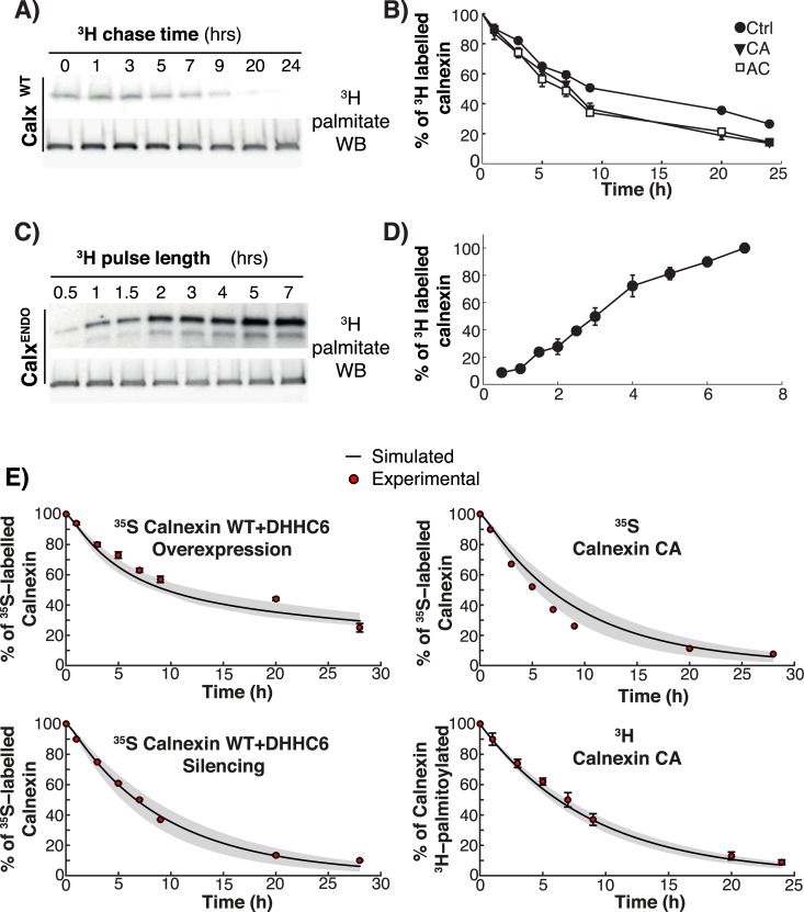
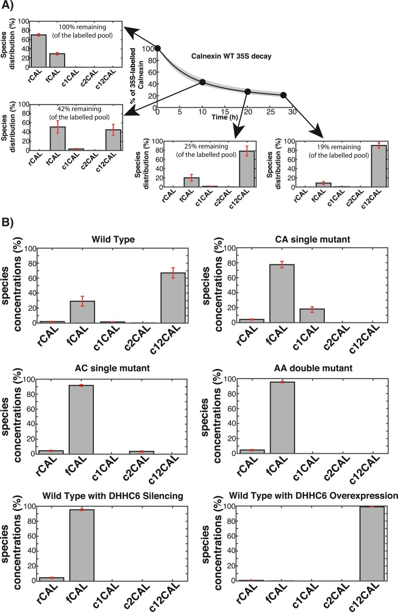
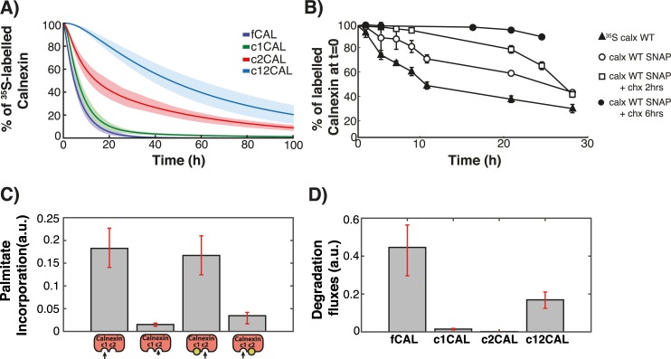
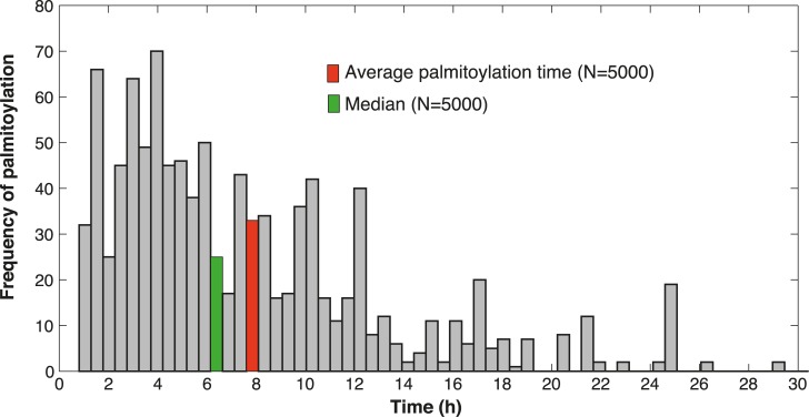
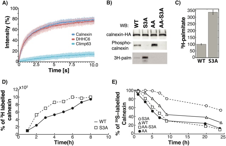
Cellular functions are largely regulated by reversible post-translational modifications of proteins which act as switches. Amongst these, S-palmitoylation is unique in that it confers hydrophobicity. Due to technical difficulties, the understanding of this modification has lagged behind. To investigate principles underlying dynamics and regulation of palmitoylation, we have here studied a key cellular protein, the ER chaperone calnexin, which requires dual palmitoylation for function. Apprehending the complex inter-conversion between single-, double- and non-palmitoylated species required combining experimental determination of kinetic parameters with extensive mathematical modelling. We found that calnexin, due to the presence of two cooperative sites, becomes stably acylated, which not only confers function but also a remarkable increase in stability. Unexpectedly, stochastic simulations revealed that palmitoylation does not occur soon after synthesis, but many hours later. This prediction guided us to find that phosphorylation actively delays calnexin palmitoylation in resting cells. Altogether this study reveals that cells synthesize 5 times more calnexin than needed under resting condition, most of which is degraded. This unused pool can be mobilized by preventing phosphorylation or increasing the activity of the palmitoyltransferase DHHC6.


Conflict of interest statement

The authors have declared that no competing interests exist.

Figures

Fig 1
Fig 1. Calnexin palmitoylation model and kinetics of decay.
A: Calnexin is first synthesized (rCAL). During the folding process the protein assumes the proper conformation (fCAL), which then can be palmitoylated twice by DHHC6. The first palmitoylation can occur on either of the two sites leading to c1CAL or c2CAL. The second palmitoylation events leads to c12CAL. We assume that both palmitoylation steps are reversible. Degradation can occur from all states. B-E: HeLa cells were transfected or not for 24h with calnexin-WT-HA, HA-calnexin-KDEL, calnexin-CA-HA, calnexin-AC-HA, calnexin-AA-HA and with or without DHHC6-myc, or additional transfected for 72h with DHHC6 siRNA. Cells were incubated 20 min pulse at 37°C with 35S-methionine/cysteine, washed and further incubated for different times at 37°C. Calnexin was immunoprecipitated and analyzed by SDS-PAGE. Autoradiography (B) and western blotting were quantified using the Typhoon Imager (Image QuantTool, GE healthcare). C: Decay profile of WT calnexin (Calx-WT) and of a calnexin mutant in which the transmembrane and cytosolic domain were removed and replaced by the KDEL sequence for ER retention (Calx-KDEL). Errors correspond to standard deviations (n = 7 for Calx-WT, n = 3 for Calx-KDEL). D: Decay profile of WT calnexin was observed under normal condition (Ctrl) and after overexpression (DHHC6 Overexpression) or silencing (DHHC6 Silencing) of DHHC6. Errors correspond to standard deviations (n = 7, 3, 3 for Crtl, DHHC6 overexpression and silencing respectively). E: Decay profile of WT calnexin (Calx-WT) and different mutants, in which site c1 (Calx-AC), site c2 (Calx-CA), or both palmitoylation sites (Calx-AA) were mutated. Errors correspond to standard deviations (n = 8, 3, 3, 3 for Calx-WT, AC, CA and AA respectively).
Fig 2
Fig 2. Experiments and modelling of Palmitoylation/depalmitoylation of calnexin.
AB: HeLa cells were transfected for 24h with calnexin-WT-HA, calnexin-CA-HA, or calnexin-AC-HA. Cells were incubated with 3H-palmitic acid for 2h, washed and further incubated for different times at 37°C in complete medium prior to immunoprecipitation using anti-calnexin or anti-HA antibodies. Immunoprecipitates were split into two, run on SDS-PAGE and analyzed either by autoradiography (3H-palmitate) or Western blotting (anti-HA). Autoradiograms were quantified using the Typhoon Imager (Image QuantTool, GE healthcare). Errors correspond to standard deviations (n = 3). CD: HeLa cells were incubated with 3H-palmitic acid for different times at 37°C, washed prior to immunoprecipitation using anti-calnexin antibodies. Immunoprecipitates were split into two, run on SDS-PAGE and analyzed either by autoradiography (3H-palmitate) or Western blotting (anti-calnexin). Errors correspond to standard deviations (n = 4). E: Validation of the model output though comparison of the in silico experiments with experimental data that was not used in the calibration of the model. Solid line is the mean of the simulations of 382 models; the shaded area is defined by the first and the third quartile of the simulations of 382 models (details of the in silico labelling experiment can be found in the Supplemental Information).
Fig 3
Fig 3. In silico analysis of the calnexin species distribution.
A: We replicated in silico the 35S pulse-chase experiment on WT calnexin. During the experiment we monitored the relative distribution of calnexin in the different palmitoylation states. Solid line is the mean of the simulations of 382 models; error bars are defined by the first and the third quartile of the simulations of 382 models (details of the in silico labelling experiment can be found in the Expanded View). B: The model was used to predict the steady state distribution of WT calnexin and of the mutants in the cell. rCAL correspond to calnexin during the synthesis, fCAL represent folded calnexin while c1CAL and c2CAL denote the two single palmitoylated species. c12CAL represents the double palmitoylated calnexin. Error bars correspond to first and third quartile of the simulations of 382 models (details of the in silico labelling experiment can be found in the Supplementary Information).
Fig 4
Fig 4. Prediction of depalmitoylation kinetics.
A: WT calnexin was labelled with 3H-palmitate in silico either for two hrs or to reach the steady state distribution of palmitoylation species. B: The kinetics of palmitate loss starting from the two distributions in A were determined. The solid lines represent the means of the simulations of 382 models and the shaded areas are defined by the first and the third quartile of the simulations of 382 models (details of the in silico labelling experiment can be found in the Expanded View). C: The rate constants for depalmitoylation from c1CAL, c2CAL and c1c2CAL where determined. Error bars correspond to first and third quartile of the simulations of 382 models.
Fig 5
Fig 5. Kinetics of calnexin palmitoylation and stability.
A: The decay curves were predicted for the different palmitoylation species: fCAL represent folded calnexin, c1CAL and c2CAL the single palmitoylated species, c12CAL double palmitoylated calnexin. Solid line is the mean of the simulations of 382 models; shaded area is defined by the first and the third quartile of the simulations of 382 models (details of the in silico labelling experiment can be found in the Expanded View). B: Decay profiles were determined for WT calnexin either by 35S Cys/Meth labelling (35S calx WT) and by SNAP labeling (Calx WT SNAP). C: The model was simulated until it reached steady state and the values of the palmitoylation rates under steady state conditions were plotted. Error bars correspond to first and third quartile of the simulations of 382 models (details of the in silico labelling experiment can be found in the Expanded View). D: The model was simulated until it reached steady state and the values of the degradation rates under steady state conditions were plotted. Error bars were determined as in (C).
Fig 6
Fig 6. Stochastic simulations reveal the average palmitoylation time for calnexin.
Single proteins were tracked in 5000 stochastic simulations of the labelling method described in Expanded View. From the simulations we estimated the average and median time requested for a single molecule of calnexin to undergo double palmitoylation.
Fig 7
Fig 7. Premature calnexin palmitoylation is prevented by serine phosphorylation.
A: Hela cells were transfected 48 hrs with Calnexin-GFP, GFP-DHHC6 or GFP-Climp63. Cells were submitted to FRAP analysis (see Material and Methods). B-E: HeLa cells were transfected for 24h with calnexin-WT-HA, calnexin-S3A-HA, calnexin-AA-HA or calnexin-AA-S3A-HA. C: Cells were incubated with 3H-palmitic acid for 2h, washed prior to immunoprecipitation using anti-HA antibodies. Immunoprecipitates were split into two, run on SDS-PAGE and analyzed either by autoradiography (3H-palmitate) or Western blotting (anti-HA or anti phospho-calnexin). Autoradiograms were quantified using the Typhoon Imager (Image QuantTool, GE healthcare). Errors correspond to standard deviations (n = 3). D: Cells were incubated with 3H-palmitic acid for different hours at 37°C, washed prior to immunoprecipitation using anti-HA antibodies. Immunoprecipitates were split into two, run on SDS-PAGE and analyzed either by autoradiography (3H-palmitate) or Western blotting and autoradiograms were quantified using the Typhoon Imager (Image QuantTool, GE healthcare). E: Cells were incubated 20 min pulse at 37°C with 35S-methionine/cysteine, washed and further incubated for different times at 37°C. Calnexin were immunoprecipitated and analyzed by SDS-PAGE. Autoradiography and western blotting were quantified using the Typhoon Imager (Image QuantTool, GE healthcare).

Similar articles

Cited by

References

    1. Tsuchiya Y, Akashi M, Matsuda M, Goto K, Miyata Y, Node K, et al. Involvement of the Protein Kinase CK2 in the Regulation of Mammalian Circadian Rhythms. Sci Signal. 2009;2(73):ra26-. 10.1126/scisignal.2000305 - DOI - PubMed
    1. Lee H-S, Hwang CY, Shin S-Y, Kwon K-S, Cho K-H. MLK3 Is Part of a Feedback Mechanism That Regulates Different Cellular Responses to Reactive Oxygen Species. Sci Signal. 2014;7(328):ra52-. 10.1126/scisignal.2005260 - DOI - PubMed
    1. Lai AZ, Cory S, Zhao H, Gigoux M, Monast A, Guiot M-C, et al. Dynamic Reprogramming of Signaling Upon Met Inhibition Reveals a Mechanism of Drug Resistance in Gastric Cancer. Sci Signal. 2014;7(322):ra38-. 10.1126/scisignal.2004839 - DOI - PubMed
    1. Pauli E-K, Chan YK, Davis ME, Gableske S, Wang MK, Feister KF, et al. The Ubiquitin-Specific Protease USP15 Promotes RIG-I-Mediated Antiviral Signaling by Deubiquitylating TRIM25. Sci Signal. 2014;7(307):ra3-. 10.1126/scisignal.2004577 - DOI - PMC - PubMed
    1. Rocks O, Peyker A, Kahms M, Verveer PJ, Koerner C, Lumbierres M, et al. An acylation cycle regulates localization and activity of palmitoylated Ras isoforms. Science. 2005;307(5716):1746–52. - PubMed

Publication types

Grants and funding

The research leading to these results has received funding from the European Research Council under the European Union's Seventh Framework Programme (FP/2007-2013) / ERC Grant Agreement n. 340260 - PalmERa'. This work was also supported by grants from the Swiss National Science Foundation (to FGvdG and to VH), and the Swiss SystemsX.ch initiative evaluated by the Swiss National Science Foundation (LipidX) (to FGvdG and to VH). TD is a recipient of an iPhD fellowship from the Swiss SystemsX.ch initiative. The funders had no role in study design, data collection and analysis, decision to publish, or preparation of the manuscript.

LinkOut - more resources