. 2016 Feb 4:4:e1663.
doi: 10.7717/peerj.1663. eCollection 2016.

LPS- or Pseudomonas aeruginosa-mediated activation of the macrophage TLR4 signaling cascade depends on membrane lipid composition

Affiliations

LPS- or Pseudomonas aeruginosa-mediated activation of the macrophage TLR4 signaling cascade depends on membrane lipid composition

Axel Schoeniger et al. PeerJ. .

Abstract

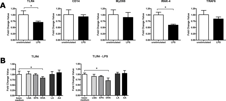
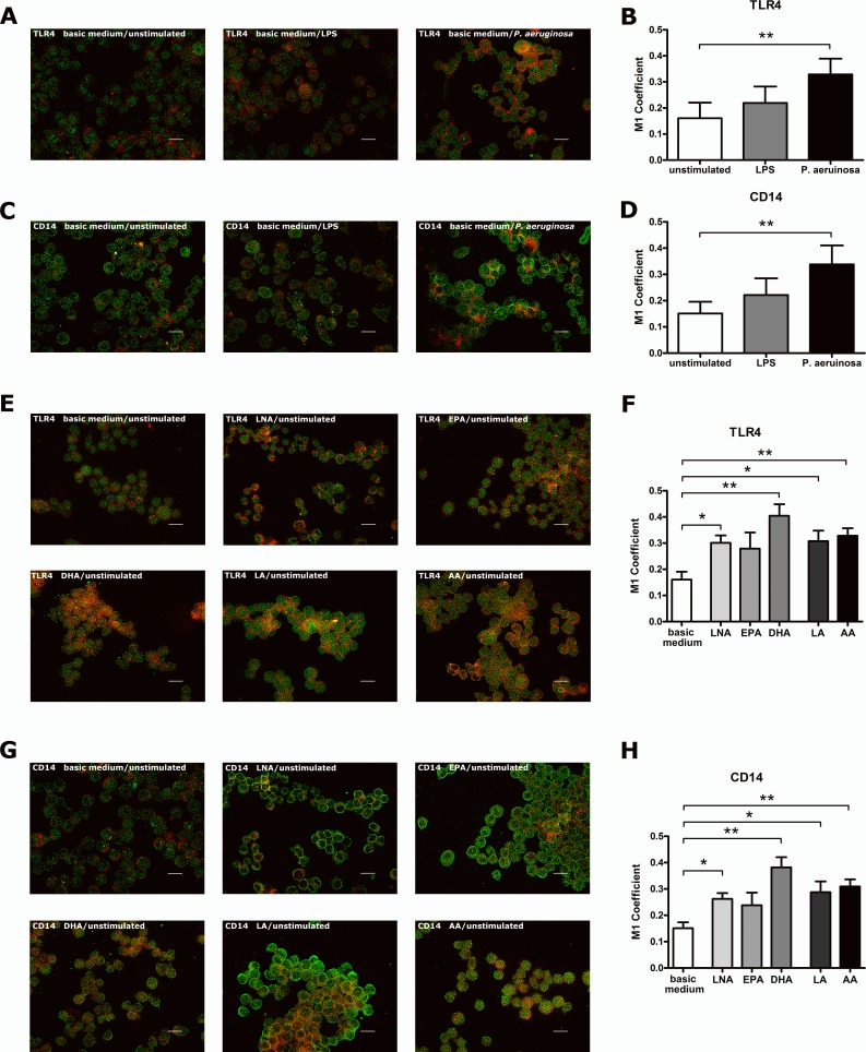
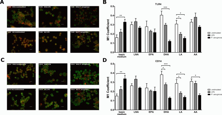
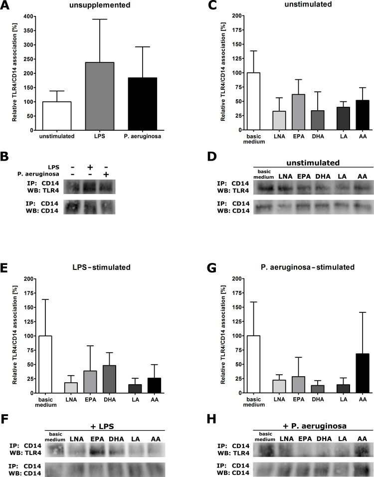
It is well known that PUFA impede the LPS-mediated activation of the transcription factor NFkappaB. However, the underlying mode of action has not been clarified yet. To address this issue in a comprehensive approach, we used the monocyte/macrophage cell line RAW264.7 to investigate the consequences of a PUFA supplementation on the TLR4 pathway with a focus on (i) the gene expression of TLR4 itself as well as of its downstream mediators, (ii) the membrane microdomain localization of TLR4 and CD14, (iii) the stimulation-induced interaction of TLR4 and CD14. Our data indicate that the impairment of the TLR4-mediated cell activation by PUFA supplementation is not due to changes in gene expression of mediator proteins of the signaling cascade. Rather, our data provide evidence that the PUFA enrichment of macrophages affects the TLR4 pathway at the membrane level. PUFA incorporation into membrane lipids induces a reordering of membrane microdomains thereby affecting cellular signal transduction. It is important to note that this remodeling of macrophage rafts has no adverse effect on cell viability. Hence, microdomain disruption via macrophage PUFA supplementation has a potential as non-toxic strategy to attenuate inflammatory signaling.

Keywords: CD14; Lipid rafts; Macrophages; PUFA; TLR4.


Conflict of interest statement

The authors declare there are no competing interests.

Figures

Figure 1
Figure 1. LPS- or PUFA-mediated modulation of gene expression of TLR4, CD14, MyD88, IRAK-4 and TRAF6 in RAW264.7 macrophages.
Gene expression was determined by quantitative real-time PCR of total RNA isolated from RAW264.7 macrophages using the housekeeping gene CASC3 for normalization of mRNA expression levels. Data are expressed as mean ± S.D. (N = 3, n = 3). Asterisks indicate a statistically significant difference compared with the unstimulated or unsupplemented controls (p < 0.05, ∗∗p < 0.01). (A) Cells were cultured in basic medium (RPMI 1640 containing 4.5 g/L glucose, 5% v/v FCS, 0.2% v/v ethanol) and stimulated with LPS (1 µg/mL) for 24 h. (B) Cells were cultured in basic medium supplemented with alpha-linolenic acid (LNA), eicosapentaenoic acid (EPA), docosahexaenoic acid (DHA), linoleic acid (LA) or arachidonic acid (AA) in a concentration of 15 µM for 72 h. Cell stimulation was performed by addition of LPS (1 µg/mL) to the culture medium in the last 24 h of incubation.
Figure 2
Figure 2. Stimulus- or PUFA-mediated modulation of the co-localization of TLR4 or CD14 with the raft marker GM1 on RAW264.7 macrophages.
Co-localization of TLR4 or CD14 with the raft marker GM1 on RAW264.7 macrophages was analyzed by indirect immunofluorescence microscopy. Cells were cultured in basic medium (RPMI 1640 containing 4.5 g/L glucose, 5% v/v FCS, 0.2% v/v ethanol). Stimulation was performed by supplementation of basic medium with LPS (1 µg/mL) or viable P. aeruginosa (MOI 1) for 24 h. PUFA enrichment was performed by supplementation of basic medium with alpha-linolenic acid (LNA), eicosapentaenoic acid (EPA), docosahexaenoic acid (DHA), linoleic acid (LA) or arachidonic acid (AA) in a concentration of 15 µM for 72 h. Cells, grown on coverslips, were fixed in 4% paraformaldehyde, permeabilized in 0.1% Triton X-100 and subsequently immunostained using specific primary antibodies against TLR4, CD14 and GM1, respectively, as well as Alexa Fluor-labeled secondary antibodies. Representative images from 4 independent experiments were captured. GM1 is labeled in red; TLR4 and CD14 are labeled in green. Scale bar represents 20 µm. Quantification of TLR4 or CD14 co-localization with GM1 was performed by calculating the Manders‘ (M1) coefficient using the JACoP plugin of ImageJ. Data are expressed as mean ± S.D. (N = 4, n = 3). Asterisks indicate a statistically significant difference compared with the unstimulated or unsupplemented controls (p < 0.05, ∗∗p < 0.01). (A) Representative images showing the stimulus effects on TLR4-GM1 co-localization. High resolution images can be found in Figs. S2–S4. (B) Graph showing the stimulus effects on TLR4-GM1 co-localization. (C) Representative images showing the stimulus effects on CD14-GM1 co-localization. High resolution images can be found in Figs. S5–S7. (D) Graph showing the stimulus effects on CD14-GM1 co-localization. (E) Representative images showing the PUFA effects on TLR4-GM1 co-localization. High resolution images can be found in Figs. S8–S13. (F) Graph showing the PUFA effects on TLR4-GM1 co-localization. (G) Representative images showing the PUFA effects on CD14-GM1 co-localization. High resolution images can be found in Figs. S14–S19. (H) Graph showing the PUFA effects on CD14-GM1 co-localization.
Figure 3
Figure 3. Co-localization of TLR4 or CD14 with the raft marker GM1 on PUFA-enriched and stimulated RAW264.7 macrophages.
Co-localization of TLR4 or CD14 with the raft marker GM1 on RAW264.7 macrophages was analyzed by indirect immunofluorescence microscopy. Cells were cultured in basic medium (RPMI 1640 containing 4.5 g/L glucose, 5% v/v FCS, 0.2% v/v ethanol) supplemented with alpha-linolenic acid (LNA), eicosapentaenoic acid (EPA), docosahexaenoic acid (DHA), linoleic acid (LA) or arachidonic acid (AA) in a concentration of 15 µM for 72 h. Cell stimulation was performed by addition of LPS (1 µg/mL) or viable P. aeruginosa (MOI 1) to the culture medium in the last 24 h of incubation. Cells grown on coverslips were fixed in 4% paraformaldehyde, permeabilized in 0.1% Triton X-100 and subsequently immunostained using specific primary antibodies against TLR4, CD14 and GM1, respectively, as well as Alexa Fluor-labeled secondary antibodies. Representative images from 4 independent experiments were captured. GM1 is labeled in red; TLR4 and CD14 are labeled in green. Scale bar represents 20 µm. Quantification of TLR4 or CD14 co-localization with GM1 was performed by calculating the Manders‘ (M1) coefficient using the JACoP plugin of ImageJ. Data are expressed as mean ± S.D. (N = 4, n = 3). Asterisks indicate a statistically significant difference compared with the unstimulated controls (p < 0.05, ∗∗p < 0.01, ∗∗∗p < 0.001). (A) Representative images showing the stimulus effects on TLR4-GM1 co-localization of PUFA-supplemented RAW264.7. High resolution images can be found in Figs. S20–S25. (B) Graph showing the stimulus effects on TLR4-GM1 co-localization of PUFA-supplemented RAW264.7. (C) Representative images showing the stimulus effects on CD14-GM1 co-localization of PUFA-supplemented RAW264.7. High resolution images can be found in Figs. S26–S31. (D) Graph showing the stimulus effects on CD14-GM1 co-localization of PUFA-supplemented RAW264.7.
Figure 4
Figure 4. Receptor association of TLR4 and CD14 of PUFA-enriched and stimulated RAW264.7 macrophages.
Formation of TLR4-CD14 complexes was determined by co-immunoprecipitation (IP) and visualized by Western blot (WB). Cells were cultured in basic medium (RPMI 1640 containing 4.5 g/L glucose, 5% v/v FCS, 0.2% v/v ethanol) supplemented with alpha-linolenic acid (LNA), eicosapentaenoic acid (EPA), docosahexaenoic acid (DHA), linoleic acid (LA) or arachidonic acid (AA) in a concentration of 15 µM for 72 h. Cell stimulation was performed by addition of LPS (1 µg/mL) or viable P. aeruginosa (MOI 1) to the culture medium in the last 24 h of incubation. TLR4-CD14 complexes from cell lysates were co-immunoprecipitated using an appropriate anti-mouse CD14 antibody and immunoblotted using an appropriate anti-mouse TLR4 antibody. Band size was checked using a protein ladder, and band intensities were analyzed densiometrically. The IP:CD14/WB:CD14 blot represents the lower part of the IP membrane and serves as loading control used for data normalization. To account for inter-assay variability, the stimulation/supplementation conditions compared were run on the same gel. Data from unstimulated (A) or unsupplemented (C+E+G) cells were set 100%, and values from stimulated (A) or supplemented (C+E+G) cells were expressed relative to this control. Four independent experiments were performed in duplicate for each combination of PUFA supplementation and stimulation of cells (N = 4, n = 2). (A) Graph showing the stimulus effects on TLR4-CD14 association of unsupplemented RAW264.7. (B) Representative Western blots showing the stimulus effects on TLR4-CD14 association of unsupplemented RAW264.7. (C) Graph showing the PUFA effects on TLR4-CD14 association of unstimulated RAW264.7. (D) Representative Western blots showing the PUFA effects on TLR4-CD14 association of unstimulated RAW264.7. (E) Graph showing the PUFA effects on TLR4-CD14 association of LPS-stimulated RAW264.7. (F) Representative Western blots showing the PUFA effects on TLR4-CD14 association of LPS-stimulated RAW264.7. (G) Graph showing the PUFA effects on TLR4-CD14 association of P. aeruginosa-stimulated RAW264.7. (H) Representative Western blots showing the PUFA effects on TLR4-CD14 association of P. aeruginosa-stimulated RAW264.7.

Similar articles

Cited by

References

    1. Adolph S, Fuhrmann H, Schumann J. Unsaturated fatty acids promote the phagocytosis of P. aeruginosa and R. equi by RAW264.7 macrophages. Current Microbiology. 2012;65(6):649–655. doi: 10.1007/s00284-012-0207-3. - DOI - PubMed
    1. Adolph S, Schoeniger A, Fuhrmann H, Schumann J. Unsaturated fatty acids as modulators of macrophage respiratory burst in the immune response against Rhodococcus equi and Pseudomonas aeruginosa. Free Radical Biology & Medicine. 2012;52(11-12):2246–2253. doi: 10.1016/j.freeradbiomed.2012.04.001. - DOI - PubMed
    1. Ambrozova G, Pekarova M, Lojek A. Effect of polyunsaturated fatty acids on the reactive oxygen and nitrogen species production by raw 264.7 macrophages. European Journal of Nutrition. 2010;49(3):133–139. doi: 10.1007/s00394-009-0057-3. - DOI - PubMed
    1. Basiouni S, Stoeckel K, Fuhrmann H, Schumann J. Polyunsaturated fatty acid supplements modulate mast cell membrane microdomain composition. Cellular Immunology. 2012;275(1–2):42–46. doi: 10.1016/j.cellimm.2012.03.004. - DOI - PubMed
    1. Chang C, Sun H, Lii C, Chen H, Chen P, Liu K. Gamma-linolenic acid inhibits inflammatory responses by regulating NF-kappa B and AP-1 Activation in Lipopolysaccharide-Induced RAW 264.7 Macrophages. Inflammation. 2010;33(1):46–57. doi: 10.1007/s10753-009-9157-8. - DOI - PubMed

Grants and funding

This work has been granted by the Deutsche Forschungsgemeinschaft (DFG, http://www.dfg.de), grant number SCHU 2586/1-2. The funders had no role in study design, data collection and analysis, decision to publish, or preparation of the manuscript.

LinkOut - more resources