. 2016 Apr 20;44(7):3330-50.
doi: 10.1093/nar/gkw061. Epub 2016 Feb 4.

Kissing-loop interaction between 5' and 3' ends of tick-borne Langat virus genome 'bridges the gap' between mosquito- and tick-borne flaviviruses in mechanisms of viral RNA cyclization: applications for virus attenuation and vaccine development

Affiliations

Kissing-loop interaction between 5' and 3' ends of tick-borne Langat virus genome 'bridges the gap' between mosquito- and tick-borne flaviviruses in mechanisms of viral RNA cyclization: applications for virus attenuation and vaccine development

Konstantin A Tsetsarkin et al. Nucleic Acids Res. .

Abstract

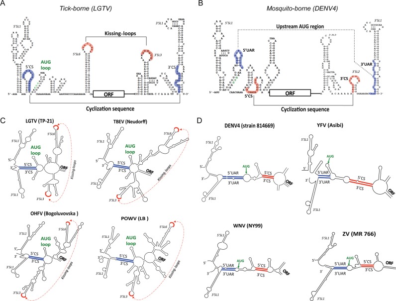
Insertion of microRNA target sequences into the flavivirus genome results in selective tissue-specific attenuation and host-range restriction of live attenuated vaccine viruses. However, previous strategies for miRNA-targeting did not incorporate a mechanism to prevent target elimination under miRNA-mediated selective pressure, restricting their use in vaccine development. To overcome this limitation, we developed a new approach for miRNA-targeting of tick-borne flavivirus (Langat virus, LGTV) in the duplicated capsid gene region (DCGR). Genetic stability of viruses with DCGR was ensured by the presence of multiple cis-acting elements within the N-terminal capsid coding region, including the stem-loop structure (5'SL6) at the 3' end of the promoter. We found that the 5'SL6 functions as a structural scaffold for the conserved hexanucleotide motif at its tip and engages in a complementary interaction with the region present in the 3' NCR to enhance viral RNA replication. The resulting kissing-loop interaction, common in tick-borne flaviviruses, supports a single pair of cyclization elements (CYC) and functions as a homolog of the second pair of CYC that is present in the majority of mosquito-borne flaviviruses. Placing miRNA targets into the DCGR results in superior attenuation of LGTV in the CNS and does not interfere with development of protective immunity in immunized mice.


Figures

Figure 1.
Figure 1.
Growth of LGTV with DCGR in Vero cells. (A) Schematic representation of viral genomes used in this study. Target sequence for mir-124 is indicated as red box, and sequence of 2A protease from FMDV is highlighted as yellow box. Cleavage site for 2A protease is indicated by black arrow. Gray areas represent codon optimized C protein gene sequence. C17 AA, C27 AA, C68 AA and C89 AA boxes represent the duplicated regions of 5′ end of C protein gene encoding 17, 27, 68 and 89 AA of truncated C protein, respectively. (B) Growth kinetics of infectious virus recovered after plasmid DNA transfection into Vero cells. Individual samples for each time point from one biological replica were titrated in Vero cells in duplicate, and results are presented as an average ± standard deviation (shown as error bars). The dashed line indicates the limit of virus detection [0.7 log10(pfu/ml)]. (C) Immunofluorescence analysis. Representative images show the expression of LGTV antigens in Vero cells on day 5 post-transfection with indicated pDNA construct.
Figure 2.
Figure 2.
Growth and genetic stability in Vero cells of LGTV with ORF shifting mutations. (A) Top: stem-loop structure for the 5′ end of LGTV TP-21 genome. Middle: schematic representation of viral genomes used in the study. Positions of ORF-shifting insertion (+1 nt) of a single A nucleotide (C8 and C19) and position of reading frame restoration (−1 nt at C68 position) are indicated. Bottom: diagram indicating relative position of primers used for RT-PCR analysis of LGTV genomes. (B) Growth kinetics of infectious virus recovered after plasmid DNA transfection into Vero cells. Aliquots of cell culture samples harvested at indicated time from one biological replica were titrated in Vero cells in duplicate. Results are presented as an average ± standard deviation. (C) Top: results of RT-PCR analysis of indicated virus genomes isolated after ten passages in Vero cells. White arrow indicates the position of DNA fragment produced by RT-PCR and corresponding to the size of DNA fragment derived from unmodified parental LGTV genome. Bottom: results of sequence analysis of mir-124 target region for C68 FrSh1 and C68 FrSh2 virus amplicons that are shown above. (D) Top: schematic representation of hypothetical reversion of C68 FrSh1 and C68 FrSh2 genomes to wt-like LGTV containing chimeric C protein gene sequence. Viral genomes (wtC8/opt, wtC19/opt and wtC42/opt) containing 8, 19 or 42 AA codons of wt LGTV C protein gene and the rest of codon-optimized C protein gene are shown. A wt sequence is indicated in white, and a codon-optimized sequence is in grey. Bottom: growth kinetics of recovered LGTV with chimeric capsid gene sequences in Vero cells. The dashed line indicates the limit of virus detection [0.7 log10(pfu/ml)].
Figure 3.
Figure 3.
Mapping of a minimal region of C gene that is required for efficient LGTV growth in Vero cells. (A) Top: stem-loop structure of the 5′-end of TP-21 genome. Colored arrows indicate positions of fusion sites between wt C gene sequences and target sequence for mir-124 (red box). Bottom: schematic representation of viral genomes carrying the indicated portion of truncated C gene. (B and C) Growth kinetics of recovered viruses in Vero cells.
Figure 4.
Figure 4.
Role of sequence between C15 AA and C24 AA of C gene on growth of LGTV in Vero cells. (A) Top: stem-loop structure of the 5′-end of TP-21 genome. Orange arrows indicate the end of C15 AA and C24 AA regions in the C gene sequence. Target sequence for mir-124 is highlighted as red box. Bottom: schematic representation of C68 genomes and its derivatives used in the study. Green boxes indicate the position and nt length of inserted sequences. (B) Growth kinetics of recovered viruses in Vero cells. (C) Immunofluorescence analysis of LGTV antigen expression in Vero cells on day 5 post-transfection with indicated DNA constructs.
Figure 5.
Figure 5.
Significance of stem-loop organization of the 5′SL6 for virus growth in Vero cells. (A) The predicted secondary structure of 5′SL6 in C68 genome was destabilized by mutations (highlighted in red) in the 3′ strand to create the C68/124(T) virus. The 5′SL6-like secondary structure was restored in C68/124(T) by introducing a set of compensatory complementary mutations in the 5′ strand of 5′SL6 to create the C68/124(T)* virus. (B) Growth kinetics of rescued viruses in Vero cells. Aliquots of viruses in cell cultures were harvested at indicated time points from one biological replica, and virus titers were determined in Vero cells in duplicate. (C) Results of immunofluorescence assay for LGTV antigens in Vero cells on day 5 post-transfection are shown.
Figure 6.
Figure 6.
Kissing-loop interaction between 5′ and 3′ ends of LGTV genome. (A) Top: secondary structures and predicted interactions between the 5′SL6 and 3′SL3 (3′NCR) in the C68 genome. Nucleotide residues involved in complementary interactions are in blue circles connected by dashed lines. Bottom: hexanucleotide motifs at the tip of 5′SL6 and/or 3′SL3 C68 virus were substituted with 5′mut1 or 3′mut1 sequences (highlighted in red) to generate C68/5′mut1, C68/3′mut1 or C68/5′3′mut1 viruses. Potential kissing-loop contact is represented by blue dashed line, which can be destabilized by mut1 substitutions (crossed blue dashed line). (B and E) Growth kinetics of recovered viruses after DNA transfection into Vero cells. (C and F) Immunofluorescence analysis of LGTV antigen expression in Vero cells on day 5 post-transfection. (D) Schematic representation of mutations that disrupted or restored interaction between the sequences at the tips of 5′SL6 and 3′SL3 in the C68 genome. Introduced substitutions (5′mut2 or 3′mut2 sequences) are highlighted in red.
Figure 7.
Figure 7.
Kissing-loop interaction between 5′SL6 and 3′SL3 regulates LGTV replication but does not affect viral RNA translation. (A) Genetic organization of the RepC58 replicon that was constructed on the basis of C58 virus genome (Figure 3A). Regions of nLuc gene insertion and deletion of cleavage site for cellular furin protease are indicated. To generate RepC58/5′mut2, RepC58/3′mut2 or RepC58/5′3′mut2 replicons, hexanucleotide motifs at the tip of 5′SL6 and/or 3′SL3 of RepC58 replicon were substituted with 5′mut2 and/or 3′mut2 sequences (Figure 6A and D). Red arrows and dashed line indicate the region that was deleted to generate non-replicating replicons. Target sequence for mir-124 and 2A protease gene sequence from FMDV are indicated as red and yellow boxes, respectively. Positions of ORF shift insertion (+1 nt after a C8 AA codon) and frame shift restoration (−1 nt in a C58 codon) are shown. (B) Vero cells were mock-transfected or transfected with RNA transcripts of replication-competent replicons (a set of RepC58). (C) Vero cells were transfected with RNA transcripts of non-replicating versions of replicons (a set of Rep*C58). (B and C) Normalized kinetics of relative luminescence are presented as the mean values ± SD (shown as error bars) of two reads for 3 biological replicates of RNA-transfected Vero cell extracts.
Figure 8.
Figure 8.
Compensatory mutations in the 3′CYC element of LGTV compensate for dysfunctional kissing-loop contact. (A) Mutations that were identified in the genomes of C68/5′mut2 and C68/3′mut2 viruses after 10 passages in Vero cells. (B) Top: predicted RNA structure of 5′- and 3′-ends of LGTV genome. Cyclization element (CYC), a region of complementarity between 5′ cyclization sequence (CS) and 3′CS of CYC element, is highlighted in pink. Kissing-loop contact is shown as black dashed line. The blue star shows location of U→C substitution common for both Vero cell passaged viruses. Bottom: the same U→C substitution is shown in context of nt base pairing of 5′CS and 3′CS in the CYC element. Predicted ΔG values are given for the CYC structure with and without U→C substitution in the 3′CS. (C and D) Effect of U→C substitution (CS) on growth of viruses with unmodified (C68) and restored (C68/5′3′mut2) kissing-loop contacts (C) or viruses with impaired (C68/5′mut2, C68/3′mut28) kissing-loop contacts (D) in Vero cells. Growth kinetics of engineered C68 and C68/5′3′mut2 (C), or C68/5′mut2 and C68/3′mut2 (D) viruses with (shown as dashed lines) and without (shown as solid lines) U→C substitution in the 3′CS.
Figure 9.
Figure 9.
Functional kissing-loop contact between 5′and 3′ ends of LGTV genome can occur if hexanucleotide motif from the tip of 5′SL6 is relocated to the neighboring stem loop structure. (A) Schematic representations of viral genomes with nt mutations or 40 nt insertion that were engineered in 5′SL6 and/or 5′SL7 sequences of C68 virus in order to relocate the position of 5′ kissing-loop sequence. (B) Growth kinetics of parental C68 and its mutant derivatives in transfected Vero cells.
Figure 10.
Figure 10.
Effect of brain-specific miRNA target insertions into the DCGR on growth of LGTV in CNS of newborn mice. (A) Schematic representation of viruses with a single copy of mir-124 target or its scrambled sequence as well as with miRNA targets for mir-1, mir-9, mir-128, mir-132, mir-137 and mir-139. (B and C). Growth kinetics of miRNA-targeted viruses in mouse brains. Three-day-old mice were inoculated IC with 100 pfu of indicated virus, and brains from three mice in each group were collected at the indicated time points. Mean titers (±SD) of brain homogenates are shown. Differences in growth kinetics were compared using 2-way ANOVA. P-values were adjusted using Tukey's multiple comparison test.
Figure 11.
Figure 11.
Insertion of miRNA target sequences for brain-specific miRNA into the DCGR resulted in more efficient LGTV attenuation in the CNS of newborn mice as compared with target insertion into 3′NCR. (A) Schematic representation of viruses used in this study. Genome sequence encoding the nonstructural proteins in the LGTV TP-21 strain and C48 virus was replaced with the corresponding sequence (gray area) derived from infectious cDNA clone of LGTV E5 strain (35). Target sequences for mir-1, mir-9 and mir-124 miRNAs or modified (scramble) miRNA targets were introduced in DCGR or 3′NCR as indicated. In the 3′NCR, a set of mir-1, mir-9 and mir-124 targets and a second copy of mir-124 target were inserted after nt 10 and 245, respectively. (B and C) Growth kinetics of miRNA-targeted viruses in mouse brains. Three-day-old mice were inoculated IC with a 100 pfu of each indicated virus. Mean titers (±SD) of three brain homogenates are shown for each time point. Differences in growth kinetics were compared between 124(2)/9/1 and miRNA targeted viruses (B), or between C48–124(2)/9/1-E5 and 3UTR-124(2)/9/1-E5 (C) using 2-way ANOVA. P-values were adjusted using Tukey's multiple comparison test.
Figure 12.
Figure 12.
Structural comparison of tick-borne and mosquito-borne flavivirus genomic regions involved in viral RNA cyclization. (A and B) Stem-loop structures in 5′ and 3′ termini of LGTV (A) and dengue type 4 virus (B) genomes are shown in a linear configuration. Regions potentially involved in putative interactions between 5′ and 3′ termini of RNA genome are highlighted as blue and red sequences. (C and D) Predicted structures of genomic RNAs are given in the circular configurations for representative members of tick-borne (C) and mosquito-borne (D) flaviviruses. Secondary RNA structures were predicted using m-fold web server for nucleic acid folding as described in materials and methods. Note: 5′UAR is the 5′upstream AUG codon region, which is complementary to the 3′end region of the virus genome (3′UAR).

Similar articles

Cited by

References

    1. Lasala P.R., Holbrook M. Tick-borne flaviviruses. Clin. Lab. Med. 2010;30:221–235. - PubMed
    1. Gritsun T.S., Lashkevich V.A., Gould E.A. Tick-borne encephalitis. Antiviral Res. 2003;57:129–146. - PubMed
    1. Smith C.E. A virus resembling Russian spring-summer encephalitis virus from an ixodid tick in Malaya. Nature. 1956;178:581–582. - PubMed
    1. Pletnev A.G., Men R. Attenuation of the Langat tick-borne flavivirus by chimerization with mosquito-borne flavivirus dengue type 4. Proc. Natl. Acad. Sci. U.S.A. 1998;95:1746–1751. - PMC - PubMed
    1. Price W.H., Thind I.S., Teasdall R.D., O'Leary W. Vaccination of human volunteers against Russian spring-summer (RSS) virus complex with attenuated Langat E5 virus. Bull. World Health Organ. 1970;42:89–94. - PMC - PubMed

Publication types

MeSH terms