. 2016 Jan 27;36(4):1362-72.
doi: 10.1523/JNEUROSCI.5090-14.2016.

Conformational Changes in Transmembrane Domain 4 of Presenilin 1 Are Associated with Altered Amyloid-β 42 Production

Affiliations

Conformational Changes in Transmembrane Domain 4 of Presenilin 1 Are Associated with Altered Amyloid-β 42 Production

Aya Tominaga et al. J Neurosci. .

Abstract

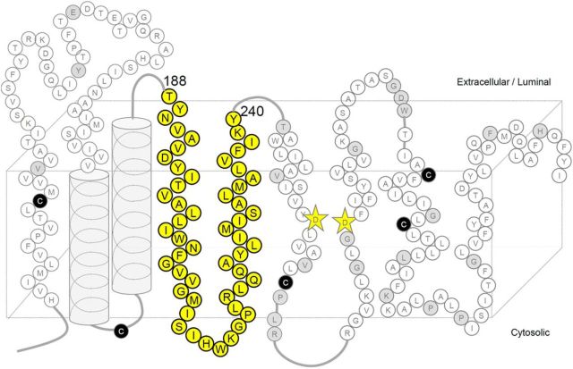
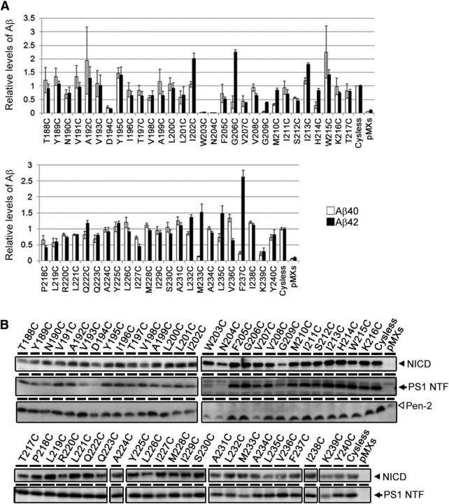
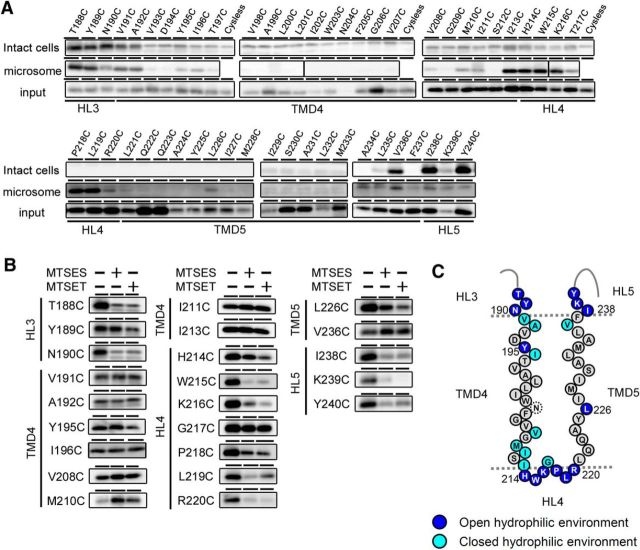
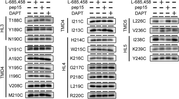
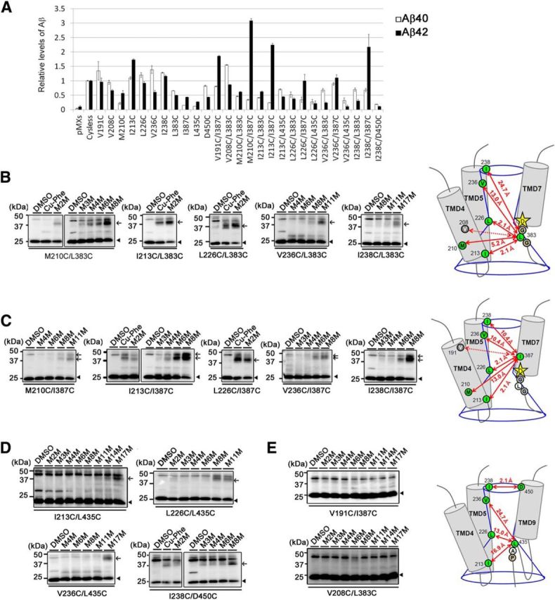
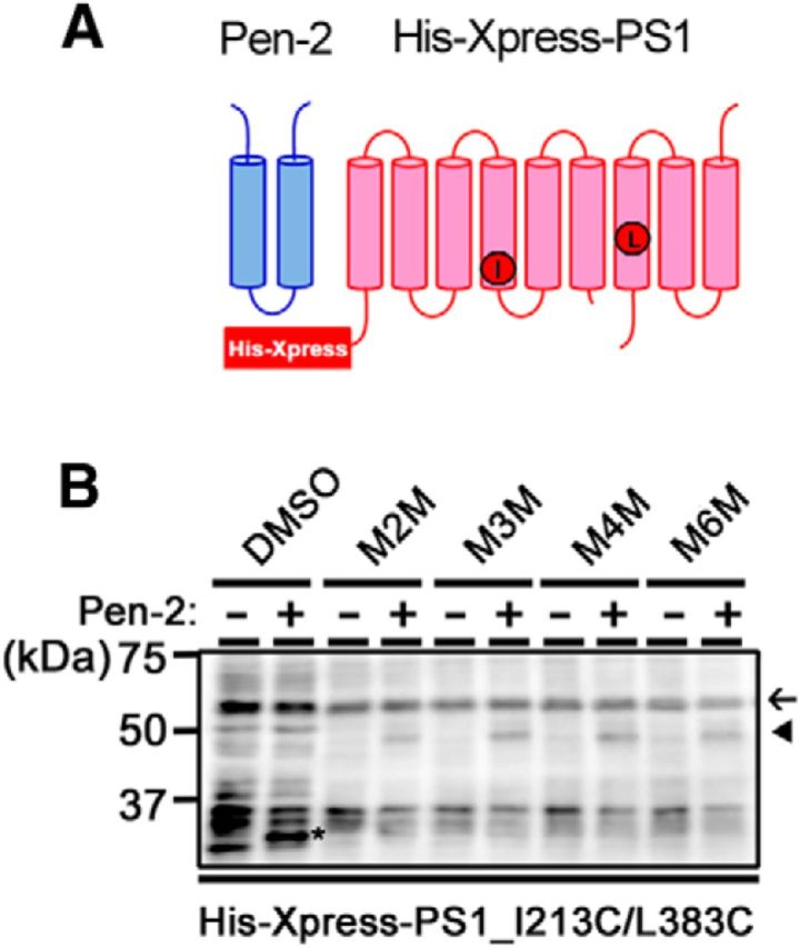
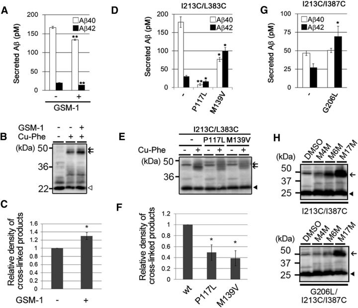
γ-Secretase is an intramembrane-cleaving protease that produces amyloid-β peptide 42 (Aβ42), which is the toxic and aggregation-prone species of Aβ that causes Alzheimer's disease. Here, we used the substituted cysteine accessibility method to analyze the structure of transmembrane domains (TMDs) 4 and 5 of human presenilin 1 (PS1), a catalytic subunit of γ-secretase. We revealed that TMD4 and TMD5 face the intramembranous hydrophilic milieu together with TMD1, TMD6, TMD7, and TMD9 of PS1 to form the catalytic pore structure. Notably, we found a correlation in the distance between the cytosolic sides of TMD4/TMD7 and Aβ42 production levels, suggesting that allosteric conformational changes of the cytosolic side of TMD4 affect Aβ42-generating γ-secretase activity. Our results provide new insights into the relationship between the structure and activity of human PS1.

Significance statement: Modulation of γ-secretase activity to reduce toxic amyloid-β peptide species is one plausible therapeutic approaches for Alzheimer's disease. However, precise mechanistic information of γ-secretase still remains unclear. Here we identified the conformational changes in transmembrane domains of presenilin 1 that affect the proteolytic activity of the γ-secretase. Our results highlight the importance of understanding the structural dynamics of presenilin 1 in drug development against Alzheimer's disease.

Keywords: allosteric change; amyloid; enzyme; membrane protein; protease; secretase.


Figures

Figure 1.
Figure 1.
Locations of the Cys mutations of PS1 used in this study. Schematic diagram of human PS1. Endogenous Cys that were replaced with Ser in PS1/Cys(−) are shown as black circles. Amino acid residues substituted to Cys in this and previous studies are shown as circles with their single-letter character representing the original amino acids. Residues analyzed in this study are shown as yellow circles. Residues in which Cys substitution resulted in a loss of γ-secretase activity are shown as gray circles. Catalytic aspartates are shown as yellow stars.
Figure 2.
Figure 2.
γ-Secretase activity of single-Cys mt PS1 used in this study. A, Sandwich ELISA of secreted Aβ from APPNL-stable DKO cells transiently transfected with single-Cys mt PS1 (n = 3–12, means ± SEs). The levels of secreted Aβ in the conditioned media were normalized by that of cells transfected with PS1/Cys(−). Amounts of Aβ40 production are shown as white bars, and amounts of Aβ42 production are shown as black bars. B, NICD generation from NotchΔE-stable DKO cells transiently transfected with single-Cys mt PS1 was detected by immunoblot analysis using an anti-cleaved Notch1 antibody (top panels). The levels of expressed PS1 NTF and Pen-2 were detected by immunoblot analysis using anti-PS1 NTF and Pen-2 antibodies (bottom panels).
Figure 3.
Figure 3.
SCAM analysis of single-Cys mt PS1s mutated around TMD4 and TMD5. A, Biotinylation experiment using MTSEA-biotin in intact cells (top panels) and microsomes (middle panels). Amounts of PS1 NTF in the input fraction are shown in the bottom panels. Putative domains are indicated below the panels. B, Labeling competition of single-Cys mt PS1s that were labeled by MTSEA-biotin in A was performed after preincubation with negatively charged MTSES or positively charged MTSET. Putative domains are indicated at the left of the panels. C, Summary of the biotinylation experiments using MTSEA-biotin and competition experiments using charged MTS reagents. All charged reagents were accessible to the Cys-substituted amino acids shown as dark blue circles (open hydrophilic environment). Residues in which labeling were not competed by MTSES or MTSET are shown as light blue circles (closed hydrophilic environment). Residues that were not labeled by MTSEA-biotin are shown as black letters in gray circles. N204C (dotted circle) was not endoproteolyzed.
Figure 4.
Figure 4.
Labeling competition by GSIs. Labeling of single-Cys mt PS1s by MTSEA-biotin was performed after preincubation with l-685,458, pep15, or DAPT. Putative domains are indicated at the left of the panels.
Figure 5.
Figure 5.
Cross-linking experiments using catalysts or MTS cross-linkers. A, Sandwich ELISA of secreted Aβ from APPNL-stable DKO cells transiently transfected with double-Cys mt PS1 (n = 3, mean ± SEs). Cross-linking experiments of double-Cys mt PS1 with Cys mutations in TMD4 or TMD5 and L383C (B), I387C (C), L435C, or D450C (D). Immunoblot analysis was performed using an anti-G1Nr5 antibody. PS1 NTFs and cross-linked products (NTF–CTF heterodimers) are shown by black arrowheads and black arrows, respectively. Double-Cys mt PS1s that failed to be cross-linked were shown in E. The predicted maximum lengths between two residues are indicated as red lines in the schematic illustrations on the right. Residues mutated to Cys are shown by circles. Positions of cross-linked cysteines were indicated by green circles. Predicted structure of the catalytic pore and distances between cross-linked residues were indicated by blue lines and red arrows, respectively. Positions of catalytic aspartates and conserved motifs were shown by stars and white circles, respectively.
Figure 6.
Figure 6.
Conformational changes in TMD4 are associated with γ-secretase complex assembly. A, Schematic diagram of Pen-2 and His-Xpress-PS1 used in this study. Positions of I213 and L383 were shown as red circles. B, Cross-linking experiments of double-Cys mt PS1 with I213C and L383C mutations, in P2KO cells with or without Pen-2 overexpression. Western blotting was performed using the anti-Xpress antibody. His-Xpress-PS1 NTF (asterisk) appeared only in the Pen-2-expressing cells. His-Xpress-PS1 holoprotein, NTFs and cross-linked products (NTF–CTF heterodimers) are shown by black arrow, asterisk, and arrowhead, respectively. Note that the cross-linked product also appeared only in the Pen-2-expressing cells.
Figure 7.
Figure 7.
Conformational changes in TMD4 associated with altered Aβ42 ratio. A, Sandwich ELISA of secreted Aβ from APPNL-stable DKO cells expressing I213C/L383C mt PS1/Cys(−) after preincubation with 30 μm GSM-1 (n = 3, means ± SEs; **p < 0.01 by Student's t test). B, Cross-linking experiments of I213C/L383C mt PS1 with a catalyst (Cu-Phe) after preincubation with GSM-1 (30 μm). Immunoblot analysis was performed using an anti-M5 antibody. PS1 CTFs and cross-linked products (NTF–CTF heterodimers) are indicated by the white arrowhead and black arrows, respectively. Densitometric analysis of the results are shown in C (n = 3, means ± SEs; *p < 0.05 by Student's t test). D, Sandwich ELISA of secreted Aβ from APPNL-stable DKO cells expressing I213C/L383C mt PS1 with an FAD-linked mutation (I213C/L383C/P117L or I213C/L383C/M139V; n = 3, means ± SEs; *p < 0.05, **p < 0.01 by Student's t test). E, Cross-linking experiments of I213C/L383C mt PS1 with or without FAD-linked mutations and a catalyst (Cu-Phe). Immunoblot analysis was performed using an anti-G1Nr5 antibody. PS1 NTFs and cross-linked products are indicated by a black arrowhead and black arrows, respectively. Densitometric analysis of the results are shown in F (n = 3, means ± SEs; *p < 0.05 by Student's t test). G, Sandwich ELISA of secreted Aβ from APPNL-stable DKO cells expressing I213C/I387C or G206L/I213C/I387C mt PS1 (n = 3, means ± SEs; *p < 0.05 by Student's t test). H, Cross-linking experiments of I213C/L383C or G206L/I213C/I387C mt PS1 with MTS cross-linkers. Immunoblot analysis was performed using an anti-G1Nr5 antibody. PS1 NTFs, and cross-linked products are indicated by black arrowheads and black arrows, respectively.
Figure 8.
Figure 8.
Structure and function of TMD4 and TMD5 of PS1. A, Summary of SCAM analyses and cross-linking experiments. The cytosolic residues of TMD4, TMD5, and L383, which face the catalytic pore, are shown as green circles. Catalytic aspartate on TMD7 is shown by stars. Conserved glycines in the catalytic motif are denoted as gray circles. GSM-1 treatment reduced the Aβ42 production and the distance between I213 and L383 in the catalytic site (left). In contrast, FAD-linked mutation increased the Aβ42 levels and the distance (right), suggesting that the proximity between the cytosolic sides of TMD4 and TMD7 correlates with Aβ42-generating activity. B, Schematic diagram of TMD4 and TMD5 (shown in yellow) in the PS1 model (Li et al., 2013). Residues facing the hydrophilic environment are shown as blue spheres and circles. Catalytic aspartates are shown as red spheres. Conserved glycines and the residues involved in the binding with l-685,458 are shown as pink and green circles, respectively. The catalytic pore is indicated by blue lines. C, Alignment of primary sequences of PS1 or its homolog TMD4 of Homo sapiens, Xenopus laevis, Drosophila melanogaster, Caenorhabditis elegans, and M. marisnigri. Positions of conserved glycines and isoleucine are indicated in pink and green, respectively.

Similar articles

Cited by

References

    1. Abramson J, Smirnova I, Kasho V, Verner G, Kaback HR, Iwata S. Structure and mechanism of the lactose permease of Escherichia coli. Science. 2003;301:610–615. doi: 10.1126/science.1088196. - DOI - PubMed
    1. Aller SG, Yu J, Ward A, Weng Y, Chittaboina S, Zhuo R, Harrell PM, Trinh YT, Zhang Q, Urbatsch IL, Chang G. Structure of P-glycoprotein reveals a molecular basis for poly-specific drug binding. Science. 2009;323:1718–1722. doi: 10.1126/science.1168750. - DOI - PMC - PubMed
    1. Asami-Odaka A, Ishibashi Y, Kikuchi T, Kitada C, Suzuki N. Long amyloid beta-protein secreted from wild-type human neuroblastoma IMR-32 cells. Biochemistry. 1995;34:10272–10278. doi: 10.1021/bi00032a022. - DOI - PubMed
    1. Bai XC, Rajendra E, Yang G, Shi Y, Scheres SH. Sampling the conformational space of the catalytic subunit of human gamma-secretase. Elife. 2015a doi: 10.7554/eLife.11182. doi: 10.7554/eLife.11182. Advance online publication. Retrieved December 28, 2015. - DOI - DOI - PMC - PubMed
    1. Bai XC, Yan C, Yang G, Lu P, Ma D, Sun L, Zhou R, Scheres SH, Shi Y. An atomic structure of human gamma-secretase. Nature. 2015b;525:212–217. doi: 10.1038/nature14892. - DOI - PMC - PubMed

Publication types

MeSH terms

LinkOut - more resources