

Hepatocyte mitochondrial DNA drives nonalcoholic steatohepatitis by activation of TLR9
- PMID: 26808498
- PMCID: PMC4767345
- DOI: 10.1172/JCI83885
Hepatocyte mitochondrial DNA drives nonalcoholic steatohepatitis by activation of TLR9
Abstract
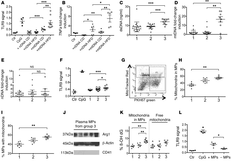
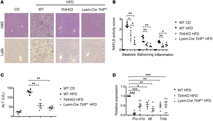
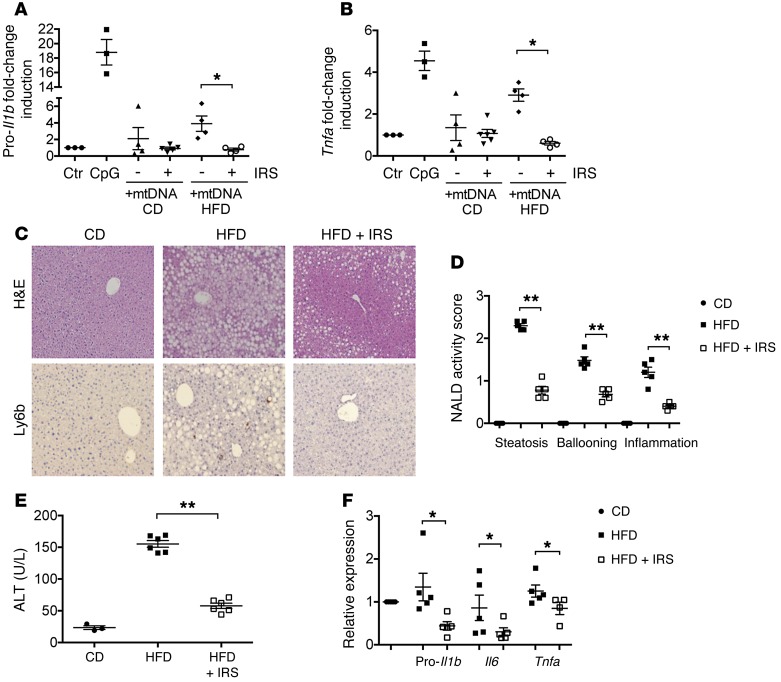
Nonalcoholic steatohepatitis (NASH) is the most common liver disease in industrialized countries. NASH is a progressive disease that can lead to cirrhosis, cancer, and death, and there are currently no approved therapies. The development of NASH in animal models requires intact TLR9, but how the TLR9 pathway is activated in NASH is not clear. Our objectives in this study were to identify NASH-associated ligands for TLR9, establish the cellular requirement for TLR9, and evaluate the role of obesity-induced changes in TLR9 pathway activation. We demonstrated that plasma from mice and patients with NASH contains high levels of mitochondrial DNA (mtDNA) and intact mitochondria and has the ability to activate TLR9. Most of the plasma mtDNA was contained in microparticles (MPs) of hepatocyte origin, and removal of these MPs from plasma resulted in a substantial decrease in TLR9 activation capacity. In mice, NASH development in response to a high-fat diet required TLR9 on lysozyme-expressing cells, and a clinically applicable TLR9 antagonist blocked the development of NASH when given prophylactically and therapeutically. These data demonstrate that activation of the TLR9 pathway provides a link between the key metabolic and inflammatory phenotypes in NASH.
Figures
Comment in
-
NASH: NASH and TLR9.Nat Rev Gastroenterol Hepatol. 2016 Mar;13(3):124. doi: 10.1038/nrgastro.2016.20. Epub 2016 Feb 10. Nat Rev Gastroenterol Hepatol. 2016. PMID: 26860268 No abstract available.
-
Hepatocyte mitochondrial DNA released in microparticles and toll-like receptor 9 activation: A link between lipotoxicity and inflammation during nonalcoholic steatohepatitis.Hepatology. 2016 Aug;64(2):669-71. doi: 10.1002/hep.28666. Epub 2016 Jun 24. Hepatology. 2016. PMID: 27239763 Free PMC article. No abstract available.
Similar articles
-
Mitochondrial DNA from hepatocytes induces upregulation of interleukin-33 expression of macrophages in nonalcoholic steatohepatitis.Dig Liver Dis. 2020 Jun;52(6):637-643. doi: 10.1016/j.dld.2020.03.021. Epub 2020 Apr 29. Dig Liver Dis. 2020. PMID: 32360132
-
Mitochondrial DNA from hepatocytes as a ligand for TLR9: Drivers of nonalcoholic steatohepatitis?World J Gastroenterol. 2016 Aug 21;22(31):6965-71. doi: 10.3748/wjg.v22.i31.6965. World J Gastroenterol. 2016. PMID: 27610009 Free PMC article.
-
TLR9 is up-regulated in human and murine NASH: pivotal role in inflammatory recruitment and cell survival.Clin Sci (Lond). 2017 Jul 24;131(16):2145-2159. doi: 10.1042/CS20160838. Print 2017 Aug 15. Clin Sci (Lond). 2017. PMID: 28687713
-
TLR9 in MAFLD and NASH: At the Intersection of Inflammation and Metabolism.Front Endocrinol (Lausanne). 2021 Jan 29;11:613639. doi: 10.3389/fendo.2020.613639. eCollection 2020. Front Endocrinol (Lausanne). 2021. PMID: 33584545 Free PMC article. Review.
-
Pathogenesis of Nonalcoholic Steatohepatitis.Gastroenterology. 2016 Jun;150(8):1769-77. doi: 10.1053/j.gastro.2016.02.066. Epub 2016 Feb 27. Gastroenterology. 2016. PMID: 26928243 Free PMC article. Review.
Cited by
-
Intraperitoneal injection of class A TLR9 agonist enhances anti-PD-1 immunotherapy in colorectal peritoneal metastases.JCI Insight. 2022 Oct 24;7(20):e160063. doi: 10.1172/jci.insight.160063. JCI Insight. 2022. PMID: 36278484 Free PMC article.
-
Large Extracellular Vesicles: Have We Found the Holy Grail of Inflammation?Front Immunol. 2018 Dec 13;9:2723. doi: 10.3389/fimmu.2018.02723. eCollection 2018. Front Immunol. 2018. PMID: 30619239 Free PMC article. Review.
-
Oxidized Mitochondrial DNA Engages TLR9 to Activate the NLRP3 Inflammasome in Myelodysplastic Syndromes.Int J Mol Sci. 2023 Feb 15;24(4):3896. doi: 10.3390/ijms24043896. Int J Mol Sci. 2023. PMID: 36835307 Free PMC article.
-
Liquid Biopsy for the Diagnosis of Viral Hepatitis, Fatty Liver Steatosis, and Alcoholic Liver Diseases.Int J Mol Sci. 2020 May 25;21(10):3732. doi: 10.3390/ijms21103732. Int J Mol Sci. 2020. PMID: 32466319 Free PMC article. Review.
-
The role of the immune system in the pathogenesis of NAFLD and potential therapeutic impacts of mesenchymal stem cell-derived extracellular vesicles.Stem Cell Res Ther. 2022 Jun 7;13(1):242. doi: 10.1186/s13287-022-02929-6. Stem Cell Res Ther. 2022. PMID: 35672797 Free PMC article. Review.
References
Publication types
MeSH terms
Substances
Grants and funding
- DK P30-034989/DK/NIDDK NIH HHS/United States
- 2R56DK076674-06/DK/NIDDK NIH HHS/United States
- R56 DK076674/DK/NIDDK NIH HHS/United States
- U01 AA021912/AA/NIAAA NIH HHS/United States
- 2R42DK101221-1/DK/NIDDK NIH HHS/United States
- 5U01AA021912-02/AA/NIAAA NIH HHS/United States
- P30 DK034989/DK/NIDDK NIH HHS/United States
- K08DK092281/DK/NIDDK NIH HHS/United States
- P01 AR050256/AR/NIAMS NIH HHS/United States
- K08 DK092281/DK/NIDDK NIH HHS/United States
- R01 HD028016/HD/NICHD NIH HHS/United States
- I01 BX003259/BX/BLRD VA/United States
- 2P01AR050256/AR/NIAMS NIH HHS/United States
- R41 DK101221/DK/NIDDK NIH HHS/United States
LinkOut - more resources
Full Text Sources
Other Literature Sources
Medical
Molecular Biology Databases
