. 2016 Mar 18;291(12):6200-17.
doi: 10.1074/jbc.M115.692756. Epub 2016 Jan 20.

Squamous Cell Carcinoma-related Oncogene (SCCRO) Family Members Regulate Cell Growth and Proliferation through Their Cooperative and Antagonistic Effects on Cullin Neddylation

Affiliations

Squamous Cell Carcinoma-related Oncogene (SCCRO) Family Members Regulate Cell Growth and Proliferation through Their Cooperative and Antagonistic Effects on Cullin Neddylation

Weimin Fu et al. J Biol Chem. .

Abstract

SCCRO (squamous cell carcinoma-related oncogene; also known as DCUN1D1) is a highly conserved gene that functions as an E3 in neddylation. Although inactivation of SCCRO in yeast results in lethality, SCCRO(-/-) mice are viable. The exclusive presence of highly conserved paralogues in higher organisms led us to assess whether compensation by SCCRO paralogues rescues lethality in SCCRO(-/-) mice. Using murine and Drosophila models, we assessed the in vivo activities of SCCRO and its paralogues in cullin neddylation. We found that SCCRO family members have overlapping and antagonistic activity that regulates neddylation and cell proliferation activities in vivo. In flies, both dSCCRO and dSCCRO3 promote neddylation and cell proliferation, whereas dSCCRO4 negatively regulates these processes. Analysis of somatic clones showed that the effects that these paralogues have on proliferation serve to promote cell competition, leading to apoptosis in clones with a net decrease in neddylation activity. We found that dSCCRO and, to a lesser extent, dSCCRO3 rescue the neddylation and proliferation defects promoted by expression of SCCRO4. dSCCRO and dSCCRO3 functioned cooperatively, with their coexpression resulting in an increase in both the neddylated cullin fraction and proliferation activity. In contrast, human SCCRO and SCCRO4 promote, and human SCCRO3 inhibits, neddylation and proliferation when expressed in flies. Our findings provide the first insights into the mechanisms through which SCCRO family members cooperatively regulate neddylation and cell proliferation.

Keywords: head and neck cancer; lung cancer; oncogene; tumor suppressor gene; ubiquitylation (ubiquitination).


Figures

FIGURE 1.
FIGURE 1.
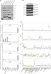
A, Western blot on GST pulldown products using the indicated constructs. SCCRO and its paralogues were probed for components of the neddylation pathway. All SCCRO paralogues bind to neddylation components. B, Western blot for Cul1 neddylation on products from in vitro neddylation assays supplemented with SCCRO or its paralogues. All paralogues promote neddylation except SCCRO3. C, results from quantitative real-time PCR showing tissue-specific expression of SCCRO and its paralogues. SCCRO expression is disproportionately higher in the testis. D, results from analysis of data from the Genotype-Tissue Expression project showing tissue-specific mRNA expression of SCCRO and its paralogues. SCCRO expression is disproportionally higher in testis compared with other paralogues in other tissues. RPKM, fragments per kilobase of exon per million reads mapped.
FIGURE 2.
FIGURE 2.
A, results from Western blot for Cul1 and Cul3 in the lysates from the indicated organs from 6-week-old SCCRO−/− and littermate control wild-type mice showing decreased neddylation in testis from SCCRO−/− mice. B, Western blots showing results from in vitro neddylation assay on lysates from testis from SCCRO−/− mice supplemented with SCCRO or SCCRO paralogues. SCCRO and SCCRO2 can rescue neddylation defects in SCCRO−/− mice. The addition of SCCRO-D241N, a neddylation-dead mutant of SCCRO, to the lysates served as a control for these experiments. C, live cell fluorescence of HeLa cells transfected with GFP-tagged SCCRO or its paralogues showing pan-cellular localization of SCCRO and SCCRO2, membrane localization of SCCRO3, and nuclear localization of SCCRO4 and SCCRO5. D, photomicrographs of eye imaginal discs from flies engineered to express HA- or Myc-tagged dSCCRO or its paralogues. Shown are the results of immunostaining using antibodies against HA- and Myc (first column), cellular markers (Elav (nuclear) and Dlg (membrane; middle column), and merged images (last column). Subcellular localization of SCCRO orthologues is preserved in flies.
FIGURE 3.
FIGURE 3.
A, images of glass jars showing upward migration of flies 30 min after flies were shaken to the bottom of the jar. Note the decrease in the migration of dSCCRO mutants (J34) but not dSCCRO3 mutants (J155). The migration defect in dSCCRO mutants could be rescued by re-expression of dSCCRO (j34-Res). B, graph showing results form touch sensitivity test (means ± S.E.). A significant decrease (asterisks indicate p < 0.01) in touch sensitivity was seen in two different dSCCRO mutants (J156 and J34) compared with control wild-type flies; this was rescued by re-expression of dSCCRO (J156-Res and J34-Res). C, graph showing actuarial survival in wild-type (Control) and mutant female flies (n = 20 for each). Survival was decreased in dSCCRO (J34) and dSCCRO3 (J155) mutant flies. Survival was further reduced in dSCCRO and SCCRO3 double mutants (J155/J34). D, graph showing results from breeding female mutants with wild-type males. A significant decrease (the asterisks indicates p < 0.05) in the number of F1 offspring was seen in both dSCCRO (J34) and dSCCRO3 (J155); double mutants (J155/J34) were increased. E, images showing representative same-age third instar larvae from control (wild-type) and dSCCRO3 mutant (J155) flies. Note the smaller size of the dSCCRO3 mutants.
FIGURE 4.
FIGURE 4.
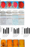
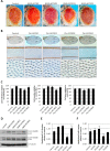
A, representative images showing a magnified view of the eyes of control flies and flies expressing RNAi against the indicated dSCCRO paralogues. There is a decrease in size in flies expressing RNAi under the control of an eye-specific promoter (Ey-GMR-Gal4) against dSCCRO and dSCCRO3 and an increase in those expressing RNAi against dSCCRO4. Eyes from Ey-GMR-Gal4/− flies are shown as a control for these experiments. B, representative images of the whole wing (top row) and a magnified view of the middle wing patch (middle row) and cells from the middle wing patch (bottom row) from flies expressing Ptc>dSCCRO RNAi, Ptc>dSCCRO3 RNAi, or Ptc>dSCCRO4 RNAi, and controls (Ptc-Gal4/−). There is a decrease in the width of the middle wing patch in flies expressing RNAi against dSCCRO and an increase in those expressing RNAi against dSCCRO3 or dSCCRO4. C, bar graph showing the mean L3-L4 distance (left plot), cell number (middle plot), and cell size (right plot) in control vector and RNAi-expressing flies (lines indicate ± S.E.; asterisks indicate p ≤ 0.05). Changes in the middle wing patch are related to an increase in cell number but not cell size in flies expressing RNAi against dSCCRO, an increase in cell size but not cell number in flies expressing RNAi against dSCCRO3, and an increase in cell number but not cell size in flies expressing RNAi against dSCCRO4. D, representative magnified images showing macrochaetae on the notum from control and RNAi-expressing flies as indicated. The circled area in the second panel shows an area shortened and missing macrochaetae in flies expressing RNAi against dSCCRO.
FIGURE 5.
FIGURE 5.
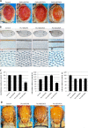
A, representative images showing a magnified view of eyes from flies expressing four copies of the indicated constructs under the control of an eye-specific promoter (Ey-GMR-Gal4). Expression of dSCCRO and dSCCRO3 caused larger and rough eyes, whereas expression of dSCCRO4 caused smaller eyes. Eyes from Ey-GMR-Gal4/− flies are shown as a control for these experiments. B, representative images of the whole wing (top row) and a magnified view of the middle wing patch (middle row), and cells from the middle wing patch (bottom row) from flies expressing Ptc>2x UAS-dSCCRO, Ptc>2x UAS-dSCCRO3, Ptc>1x SCCRO4, or wild-type controls (Ptc-gal4/−). C, bar graph showing mean L3-L4 distance (left plot), cell number (middle plot), and cell size (right plot) in control vector and flies expressing dSCCRO and its paralogues (lines indicate ± S.E.; asterisks indicate p ≤ 0.05). Note that the increase in cell number associated with expression of dSCCRO and dSCCRO3 did not affect the distance between L3-L4, as it also resulted in a decrease in cell size. The decrease in the distance between L3 and L4 in flies expressing dSCCRO4 was due to a decrease in both the total number and size of cells. D, representative magnified images showing macrochaetae on the notum from flies expressing the indicated dSCCRO paralogues or controls. The circled areas show extra macrochaetae in dSCCRO- and dSCCRO3-expressing flies; there were missing macrochaetae in dSCCRO4-expressing flies.
FIGURE 6.
FIGURE 6.
A, representative images showing a magnified view of eyes with clones from flies of the FRT80B control, dSCCRO−/− (J34), wild-type control (w1118), and dSCCRO−/− (J34) with the whole eye clone, FRT42 control, and dSCCRO3−/− (J155). Control flies maintain an equal proportion of both clones (red and white). In contrast, both dSCCRO−/− and dSCCRO3−/− clones (white patch) are smaller than wild-type twin spots (dark red), suggesting that the mutant cells have a defect in proliferation (second panel). Although eye size is normal in dSCCRO−/− somatic clones, it is markedly reduced in dSCCRO−/− whole eye clones (compare with w1118 eye). B, representative fluorescent microscopic images showing Caspase 3 (red) and GFP (green) expression in dSCCRO−/− somatic clones (left) and dSCCRO overexpressing flip out clones (right). Note the presence of apoptotic cells (red) in dSCCRO−/− somatic GFP-negative clones that border SCCRO wild-type GFP-positive (green) clones. In the dSCCRO overexpression clones (right panel); however, apoptosis (red) is primarily present in GFP-negative cells, SCCRO wild-type cells that border SCCRO-overexpressing GFP-positive cells (green). C, fluorescent microscopic images showing Caspase 3 (red) and GFP (green) expression in dSCCRO4 flip out clones. Apoptotic (red) cells were primarily observed in cells overexpressing dSCCRO4 (GFP-positive cells).
FIGURE 7.
FIGURE 7.
A, representative images showing a magnified view of eyes from flies expressing the indicated SCCRO paralogues and selected neddylation-dead mutants alone or in combination under the control of an eye-specific promoter (Ey-GMR-Gal4). GMR-Gal4/− served as control flies for these experiments. Note the smaller eyes in dSCCRO4-overexpressing mutant clones but not dSCCRO-DAD mutant clones. Coexpression of dSCCRO, but not dSCCRO-DAD, rescued the size defect seen with dSCCRO4 expression. Coexpression of dSCCRO3 only partially rescued the size defect seen with dSCCRO4 expression. Coexpression of dSCCRO and dSCCRO3 caused larger, rough eyes. B, representative images of the whole wing (top row) and a magnified view of the middle wing patch (middle row), and cells from the middle wing patch (bottom row) from flies expressing dSCCRO paralogues or mutants and controls. C, bar graph showing mean L3-L4 distance (left plot), cell number (middle plot), and cell size (right plot) in control vector and RNAi-expressing flies (lines indicate ± S.E.; asterisks indicate p ≤ 0.05).
FIGURE 8.
FIGURE 8.
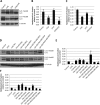
A, Westerns blots for Cul1 and Cul3 on lysates from third instar larval eye discs and brains from genomic mutants of dSCCRO3 (J156), dSCCRO (J155), or both. B, bar graphs of the results from densitometry analyses using ImageJ software showing the ratio of neddylated:nonneddylated Cul1 relative to controls (lines indicate ± S.E.; the asterisk indicates p ≤ 0.05). C, bar graphs of results from densitometry analyses using ImageJ software showing the ratio of neddylated:nonneddylated Cul3 relative to controls (lines indicate ± S.E.; the asterisk indicates p ≤ 0.05). D, Western blots for Cul1 or Cul3 on lysates from third instar larval eye discs and brains from wild-type control, SCCRO deletion mutant (J156), SCCRO deletion mutant expressing genomic rescue construct (J156-Res), and indicated overexpression clones. E and F, bar graphs of the results from densitometry analyses using ImageJ software showing the ratio of neddylated:nonneddylated Cul1 or Cul3 relative to controls (lines indicate ± S.E.; the asterisk indicates p ≤ 0.05).
FIGURE 9.
FIGURE 9.
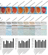
A, representative images showing a magnified view of eyes from flies expressing the indicated human SCCRO paralogues under the control of an eye-specific promoter (Ey-GMR-Gal4) and control flies (Ey-GMR-Gal4/−). B, representative images of the whole wing (top row) and a magnified view of the middle wing patch (middle row) and cells from the middle wing patch (bottom row) from flies expressing one copy of human SCCRO paralogues and control (Ptc-Gal4/−). C, bar graph showing mean L3-L4 distance (left plot), cell number (middle plot), and cell size (right plot) in control vector and RNAi-expressing flies (lines indicate ± S.E.; asterisks indicate p ≤ 0.05). D, Western blot for Cul1 and Cul3 on lysates from eye imaginal discs and brains from flies with overexpression of human SCCRO and its paralogues, as indicated. E and F, bar graphs showing the ratio of neddylated to nonneddylated Cul1 or Cul3 relative to controls (lines indicate ± S.E.; asterisks indicate p ≤ 0.05).

Similar articles

Cited by

References

    1. Hershko A., and Ciechanover A. (1998) The ubiquitin system. Annu. Rev. Biochem. 67, 425–479 - PubMed
    1. Finley D., and Chau V. (1991) Ubiquitination. Annu. Rev. Cell Biol. 7, 25–69 - PubMed
    1. Pickart C. M. (2001) Mechanisms underlying ubiquitination. Annu. Rev. Biochem. 70, 503–533 - PubMed
    1. Berndsen C. E., and Wolberger C. (2014) New insights into ubiquitin E3 ligase mechanism. Nat. Struct. Mol. Biol. 21, 301–307 - PubMed
    1. Osaka F., Saeki M., Katayama S., Aida N., Toh-E A., Kominami K., Toda T., Suzuki T., Chiba T., Tanaka K., and Kato S. (2000) Covalent modifier NEDD8 is essential for SCF ubiquitin-ligase in fission yeast. EMBO J. 19, 3475–3484 - PMC - PubMed

Publication types

Substances