. 2016 Jan 26;14(3):547-559.
doi: 10.1016/j.celrep.2015.12.055. Epub 2016 Jan 7.

The Rag-Ragulator Complex Regulates Lysosome Function and Phagocytic Flux in Microglia

Affiliations

The Rag-Ragulator Complex Regulates Lysosome Function and Phagocytic Flux in Microglia

Kimberle Shen et al. Cell Rep. .

Abstract

Microglia are resident macrophages of the CNS that are essential for phagocytosis of apoptotic neurons and weak synapses during development. We show that RagA and Lamtor4, two components of the Rag-Ragulator complex, are essential regulators of lysosomes in microglia. In zebrafish lacking RagA function, microglia exhibit an expanded lysosomal compartment, but they are unable to properly digest apoptotic neuronal debris. Previous biochemical studies have placed the Rag-Ragulator complex upstream of mTORC1 activation in response to cellular nutrient availability. Nonetheless, RagA and mTOR mutant zebrafish have distinct phenotypes, indicating that the Rag-Ragulator complex has functions independent of mTOR signaling. Our analysis reveals an essential role of the Rag-Ragulator complex in proper lysosome function and phagocytic flux in microglia.


Figures

Figure 1
Figure 1. Mutational analysis demonstrates that rraga and lamtor4 are essential for microglia development
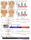
(A–F) Comparison of microglia numbers in rragast77/st77 mutants, lamtor4st99/st99 mutants, and wildtype siblings. (A, B, D, E) Neutral red stained larvae of the indicated genotypes at 5 dpf. Dorsal views, scale bar = 50 μm. (C, F) Quantification of neutral red stained microglia in rraga (C) and lamtor4 (F) mutants at 3 dpf, 4 dpf and 5 dpf. (G–J) Molecular analysis of mutations in rraga. (G) Sequence chromatograms show point mutation in rragast77 mutation. (H) Sequence deleted in rragast110 mutation, showing exon-intron boundary and deleted splice donor. (I) Immunoblot showing pronounced reduction of RagA protein in rragast77/st77 mutants at 5 dpf. Actin is shown as a loading control. (J) Schematic of RagA protein, showing conserved domains and positions of the mutant lesions. (K–M) Molecular analysis of mutations in lamtor4. (K) Sequence chromatograms show point mutation in lamtor4st74 mutation, which changes the ATG initiation codon to ATA. (L) Sequence deleted in lamtor4st99 mutation, showing the reading frame in wildtype and the alteration caused by the 11 bp deletion in the mutation. (M) Schematic of the Lamtor4 protein, showing positions of the mutant lesions. Larvae shown in A, B, D, and E were genotyped by PCR after photography.
Figure 2
Figure 2. rraga acts autonomously at an early stage of microglia development
(A–B) Reduction of microglia in rragast77/st77 mutants detected by imaging the mpeg1:EGFP transgene in living wildtype (A) and mutant (B) larvae at 5dpf. Dorsal views, anterior to the top. (C–H) Analysis of other microglia and macrophage markers reveals reduction of microglia at 3 dpf. Probes for apoe (C, D), slc7a7 (E, F), and mfap4 (G, H) were detected by whole mount in situ hybridization. Boxes in G and H show region magnified in the corresponding insets. Lateral views, anterior to the left. (I) Quantification of neutral red stained microglia in rragast77/st77 mutants after transient expression of the wildtype rraga coding sequence under control of regulatory sequences from mpeg1, cldnk, and huc. Only mpeg1-rraga, which drives expression in macrophages and microglia, significantly rescued microglia in the mutants. Dotted line at 15 and 25 shows weak and strong rescue, respectively. ****, P ≤ 0.0001. All scale bars are 50μm. All larvae shown were genotyped by PCR after photography (A–H) or after visually scoring neutral red phenotypes (I).
Figure 3
Figure 3. Dysregulated lysosomal activity in microglia of rragast77/st77 mutants
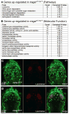
(A–B) Analysis of RNA sequencing data revealed that genes associated with lysosomal activity are significantly upregulated genes in rragast77/st77 mutants at 5 dpf. Using the DAVID Bioinformatics Resources, genes showing at least a 1.5-fold upregulation were classified accord (A) Kegg Pathway terms and (B) molecular functions (GOTERM_MF_FAT). (C–D) Visualization of Lysotracker Red and Tg(mpeg1:EGFP) in wildtype (C, n = 4) and rragast77/st77 mutants (D, n = 4) reveals that lysosomes are increased in microglia in the mutants. Larvae shown in C and D were genotyped by PCR after photography.
Figure 4
Figure 4. Abnormal vacuolar organelles in microglia and uncleared apoptotic neurons in rragast77/st77 mutants
Transmission electron micrographs of the dorsal midbrain of (A) WT, (B, B′) rragast77/st77, and (C) irf8st95/st95 larvae at 4 dpf. Boxed region in B is shown at higher magnification in B′. Microglia in the rragast77/st77 mutants have a striking accumulation of vacuolar organelles that appear to contain undigested material (white arrows). Microglia were absent in the irf8 mutant. In both mutants uncleared corpses of apoptotic neurons are present (white arrowheads). These phenotypes were also evident in mutants stained with acridine orange (Supp. Fig. 3). Toluidene blue stained sections of the same larvae are shown in Supp. Fig 4. Larvae were genotyped by PCR from tail biopsies collected immediately prior to fixation.
Figure 5
Figure 5. Accumulation of undigested neuronal material in microglia of In rragast77/st77 mutants
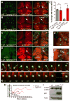
(A–D, G, H) Confocal images of living larvae bearing transgenes that label neurons Tg(nbt:DsRed) and microglia Tg(mpeg1:EGFP). In wildtype (A, C) and rragast77/st77 mutant (B, D) larvae, arrows indicate microglia that contain neuronal material. In wildtype larvae, more microglia contain neuronal material at 4 dpf (A, E) than at 6 dpf (C, C′, E), whereas most microglia in mutants contained neuronal material at both 4 dpf and 6 dpf (B, D, D′, E). Each point in E represents the data from one larva. (F, G) Frames from timelapse movies of WT (F) and rragast77/st77 (G) mutant larvae starting at 4 dpf, with timepoints indicated in minutes. (H) Quantification of the number of neuronal puncta inside microglia in timelapse movies of wildtype and rragast77/st77 mutant larvae. Each line represents a single microglia (WT: n = 4 cells from 3 larvae; mutant: n = 4 cells from 3 larvae). (I) Immunoblot blot of whole-animal protein lysates at 5 dpf shows normal LC3-I to LC3-II conversion, but an accumulation of p62 in rragast77/st77 mutants. Actin is shown as a loading control. All larvae shown in A–H were genotyped by PCR after photography. Larvae analyzed in the immunoblot (I) were genotyped as described in Experimental Procedures.
Figure 6
Figure 6. Distinct phenotypes of mtorxu015/xu015 and rragast77/st77 mutants
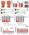
(A–B) Images of living neutral red stained (A) WT and (B) mtor mutant larvae at 5 dpf. Dorsal views, anterior to the top. (C, D) There was no significant reduction of the number of microglia at 5 dpf in mtor mutants or wildtype animals treated with the mTOR inhibitor Torin1. (E) Immunoblots blots of 5 dpf whole animal protein lysates showing reduction of phospho-S6p levels after Torin1 treatment compared to control DMSO treatment. The level of phospho-S6p was similar in rragast77/st77 mutants and their wildtype siblings. Actin is shown as a loading control. (F–I) Lateral views of living larvae at 7 dpf, showing normal morphology of rragast77/st77 mutant (G) and abnormal morphology of mtorxu015/xu015 mutant (H), compared to their wildtype siblings (F and G, respectively). (J, K) Analysis of survival of (J) rragast77/st77 and (K) mtorxu015/xu015 mutants. Progeny of intercross of heterozygotes were raised, and 48 animals were genotyped by PCR for the mutant lesions at the indicated time points. Very few mtorxu015/xu015 mutants survived to 10 dpf, but most rragast77/st77 mutants survived to 12 dpf. For a homozygous viable mutation, 12 mutants would be expected at each time point on average (dotted lines). (L) Quantitative RTPCR of whole-animal RNA samples at 5 dpf showed an increase in expression of some target genes of the lysosomal transcription factor TFEB in rragast77/st77 mutants relative to their wildtype siblings. (M) Similar quantitative RTPCR did not detect any increased expression of the genes analyzed in mtorxu015/xu015 mutants, and many were significantly reduced. Error bars show s.e.m. of samples analyzed in triplicate. Statistical significance determined by a two-tailed t-test (* p<0.05, ** p<0.01, *** p<0.005). All scale bars are 50μm. All larvae analyzed in A–C and E–J were genotyped by PCR. rragast77/st77 mutants analyzed in the immunoblot (E) were genotyped as described in Experimental Procedures. rraga mutants analyzed by RT-PCR were identified by neutral red staining prior to RNA isolation.

Similar articles

Cited by

References

    1. Aguzzi A, Barres BA, Bennett ML. Microglia: Scapegoat, Saboteur, or Something Else? Science. 2013;339(6116):156–161. - PMC - PubMed
    1. Appelqvist H, Wäster P, Kågedal K, Öllinger K. The lysosome: from waste bag to potential therapeutic target. Journal of Molecular Cell Biology. 2013;5(4):214–226. - PubMed
    1. Bae M, Patel N, Xu H, Lee M, Tominaga-Yamanaka K, Nath A, Haughey NJ. Activation of TRPML1 clears intraneuronal Aβ in preclinical models of HIV infection. The Journal of Neuroscience. 2014;34(34):11485–503. - PMC - PubMed
    1. Ballabio A, Gieselmann V. Lysosomal disorders: From storage to cellular damage. Biochimica et Biophysica Acta (BBA) - Molecular Cell Research. 2009;1793(4):684–696. - PubMed
    1. Bar-Peled L, Schweitzer LD, Zoncu R, Sabatini DM. Ragulator is a GEF for the rag GTPases that signal amino acid levels to mTORC1. Cell. 2012;150(6):1196–208. - PMC - PubMed

Publication types

Substances

LinkOut - more resources