

SDPR functions as a metastasis suppressor in breast cancer by promoting apoptosis
- PMID: 26739564
- PMCID: PMC4725521
- DOI: 10.1073/pnas.1514663113
SDPR functions as a metastasis suppressor in breast cancer by promoting apoptosis
Abstract
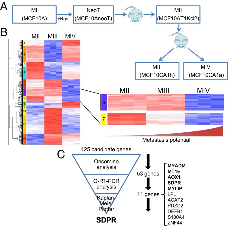
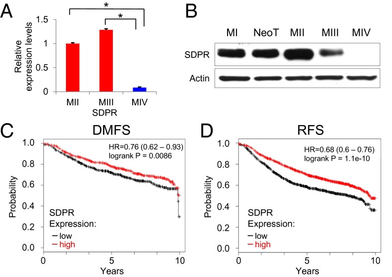
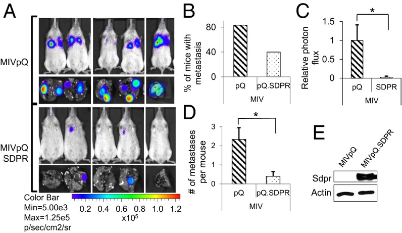
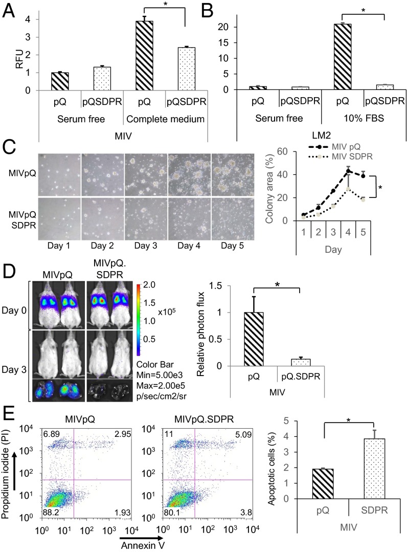
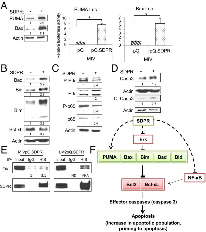
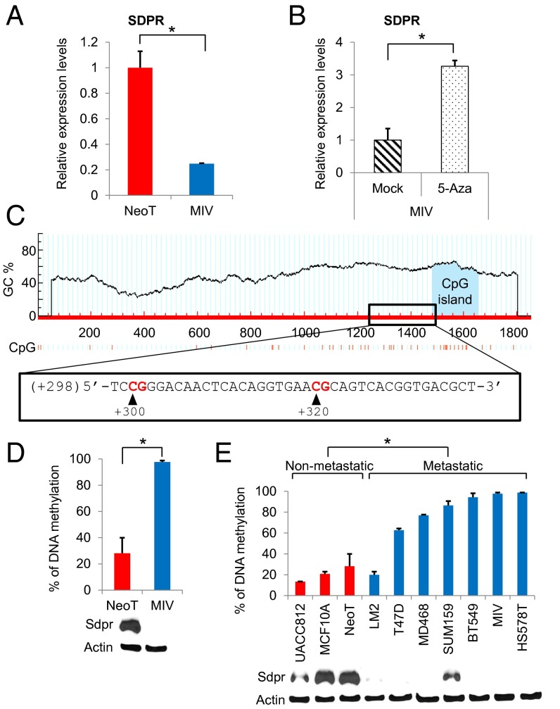
Metastatic dissemination of breast cancer cells represents a significant clinical obstacle to curative therapy. The loss of function of metastasis suppressor genes is a major rate-limiting step in breast cancer progression that prevents the formation of new colonies at distal sites. However, the discovery of new metastasis suppressor genes in breast cancer using genomic efforts has been slow, potentially due to their primary regulation by epigenetic mechanisms. Here, we report the use of model cell lines with the same genetic lineage for the identification of a novel metastasis suppressor gene, serum deprivation response (SDPR), localized to 2q32-33, a region reported to be associated with significant loss of heterozygosity in breast cancer. In silico metaanalysis of publicly available gene expression datasets suggests that the loss of expression of SDPR correlates with significantly reduced distant-metastasis-free and relapse-free survival of breast cancer patients who underwent therapy. Furthermore, we found that stable SDPR overexpression in highly metastatic breast cancer model cell lines inhibited prosurvival pathways, shifted the balance of Bcl-2 family proteins in favor of apoptosis, and decreased migration and intravasation/extravasation potential, with a corresponding drastic suppression of metastatic nodule formation in the lungs of NOD/SCID mice. Moreover, SDPR expression is silenced by promoter DNA methylation, and as such it exemplifies epigenetic regulation of metastatic breast cancer progression. These observations highlight SDPR as a potential prognostic biomarker and a target for future therapeutic applications.
Keywords: SDPR; breast cancer; epigenetics; metastasis; metastasis suppressor.
Conflict of interest statement
The authors declare no conflict of interest.
Figures
Similar articles
-
Targeting IL13Ralpha2 activates STAT6-TP63 pathway to suppress breast cancer lung metastasis.Breast Cancer Res. 2015 Jul 25;17(1):98. doi: 10.1186/s13058-015-0607-y. Breast Cancer Res. 2015. PMID: 26208975 Free PMC article.
-
ITIH5 mediates epigenetic reprogramming of breast cancer cells.Mol Cancer. 2017 Feb 23;16(1):44. doi: 10.1186/s12943-017-0610-2. Mol Cancer. 2017. PMID: 28231808 Free PMC article.
-
Serum deprivation response functions as a tumor suppressor gene in papillary thyroid cancer.Clin Genet. 2019 Nov;96(5):418-428. doi: 10.1111/cge.13609. Epub 2019 Jul 31. Clin Genet. 2019. PMID: 31334828
-
Metastasis suppressor genes: basic biology and potential clinical use.Clin Breast Cancer. 2003 Apr;4(1):51-62. doi: 10.3816/cbc.2003.n.012. Clin Breast Cancer. 2003. PMID: 12744759 Review.
-
Epigenetic regulation of breast cancer metastasis.Cancer Metastasis Rev. 2024 Jun;43(2):597-619. doi: 10.1007/s10555-023-10146-7. Epub 2023 Oct 19. Cancer Metastasis Rev. 2024. PMID: 37857941 Review.
Cited by
-
A six-gene panel to label follicular adenoma, low- and high-risk follicular thyroid carcinoma.Endocr Connect. 2018 Jan;7(1):124-132. doi: 10.1530/EC-17-0261. Endocr Connect. 2018. PMID: 29298844 Free PMC article.
-
Targeting IL13Ralpha2 activates STAT6-TP63 pathway to suppress breast cancer lung metastasis.Breast Cancer Res. 2015 Jul 25;17(1):98. doi: 10.1186/s13058-015-0607-y. Breast Cancer Res. 2015. PMID: 26208975 Free PMC article.
-
Cavin3 Suppresses Breast Cancer Metastasis via Inhibiting AKT Pathway.Front Pharmacol. 2020 Sep 30;11:01228. doi: 10.3389/fphar.2020.01228. eCollection 2020. Front Pharmacol. 2020. PMID: 33101009 Free PMC article.
-
Transcriptomic signature associated with carcinogenesis and aggressiveness of papillary thyroid carcinoma.Theranostics. 2018 Jul 30;8(16):4345-4358. doi: 10.7150/thno.26862. eCollection 2018. Theranostics. 2018. PMID: 30214625 Free PMC article.
-
Downregulation of lncRNA SDPR-AS is associated with poor prognosis in renal cell carcinoma.Onco Targets Ther. 2017 Jun 19;10:3039-3047. doi: 10.2147/OTT.S137641. eCollection 2017. Onco Targets Ther. 2017. PMID: 28790838 Free PMC article.
References
-
- Leone A, Flatow U, VanHoutte K, Steeg PS. Transfection of human nm23-H1 into the human MDA-MB-435 breast carcinoma cell line: Effects on tumor metastatic potential, colonization and enzymatic activity. Oncogene. 1993;8(9):2325–2333. - PubMed
-
- Montagner M, et al. SHARP1 suppresses breast cancer metastasis by promoting degradation of hypoxia-inducible factors. Nature. 2012;487(7407):380–384. - PubMed
Publication types
MeSH terms
Substances
Grants and funding
LinkOut - more resources
Full Text Sources
Other Literature Sources
Medical
Molecular Biology Databases
Miscellaneous
