. 2016 Jan 7:6:18937.
doi: 10.1038/srep18937.

Chi and dLMO function antagonistically on Notch signaling through directly regulation of fng transcription

Affiliations

Chi and dLMO function antagonistically on Notch signaling through directly regulation of fng transcription

Hui Han et al. Sci Rep. .

Abstract

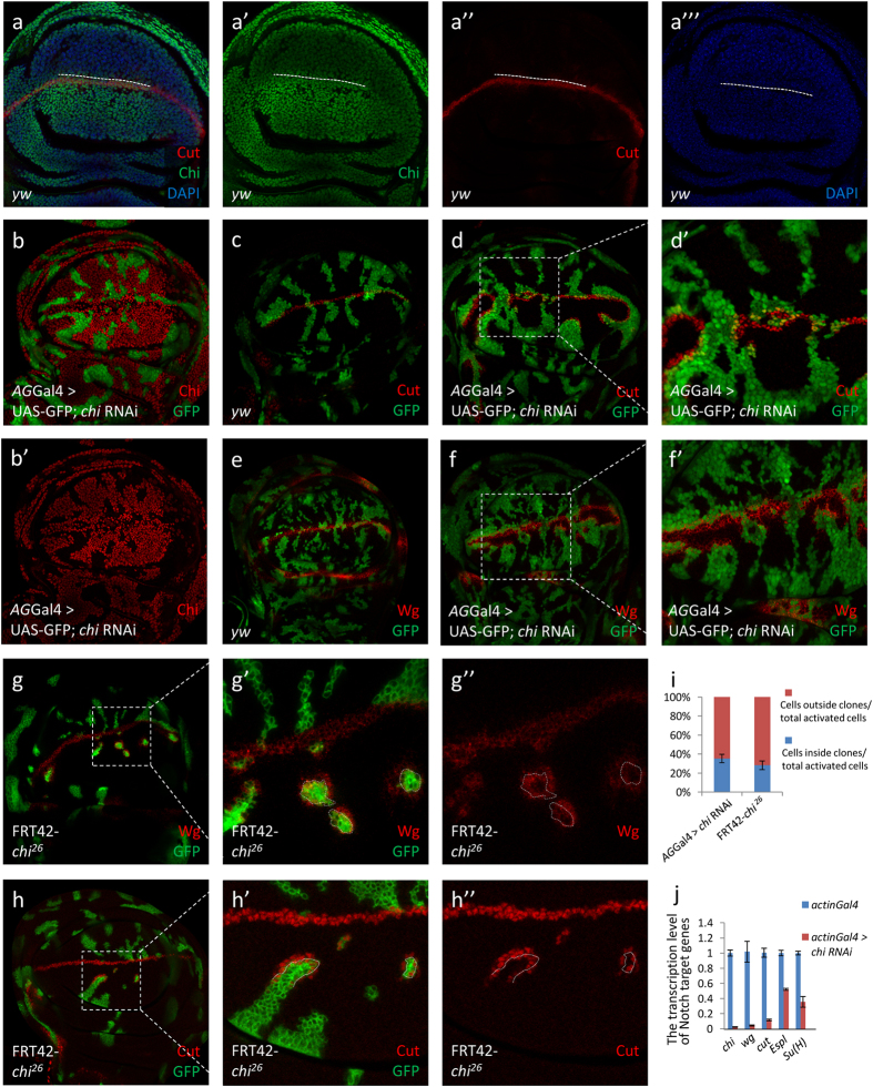
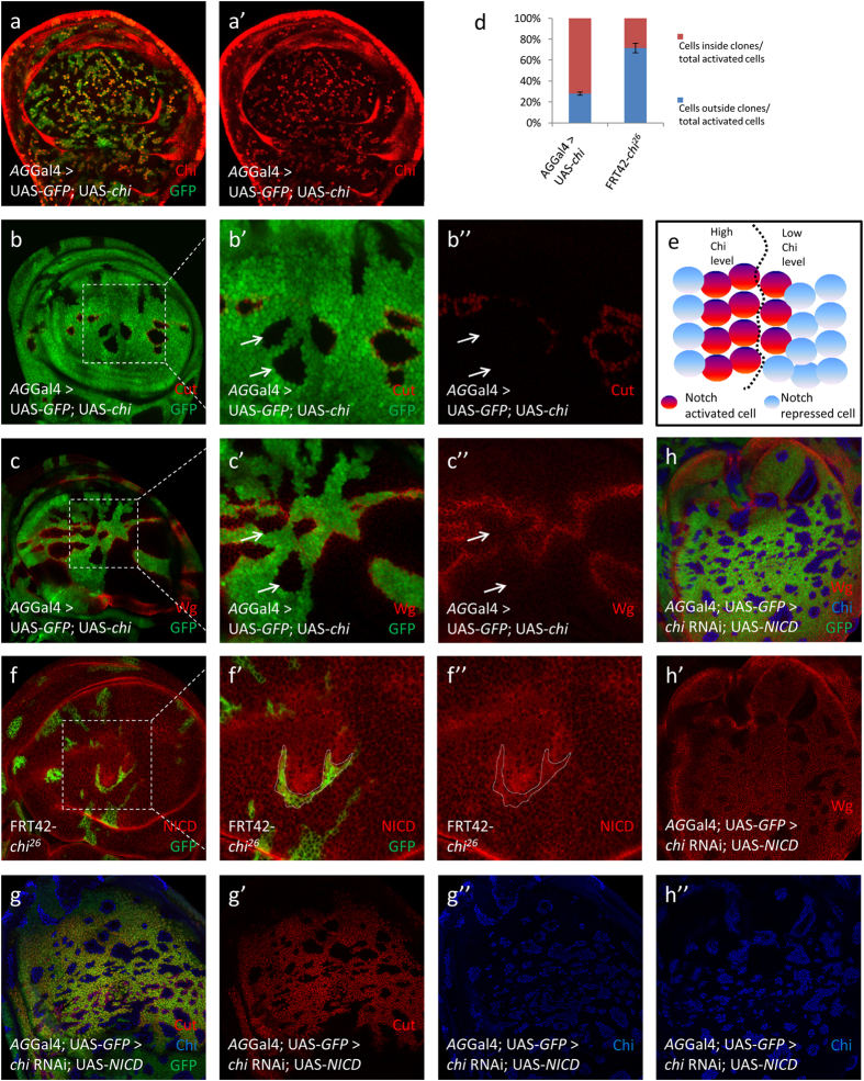
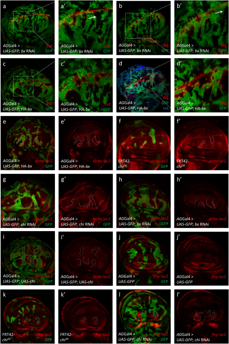
Gene apterous (ap), chip (chi) and beadex (bx) play important roles in the dorsal-ventral compartmentalization in Drosophila wing discs. Meanwhile, Notch signaling is essential to the same process. It has been reported that Ap and Chi function as a tetramer to regulate Notch signaling. At the same time, dLMO (the protein product of gene bx) regulates the activity of Ap by competing its binding with Chi. However, the detailed functions of Chi and dLMO on Notch signaling and the relevant mechanisms remain largely unknown. Here, we report the detailed functions of Chi and dLMO on Notch signaling. Different Chi protein levels in adjacent cells could activate Notch signaling mainly in the cells with higher level of Chi. dLMO could induce antagonistical phenotypes on Notch signaling compared to that induced by Chi. These processes depend on their direct regulation of fringe (fng) transcription.


Figures

Figure 1
Figure 1. Different Chi levels in adjacent cells are essential for Notch signaling activity.
(a–a”’) Immunostaining of Chi (green) in wing discs of early third instar larvae. Cells with high Cut expression (red) locate in the boundary between high and low Chi protein regions. DAPI was used to mark the cell nuclei (blue). (b-b’) Immunostaining of Chi (red) in chi RNAi wing discs. chi RNAi was overexpressed in the clones (green). (c-f’) Immunostaining of Cut (c-d’; red) and Wg (e-f’; red) in yw and chi RNAi wing discs. (d’,f’) are the enlarged pictures of (d,f). Clones are marked with GFP (green). (g-h”) Immunostaining of Wg (g-g”) and Cut (h-h”) in the chi26 wing discs. Wg (g; red) and Cut (h; red) expression are along the D/V boundary. Wg (g-g”) and Cut (h-h”) are activated in the boundary of chi26 clones. (g’) and (h’) are the enlarged pictures of (g,h). Clones are marked with green and circled with dashed line. (i) Quantitative analysis of Cut activated cells around the clones. Shown are the Means ± s.d. (j) qPCR analysis of the Notch target genes. Shown are Means ± s.d. from 3 independent experiments. In every experiment, at least 20 discs were pooled together for analysis.
Figure 2
Figure 2. The Chi overexpression induces Notch signaling activation mainly inside clones.
(a-a’) The overexpression of chi generates the boundary of high and low Chi protein (red) regions. Clones were marked with GFP (green) in all the IF results. (b-c”) Immunostaining of Cut (b-b”; red) and Wg (c-c”; red) in the discs of chi overexpression. Cut and Wg activation did not occur at the clones (arrows) near A/P boundary. (b’)and (c’)are the enlarged pictures of (b,c). (d) Quantitative analysis of Cut activated cells around the clones. Shown are the Means ± s.d. (e) A model for the role of Chi on Notch signaling. Notch signaling could be activated along the boundary of different chi expression regions. Around 70% of activated cells are cells with higher Chi protein level. (f-f”) Immunostaining of NICD (red) in the discs of chi26 discs. Clones are marked with dashed line. (g-h”) Immunostaining of Cut (g; red) and Wg (h; red) in the discs of chi RNAi and UAS-NICD overexpression. Cut and Wg are activated in the clones (green). Chi proteins (blue) are totally lost in the clones.
Figure 3
Figure 3. Chi and dLMO induce opposite functions on Notch signaling.
(a-b’) Immunostaining of Cut (a-a’; red) and Wg (b-b’; red) in the discs of bx RNAi flies. Cut and Wg activation did not occur at the clones (arrows) near A/P boundary. (a’,b’) are the enlarged pictures of (a,b). In all the IF results, clones are marked with GFP (green) and marked with dashed line. (c-d’) Immunostaining of Cut (c-c’; red) and Wg (d-d’; red) in the discs of bx overexpression flies. HA tag was marked with blue (d). (c’,d’) are the enlarged pictures of (c) and (d). (e-i’) Immunostaining of delta-lacZ (red) in the discs of indicated genotype flies. The delta was activated along the clone boundaries in the bx overexpression (e-e’) and chi loss-of-function flies (f-g’). The delta was downregulated inside the whole clones in the bx RNAi (h-h’) and chi overexpression flies (i-i’). (j-l’) Immunostaining of fng-lacZ (red) in the discs of indicated genotype flies. Compared to control (j-j’), fng was downregulated inside the whole clones in the chi26 (k-k’) and chi RNAi flies (l-l’).
Figure 4
Figure 4. Chi and dLMO form a complex to directly regulate fng transcription.
(a-b) Co-IP results of endogenous Chi and HA-dLMO with or without Dpp treatment. The images were cropped. The full-length images were presented in Supplementary Figures 6a–b”. (c) ChIP-qPCR analysis using anti-Chi antibody with chi RNAi as control at fng locus. ChIP signal levels are represented as percentage of input chromatin. Chi could bind to fng locus at three regions. (d) ChIP-qPCR analysis using anti-HA antibody from discs of ms1096Gal4 > HA-bx at fng locus. ChIP signal levels are represented as percentage of input chromatin. dLMO could bind to fng locus at three regions. (e-f’) Immunostaining of fng-lacZ (red) in the discs of indicated genotype flies. Compared to the overexpression of HA-bx (e-e’), the fng downregulation could be partially rescued by the chi overexpression (f-f’). In all IF results, clones are marked with GFP (green) and circled with dashed line. (g-h’) Immunostaining of Cut (g-g’; red) and Wg (h-h’; red) in the discs of indicated genotype flies. Compared to the overexpression of HA-bx alone (Figure 3 c–d’), Cut and Wg activation could be partially rescued by the chi overexpression. Clones are marked with GFP (green). (g’,h’) are the enlarged pictures of (g,h).

Similar articles

Cited by

References

    1. Dexter J. S. The analysis of a case of continuous variation in Drosophila by a study of its linkage relations. Am. Nat. 48, 712–758 (1914).
    1. Morgan T. H. B. & C. B. Sex-Linked Inheritance in Drosophila. PartII, 63–64 (Press of Gibson Brothers 1916).
    1. Mohr O. L. Character Changes Caused by Mutation of an Entire Region of a Chromosome in Drosophila. Genetics 4, 275–282 (1919). - PMC - PubMed
    1. Wharton K. A., Johansen K. M., Xu T. & Artavanis-Tsakonas S. Nucleotide sequence from the neurogenic locus notch implies a gene product that shares homology with proteins containing EGF-like repeats. Cell 43, 567–581 (1985). - PubMed
    1. Bray S. J. Notch signalling: a simple pathway becomes complex. Nat Rev Mol Cell Biol 7, 678–689, 10.1038/nrm2009 (2006). - DOI - PubMed

Publication types

MeSH terms

LinkOut - more resources